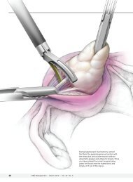
Paralytic Strabismus: Third, Fourth, and Sixth Nerve Palsy
Paralytic Strabismus: Third, Fourth, and Sixth Nerve Palsy
Paralytic Strabismus: Third, Fourth, and Sixth Nerve Palsy

Create successful ePaper yourself
Turn your PDF publications into a flip-book with our unique Google optimized e-Paper software.
806<br />
Prasad & Volpe<br />
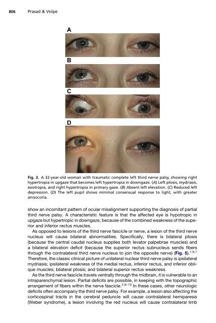
Fig. 3. A 32-year-old woman with traumatic complete left third nerve palsy, showing right<br />
hypertropia in upgaze that becomes left hypertropia in downgaze. (A) Left ptosis, mydriasis,<br />
exotropia, <strong>and</strong> right hypertropia in primary gaze. (B) Absent left elevation. (C) Reduced left<br />
depression. (D) The left pupil shows minimal consensual response to light, with greater<br />
anisocoria.<br />
show an incomitant pattern of ocular misalignment supporting the diagnosis of partial<br />
third nerve palsy. A characteristic feature is that the affected eye is hypotropic in<br />
upgaze but hypertropic in downgaze, because of the combined weakness of the superior<br />
<strong>and</strong> inferior rectus muscles.<br />
As opposed to lesions of the third nerve fascicle or nerve, a lesion of the third nerve<br />
nucleus will cause bilateral abnormalities. Specifically, there is bilateral ptosis<br />
(because the central caudal nucleus supplies both levator palpebrae muscles) <strong>and</strong><br />
a bilateral elevation deficit (because the superior rectus subnucleus sends fibers<br />
through the contralateral third nerve nucleus to join the opposite nerve) (Fig. 5). 1,6,7<br />
Therefore, the classic clinical picture of unilateral nuclear third nerve palsy is ipsilateral<br />
mydriasis; ipsilateral weakness of the medial rectus, inferior rectus, <strong>and</strong> inferior oblique<br />
muscles; bilateral ptosis; <strong>and</strong> bilateral superior rectus weakness.<br />
As the third nerve fascicle travels ventrally through the midbrain, it is vulnerable to an<br />
intraparenchymal lesion. Partial deficits are possible, in keeping with the topographic<br />
arrangement of fibers within the nerve fascicle. 2,8–10 In these cases, other neurologic<br />
deficits often accompany the third nerve palsy. For example, a lesion also affecting the<br />
corticospinal tracts in the cerebral peduncle will cause contralateral hemiparesis<br />
(Weber syndrome), a lesion involving the red nucleus will cause contralateral limb