Paralytic Strabismus: Third, Fourth, and Sixth Nerve Palsy
Paralytic Strabismus: Third, Fourth, and Sixth Nerve Palsy
Paralytic Strabismus: Third, Fourth, and Sixth Nerve Palsy

Create successful ePaper yourself
Turn your PDF publications into a flip-book with our unique Google optimized e-Paper software.
<strong>Paralytic</strong> <strong>Strabismus</strong> 805<br />
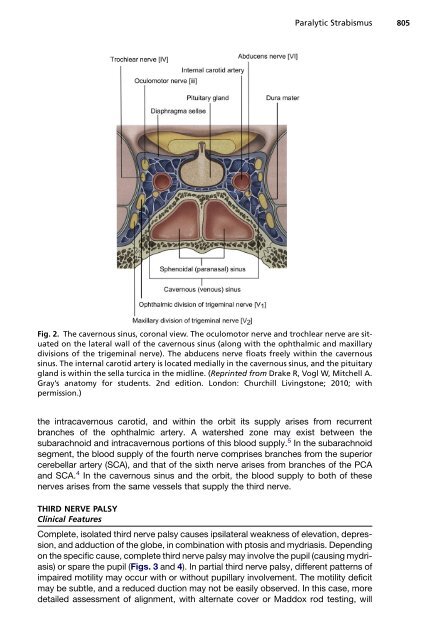
Fig. 2. The cavernous sinus, coronal view. The oculomotor nerve <strong>and</strong> trochlear nerve are situated<br />
on the lateral wall of the cavernous sinus (along with the ophthalmic <strong>and</strong> maxillary<br />
divisions of the trigeminal nerve). The abducens nerve floats freely within the cavernous<br />
sinus. The internal carotid artery is located medially in the cavernous sinus, <strong>and</strong> the pituitary<br />
gl<strong>and</strong> is within the sella turcica in the midline. (Reprinted from Drake R, Vogl W, Mitchell A.<br />
Gray’s anatomy for students. 2nd edition. London: Churchill Livingstone; 2010; with<br />
permission.)<br />
the intracavernous carotid, <strong>and</strong> within the orbit its supply arises from recurrent<br />
branches of the ophthalmic artery. A watershed zone may exist between the<br />
subarachnoid <strong>and</strong> intracavernous portions of this blood supply. 5 In the subarachnoid<br />
segment, the blood supply of the fourth nerve comprises branches from the superior<br />
cerebellar artery (SCA), <strong>and</strong> that of the sixth nerve arises from branches of the PCA<br />
<strong>and</strong> SCA. 4 In the cavernous sinus <strong>and</strong> the orbit, the blood supply to both of these<br />
nerves arises from the same vessels that supply the third nerve.<br />
THIRD NERVE PALSY<br />
Clinical Features<br />
Complete, isolated third nerve palsy causes ipsilateral weakness of elevation, depression,<br />
<strong>and</strong> adduction of the globe, in combination with ptosis <strong>and</strong> mydriasis. Depending<br />
on the specific cause, complete third nerve palsy may involve the pupil (causing mydriasis)<br />
or spare the pupil (Figs. 3 <strong>and</strong> 4). In partial third nerve palsy, different patterns of<br />
impaired motility may occur with or without pupillary involvement. The motility deficit<br />
may be subtle, <strong>and</strong> a reduced duction may not be easily observed. In this case, more<br />
detailed assessment of alignment, with alternate cover or Maddox rod testing, will