Paralytic Strabismus: Third, Fourth, and Sixth Nerve Palsy
Paralytic Strabismus: Third, Fourth, and Sixth Nerve Palsy
Paralytic Strabismus: Third, Fourth, and Sixth Nerve Palsy

Create successful ePaper yourself
Turn your PDF publications into a flip-book with our unique Google optimized e-Paper software.
820<br />
Prasad & Volpe<br />
on the amplitude of deviation <strong>and</strong> the presence of associated features such as inferior<br />
oblique overaction, superior rectus contracture, or superior oblique tendon laxity. 60 In<br />
a patient with less than 10 diopters of deviation, recession of 1 muscle may be sufficient.<br />
If the inferior oblique shows overaction, it should be selected. However, if the<br />
deviation is greater than 15 diopters, then a second muscle should also be recessed.<br />
For this purpose, the contralateral inferior rectus or the ipsilateral superior rectus may<br />
be selected. 60 Unless superior rectus contracture needs to be addressed, recession of<br />
the contralateral inferior rectus may be the superior procedure because it can be performed<br />
with adjustable sutures. 46<br />
The approach outlined earlier can be effective at reducing vertical diplopia. Surgery<br />
directly on the superior oblique should generally be avoided in this situation because it<br />
carries the risk of iatrogenic Brown syndrome (superior oblique tendon sheath insufficiency).<br />
However, with bilateral palsies, or with unilateral palsy causing significant<br />
torsional diplopia, the Harado-Ito procedure may be required, in which the anterior<br />
portion of the superior oblique is advanced toward the lateral rectus.<br />
SIXTH NERVE PALSY<br />
Clinical Features<br />
Weakness of the lateral rectus due to sixth nerve palsy leads to horizontal diplopia,<br />
worse to the affected side <strong>and</strong> at distance. Often, the abnormal duction is easily<br />
observed, but in subtle cases, an incomitant esotropia must be shown by testing<br />
binocular alignment.<br />
Differential Diagnosis<br />
Nuclear sixth nerve palsy affects the ipsilateral sixth nerve as well as the interneurons<br />
destined for the contralateral medial rectus subnucleus. This lesion causes an<br />
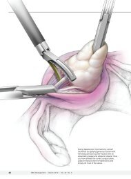
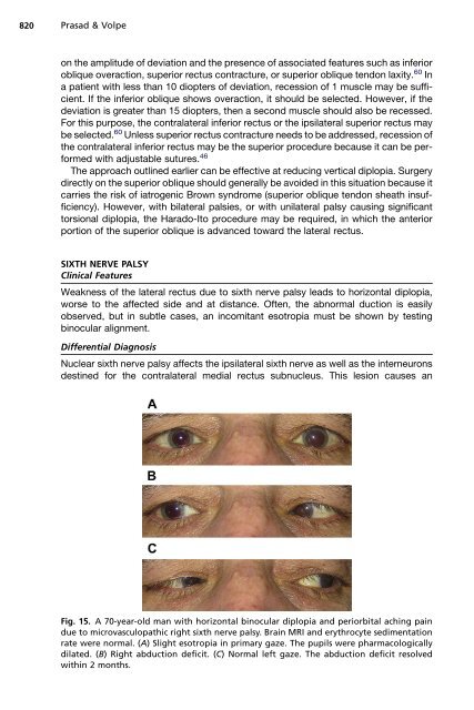
Fig. 15. A 70-year-old man with horizontal binocular diplopia <strong>and</strong> periorbital aching pain<br />
due to microvasculopathic right sixth nerve palsy. Brain MRI <strong>and</strong> erythrocyte sedimentation<br />
rate were normal. (A) Slight esotropia in primary gaze. The pupils were pharmacologically<br />
dilated. (B) Right abduction deficit. (C) Normal left gaze. The abduction deficit resolved<br />
within 2 months.