Paralytic Strabismus: Third, Fourth, and Sixth Nerve Palsy
Paralytic Strabismus: Third, Fourth, and Sixth Nerve Palsy
Paralytic Strabismus: Third, Fourth, and Sixth Nerve Palsy

You also want an ePaper? Increase the reach of your titles
YUMPU automatically turns print PDFs into web optimized ePapers that Google loves.
816<br />
Prasad & Volpe<br />
fundus examination, by assessing the position of the macula with respect to the optic<br />
disc. Excyclotorsion of the hypertropic eye suggests fourth nerve palsy because of<br />
weakened intorsion; in contrast, intorsion of the hypertropic eye occurs in skew deviation,<br />
due to decreased stimulation of the inferior oblique subnucleus.<br />
Assessing cyclotorsion <strong>and</strong> vertical misalignment in the upright <strong>and</strong> supine positions<br />
may be helpful in distinguishing a fourth nerve palsy from a skew deviation. 48<br />
The misalignment remains fairly constant between these positions in fourth nerve<br />
palsy, whereas it is mitigated in the supine position in skew deviation, possibly<br />
because the utricular imbalance that causes a skew deviation becomes reduced. 48<br />
A final clue about the cause of vertical misalignment comes from the fusional amplitude<br />
(the ability to fuse disparate images), which suggests the chronicity of strabismus.<br />
The fusional amplitude is measured by asking the patient to report double<br />
vision while progressively increased prisms are placed over 1 eye. A vertical fusional<br />
capacity greater than 8 to 10 diopters suggests the presence of higher compensatory<br />
mechanisms that occur with long-st<strong>and</strong>ing misalignment, such as a congenital lesion.<br />
Bilateral fourth nerve palsy, which most commonly results from trauma, is characterized<br />
by a unique constellation of findings. 49 Primary position vertical alignment<br />
may be fairly good because of the canceling effect from bilateral palsies. Esotropia<br />
may be present, making the initial diagnosis difficult by potentially suggesting sixth<br />
nerve palsies. However, with careful examination, bilateral fourth nerve palsies are<br />
readily identified. First, hyperdeviation alternates such that it is contralateral to the<br />
direction of gaze <strong>and</strong> ipsilateral to the side of head tilt. Second, there is esotropia<br />
greatest in downgaze (so-called V-pattern esotropia, with >15 prism diopters difference<br />
between upgaze <strong>and</strong> downgaze) because of weakened abduction in depression<br />
(the superior oblique acts as an abductor). <strong>Third</strong>, there is often a large angle of excyclotorsion<br />
(>10 ), accompanied by prominent torsional diplopia. Rarely, bilateral<br />
congenital fourth nerve palsy may occur (Fig. 11).<br />
Identifying fourth nerve palsy in the setting of concomitant third nerve palsy can be<br />
difficult, because the failure of adduction prevents complete testing of superior oblique<br />
function. In this setting, the superior oblique can be evaluated by assessing its<br />
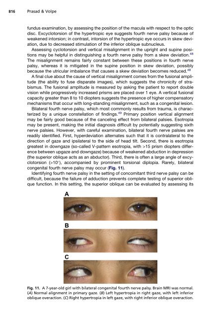
Fig. 11. A 7-year-old girl with bilateral congenital fourth nerve palsy. Brain MRI was normal.<br />
(A) Normal alignment in primary gaze. (B) Left hypertropia in right gaze, with left inferior<br />
oblique overaction. (C) Right hypertropia in left gaze, with right inferior oblique overaction.