Paralytic Strabismus: Third, Fourth, and Sixth Nerve Palsy
Paralytic Strabismus: Third, Fourth, and Sixth Nerve Palsy
Paralytic Strabismus: Third, Fourth, and Sixth Nerve Palsy

You also want an ePaper? Increase the reach of your titles
YUMPU automatically turns print PDFs into web optimized ePapers that Google loves.
804<br />
Prasad & Volpe<br />
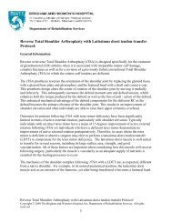
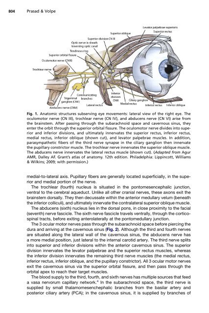
Fig. 1. Anatomic structures subserving eye movements: lateral view of the right eye. The<br />
oculomotor nerve (CN III), trochlear nerve (CN IV), <strong>and</strong> abducens nerve (CN VI) arise from<br />
the brainstem. After passing through the subarachnoid space <strong>and</strong> cavernous sinus, they<br />
enter the orbit through the superior orbital fissure. The oculomotor nerve divides into superior<br />
<strong>and</strong> inferior divisions, <strong>and</strong> ultimately innervates the superior rectus, inferior rectus,<br />
medial rectus, inferior oblique (shown cut), <strong>and</strong> levator palpebrae muscles. In addition,<br />
parasympathetic fibers of the third nerve synapse in the ciliary ganglion then innervate<br />
the pupillary constrictor muscle. The trochlear nerve innervates the superior oblique muscle.<br />
The abducens nerve innervates the lateral rectus muscle (shown cut). (Adapted from Agur<br />
AMR, Dalley AF. Grant’s atlas of anatomy. 12th edition. Philadelphia: Lippincott, Williams<br />
& Wilkins; 2009; with permission.)<br />
medial-to-lateral axis. Pupillary fibers are generally located superficially, in the superior<br />
<strong>and</strong> medial portion of the nerve.<br />
The trochlear (fourth) nucleus is situated in the pontomesencephalic junction,<br />
ventral to the cerebral aqueduct. Unlike all other cranial nerves, these axons exit the<br />
brainstem dorsally. They then decussate within the anterior medullary velum (beneath<br />
the inferior colliculi), <strong>and</strong> ultimately innervate the contralateral superior oblique muscle.<br />
The abducens (sixth) nucleus lies in the dorsal pons, in close proximity to the facial<br />
(seventh) nerve fascicle. The sixth nerve fascicle travels ventrally, through the corticospinal<br />
tracts, before exiting anterolaterally at the pontomedullary junction.<br />
The 3 ocular motor nerves pass through the subarachnoid space before piercing the<br />
dura <strong>and</strong> arriving at the cavernous sinus (Fig. 2). Although the third <strong>and</strong> fourth nerves<br />
are situated along the lateral wall of the cavernous sinus, the abducens nerve has<br />
a more medial position, just lateral to the internal carotid artery. The third nerve splits<br />
into superior <strong>and</strong> inferior divisions within the anterior cavernous sinus. The superior<br />
division innervates the levator palpebrae <strong>and</strong> the superior rectus muscles, whereas<br />
the inferior division innervates the remaining third nerve muscles (the medial rectus,<br />
inferior rectus, inferior oblique, <strong>and</strong> the pupillary constrictor). All 3 ocular motor nerves<br />
exit the cavernous sinus via the superior orbital fissure, <strong>and</strong> then pass through the<br />
orbital apex to reach their target muscles.<br />
The blood supply to the third, fourth, <strong>and</strong> sixth nerves has multiple sources that feed<br />
a vasa nervorum capillary network. 4 In the subarachnoid space, the third nerve is<br />
supplied by small thalamomesenchephalic branches from the basilar artery <strong>and</strong><br />
posterior ciliary artery (PCA); in the cavernous sinus, it is supplied by branches of