here - Health Promotion Agency
here - Health Promotion Agency
here - Health Promotion Agency

Create successful ePaper yourself
Turn your PDF publications into a flip-book with our unique Google optimized e-Paper software.
Antenatal care and antenatal classes<br />
56<br />
L ATER VISITS<br />
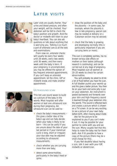
Later visits are usually shorter. Your<br />
urine and blood pressure, and often<br />
your weight, will be checked. Your<br />
abdomen will be felt to check the<br />
baby’s position and growth. And the<br />
doctor or midwife will listen to your<br />
baby’s heartbeat. You can also ask<br />
questions or talk about anything that<br />
is worrying you. Talking is as much<br />
a part of antenatal care as all the tests<br />
and examinations.<br />
From now on, antenatal checks<br />
will usually be every four weeks<br />
until 28 weeks, every two weeks<br />
until 36 weeks, and then every<br />
week until the baby is born. If<br />
your pregnancy is uncomplicated,<br />
you may be offered the option of<br />
less frequent antenatal appointments.<br />
If you can’t keep an antenatal<br />
appointment, let the clinic, GP or<br />
midwife know, and make another<br />
appointment.<br />
ULTRASOUND SCAN<br />
This test uses sound waves to build<br />
up a picture of the baby in the<br />
womb. Most hospitals will offer<br />
women at least one ultrasound scan<br />
during their pregnancy. An<br />
ultrasound scan can be used to:<br />
•<br />
check the baby’s measurements –<br />
this gives a better idea of the<br />
baby’s age and can help decide<br />
when your baby is likely to be<br />
born – this can be useful if you<br />
are unsure about the date of your<br />
last period or if your menstrual<br />
cycle is long, short or irregular;<br />
your due date may be adjusted<br />
according to ultrasound<br />
measurements;<br />
•<br />
check whether you are carrying<br />
more than one baby;<br />
•<br />
detect some abnormalities,<br />
particularly in the baby’s head or<br />
spine;<br />
• show the position of the baby and<br />
the placenta – in some cases, for<br />
example, w<strong>here</strong> the placenta is<br />
low in late pregnancy, special care<br />
may be needed at delivery or a<br />
Caesarean section may be advised;<br />
•<br />
check that the baby is growing<br />
and developing normally (this is<br />
particularly important if you are<br />
carrying twins or more).<br />
The scan is completely painless, has no<br />
known serious side-effects on<br />
mothers or their babies (although<br />
research is continuing), and can be<br />
carried out at any stage of pregnancy.<br />
Most hospitals scan all women at<br />
18–20 weeks to check for certain<br />
abnormalities.<br />
You will probably be asked to drink<br />
a lot of fluid before you have the scan.<br />
A full bladder pushes your womb up<br />
and this gives a better picture. You then<br />
lie on your back and some jelly is put<br />
on your abdomen. An instrument is<br />
passed backwards and forwards over<br />
your skin and high-frequency sound<br />
is beamed through your abdomen into<br />
the womb. The sound is reflected back<br />
and creates a picture which is shown<br />
on a TV screen. It can be very exciting<br />
to see a picture of your own baby<br />
before birth, often moving about inside.<br />
Ask for the picture to be<br />
explained to you if you can’t make<br />
it out. It may be possible for your<br />
partner to come with you and see<br />
the scan. Many couples feel that this<br />
helps to make the baby real for them<br />
both. Ask if it’s possible to have a<br />
copy of the picture (t<strong>here</strong> may be a<br />
small charge for this).<br />
If you feel doubtful about having<br />
a scan, talk it over with your GP,<br />
midwife or obstetrician.