Flexoelectric rotation of polarization in ferroelectric thin films - Nature
Flexoelectric rotation of polarization in ferroelectric thin films - Nature
Flexoelectric rotation of polarization in ferroelectric thin films - Nature

You also want an ePaper? Increase the reach of your titles
YUMPU automatically turns print PDFs into web optimized ePapers that Google loves.
ARTICLES<br />
NATURE MATERIALS DOI: 10.1038/NMAT3141<br />
substrate’s. This broad maximum corresponds to the c doma<strong>in</strong>s,<br />
which have <strong>polarization</strong> out <strong>of</strong> plane and thus a compressed<br />
<strong>in</strong>-plane lattice parameter. As α i <strong>in</strong>creases, a second peak appears<br />
at smaller angles (larger lattice parameters), correspond<strong>in</strong>g<br />
to the a doma<strong>in</strong>s, which have <strong>polarization</strong> <strong>in</strong> plane and<br />
thus horizontal elongation. As the <strong>in</strong>cident angle is further <strong>in</strong>creased,<br />
the two diffraction maxima migrate away from each other,<br />
signall<strong>in</strong>g a grow<strong>in</strong>g difference between the <strong>in</strong>-plane a and c<br />
lattice parameters.<br />
Given that <strong>in</strong>creas<strong>in</strong>g the <strong>in</strong>cidence angle <strong>in</strong>creases the penetration<br />
depth <strong>of</strong> the X-rays 37 , it is tempt<strong>in</strong>g to assign the <strong>in</strong>creased<br />
tetragonality to lattice planes that are deeper 38 . However, it makes<br />
little physical sense that the least cubic parts <strong>of</strong> the film should be<br />
those closest to the substrate. Moreover, all the change <strong>in</strong> our <strong>films</strong><br />
happens at angles above the critical <strong>in</strong>cidence one, for which the<br />
X-ray penetration depth is already larger than the entire thickness <strong>of</strong><br />
the film (as confirmed by the observation <strong>of</strong> the substrate peak). The<br />
observed evolution <strong>of</strong> the diffraction maxima with <strong>in</strong>cidence angle<br />
reflects <strong>in</strong>stead a distribution <strong>of</strong> crystallographic <strong>in</strong>cl<strong>in</strong>ation angles<br />
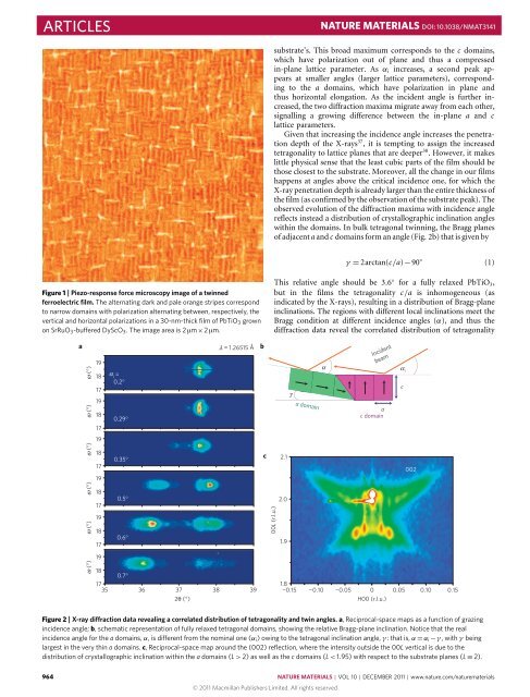
with<strong>in</strong> the doma<strong>in</strong>s. In bulk tetragonal tw<strong>in</strong>n<strong>in</strong>g, the Bragg planes<br />
<strong>of</strong> adjacent a and c doma<strong>in</strong>s form an angle (Fig. 2b) that is given by<br />
γ = 2arctan(c/a)−90 ◦ (1)<br />
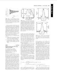
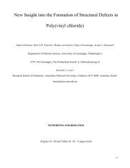
Figure 1 | Piezo-response force microscopy image <strong>of</strong> a tw<strong>in</strong>ned<br />
<strong>ferroelectric</strong> film. The alternat<strong>in</strong>g dark and pale orange stripes correspond<br />
to narrow doma<strong>in</strong>s with <strong>polarization</strong> alternat<strong>in</strong>g between, respectively, the<br />
vertical and horizontal <strong>polarization</strong>s <strong>in</strong> a 30-nm-thick film <strong>of</strong> PbTiO 3 grown<br />
on SrRuO 3 -buffered DyScO 3 . The image area is 2 µm×2 µm.<br />
This relative angle should be 3.6 ◦ for a fully relaxed PbTiO 3 ,<br />
but <strong>in</strong> the <strong>films</strong> the tetragonality c/a is <strong>in</strong>homogeneous (as<br />
<strong>in</strong>dicated by the X-rays), result<strong>in</strong>g <strong>in</strong> a distribution <strong>of</strong> Bragg-plane<br />
<strong>in</strong>cl<strong>in</strong>ations. The regions with different local <strong>in</strong>cl<strong>in</strong>ations meet the<br />
Bragg condition at different <strong>in</strong>cidence angles (α), and thus the<br />
diffraction data reveal the correlated distribution <strong>of</strong> tetragonality<br />
a<br />
ω (°) ω (°) ω (°) ω (°) ω (°) ω (°)<br />
19<br />
18<br />
17<br />
19<br />
18<br />
17<br />
19<br />
18<br />
17<br />
19<br />
18<br />
17<br />
19<br />
18<br />
17<br />
19<br />
α<br />
i =<br />
0.2°<br />
0.29°<br />
0.35°<br />
0.5°<br />
0.6°<br />
λ = 1.26515 Å<br />
18<br />
0.7°<br />
17<br />
35 36 37 38 39<br />
2θ (°)<br />
b<br />
c<br />
00L (r.l.u.)<br />
2.1<br />
2.0<br />
1.9<br />
γ<br />
a doma<strong>in</strong><br />
α<br />
a<br />
c doma<strong>in</strong><br />
Incident<br />
beam<br />
c<br />
i<br />
002<br />
1.8<br />
¬0.15 ¬0.10 ¬0.05 0 0.05 0.10 0.15<br />
H00 (r.l.u.)<br />
α<br />
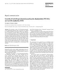
Figure 2 | X-ray diffraction data reveal<strong>in</strong>g a correlated distribution <strong>of</strong> tetragonality and tw<strong>in</strong> angles. a, Reciprocal-space maps as a function <strong>of</strong> graz<strong>in</strong>g<br />
<strong>in</strong>cidence angle; b, schematic representation <strong>of</strong> fully relaxed tetragonal doma<strong>in</strong>s, show<strong>in</strong>g the relative Bragg-plane <strong>in</strong>cl<strong>in</strong>ation. Notice that the real<br />
<strong>in</strong>cidence angle for the a doma<strong>in</strong>s, α, is different from the nom<strong>in</strong>al one (α i ) ow<strong>in</strong>g to the tetragonal <strong>in</strong>cl<strong>in</strong>ation angle, γ : that is, α = α i −γ , with γ be<strong>in</strong>g<br />
largest <strong>in</strong> the very th<strong>in</strong> a doma<strong>in</strong>s. c, Reciprocal-space map around the (002) reflection, where the <strong>in</strong>tensity outside the 00L vertical is due to the<br />
distribution <strong>of</strong> crystallographic <strong>in</strong>cl<strong>in</strong>ation with<strong>in</strong> the a doma<strong>in</strong>s (L > 2) as well as the c doma<strong>in</strong>s (L < 1.95) with respect to the substrate planes (L ≡ 2).<br />
964 NATURE MATERIALS | VOL 10 | DECEMBER 2011 | www.nature.com/naturematerials<br />
© 2011 Macmillan Publishers Limited. All rights reserved