MICRO-STRUCTURE ANALYSIS OF PLANT TISSUES - Lublin
MICRO-STRUCTURE ANALYSIS OF PLANT TISSUES - Lublin
MICRO-STRUCTURE ANALYSIS OF PLANT TISSUES - Lublin

Create successful ePaper yourself
Turn your PDF publications into a flip-book with our unique Google optimized e-Paper software.
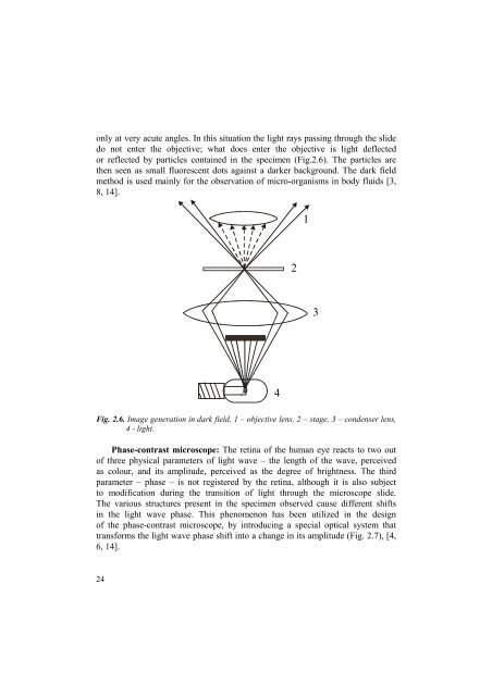
only at very acute angles. In this situation the light rays passing through the slide<br />
do not enter the objective; what does enter the objective is light deflected<br />
or reflected by particles contained in the specimen (Fig.2.6). The particles are<br />
then seen as small fluorescent dots against a darker background. The dark field<br />
method is used mainly for the observation of micro-organisms in body fluids [3,<br />
8, 14].<br />
1<br />
2<br />
3<br />
4<br />
Fig. 2.6. Image generation in dark field, 1 – objective lens, 2 – stage, 3 – condenser lens,<br />
4 - light.<br />
Phase-contrast microscope: The retina of the human eye reacts to two out<br />
of three physical parameters of light wave – the length of the wave, perceived<br />
as colour, and its amplitude, perceived as the degree of brightness. The third<br />
parameter – phase – is not registered by the retina, although it is also subject<br />
to modification during the transition of light through the microscope slide.<br />
The various structures present in the specimen observed cause different shifts<br />
in the light wave phase. This phenomenon has been utilized in the design<br />
of the phase-contrast microscope, by introducing a special optical system that<br />
transforms the light wave phase shift into a change in its amplitude (Fig. 2.7), [4,<br />
6, 14].<br />
24