MICRO-STRUCTURE ANALYSIS OF PLANT TISSUES - Lublin
MICRO-STRUCTURE ANALYSIS OF PLANT TISSUES - Lublin
MICRO-STRUCTURE ANALYSIS OF PLANT TISSUES - Lublin

Create successful ePaper yourself
Turn your PDF publications into a flip-book with our unique Google optimized e-Paper software.
that the image of a structure after the preparation is a true representation<br />
of the structure in undisturbed tissue. It is obvious that any operation on the<br />
tissue, even just a slice through the structure, disturbs the true image<br />
of the tissue. However, we should aim at such a development of the sample<br />
preparation methods that would eliminate that phenomenon.<br />
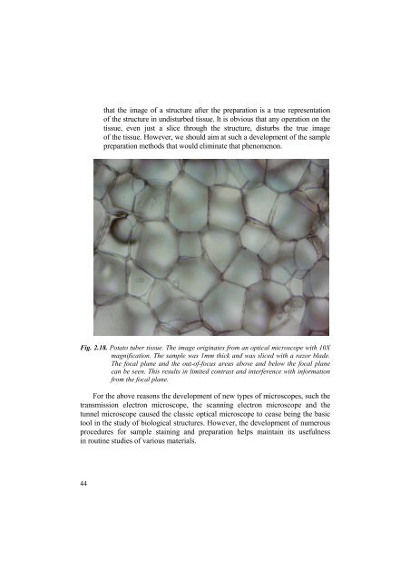
Fig. 2.18. Potato tuber tissue. The image originates from an optical microscope with 10X<br />
magnification. The sample was 1mm thick and was sliced with a razor blade.<br />
The focal plane and the out-of-focus areas above and below the focal plane<br />
can be seen. This results in limited contrast and interference with information<br />
from the focal plane.<br />
For the above reasons the development of new types of microscopes, such the<br />
transmission electron microscope, the scanning electron microscope and the<br />
tunnel microscope caused the classic optical microscope to cease being the basic<br />
tool in the study of biological structures. However, the development of numerous<br />
procedures for sample staining and preparation helps maintain its usefulness<br />
in routine studies of various materials.<br />
44