MICRO-STRUCTURE ANALYSIS OF PLANT TISSUES - Lublin
MICRO-STRUCTURE ANALYSIS OF PLANT TISSUES - Lublin
MICRO-STRUCTURE ANALYSIS OF PLANT TISSUES - Lublin

You also want an ePaper? Increase the reach of your titles
YUMPU automatically turns print PDFs into web optimized ePapers that Google loves.
Studies involving the use of top lighting, on opaque objects, are performed<br />
only in exceptional cases. The structure under study can only be perceived by the<br />
observer when there is a contrast between the structure and its surroundings,<br />
and thus the existence of such a contrast in another condition that has to be<br />
fulfilled in microscopy studies. The last condition is that the object studied must<br />
be prepared in such way that it can be easily placed within the microscope field<br />
of view [1, 2, 3, 6, 7, 12].<br />
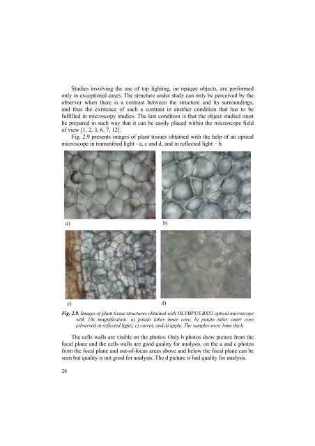
Fig. 2.9 presents images of plant tissues obtained with the help of an optical<br />
microscope in transmitted light - a, c and d, and in reflected light – b.<br />
Fig. 2.9. Images of plant tissue structures obtained with OLYMPUS BX51 optical microscope<br />
with 10x magnification: a) potato tuber inner core, b) potato tuber outer core<br />
(observed in reflected light), c) carrot, and d) apple. The samples were 1mm thick.<br />
The cells walls are visible on the photos. Only b photos show picture from the<br />
focal plane and the cells walls are good quality for analysis, on the a and c photos<br />
from the focal plane and out-of-focus areas above and below the focal plane can be<br />
seen but quality is not good for analysis. The d picture is bad quality for analysis.<br />
28