

Contact lens options for keratoconus - Optometry Today
Contact lens options for keratoconus - Optometry Today
Contact lens options for keratoconus - Optometry Today

Create successful ePaper yourself
Turn your PDF publications into a flip-book with our unique Google optimized e-Paper software.
CET CONTINUING<br />
EDUCATION<br />
& TRAINING<br />
1 FREE CET POINT<br />
Approved <strong>for</strong>: Optometrists 4 Dispensing opticians 4 CLPs 4<br />
OT CET content supports <strong>Optometry</strong> Giving Sight<br />
Having trouble signing in to take an exam?<br />
View CET FAQ Go to www.optometry.co.uk<br />
<strong>Contact</strong> <strong>lens</strong> <strong>options</strong><br />
<strong>for</strong> <strong>keratoconus</strong><br />
Course code: C-15460 O/D/CL<br />
45<br />
Dr. Narendra Kumar, BAMS, PGCR, Member IACLE<br />
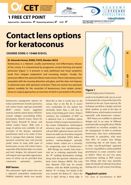
Keratoconus is a bilateral, usually asymmetrical, non-inflammatory disease<br />
of the cornea. It is characterised by progressive corneal thinning and apical<br />
protrusion (Figure 1). It presents in early adulthood and visual symptoms<br />
result from irregular astigmatism and increasing myopia. 1 Usually, the<br />
protrusion affects the axial and inferior nasal cornea. There is decreasing vision<br />
associated with the ensuing distortion and glare, and this does not improve<br />
in advanced cases with spectacle correction. There are several non-spectacle<br />
<strong>options</strong> available <strong>for</strong> the correction of <strong>keratoconus</strong>, from simple contact<br />
<strong>lens</strong>es to surgical approaches, an overview of which is presented in this article.<br />
Keratoconus management <strong>options</strong> <strong>for</strong><br />
today’s practitioners include spectacles,<br />
soft contact <strong>lens</strong>es, rigid gas permeable<br />
fitted flat so that it would stay on the<br />
cornea. Due to the flat fit, it wasn’t<br />
very com<strong>for</strong>table <strong>for</strong> the patient but<br />
(RGP) contact <strong>lens</strong>es, piggyback decidedly better than a soft <strong>lens</strong> in<br />
systems, hybrid contact <strong>lens</strong>es, INTACS,<br />
corneal collagen cross-linking (CXL),<br />
keratoplasty, Rose-K <strong>lens</strong>es, Nissel K2<br />
rigid <strong>lens</strong>es, Kerasoft IC <strong>lens</strong>es, and<br />
scleral contact <strong>lens</strong>es. With such a<br />
range of <strong>options</strong> now available, <strong>for</strong> all<br />
severities of the disease, optometric<br />
practitioners need to be aware of how<br />
best to manage their patients. Indeed,<br />
terms of providing good vision. <strong>Today</strong>’s<br />
variation, the availability of RGP 2 as<br />
a spherical <strong>lens</strong>, is a healthier option<br />
because of better oxygen transmission to<br />
the cornea. Furthermore, technological<br />
advances mean that manufacturing of<br />
soft and RGP spherical <strong>lens</strong>es and toric<br />
<strong>lens</strong>es can make use of modern computer<br />
numerical controlled (CNC) lathes to<br />
spectacle correction may well provide produce custom-designed specialty<br />
useful vision in some mild cases of<br />
<strong>keratoconus</strong>, as may soft contact <strong>lens</strong>es,<br />
especially of the thicker lathe-cut variety<br />
and toric designs, but advanced cases<br />
will require more specialist approaches,<br />
some of which can be provided from<br />
contact <strong>lens</strong>es, which are a necessity<br />
<strong>for</strong> progressing cases of <strong>keratoconus</strong>. 3<br />
RGP fittings are usually done from a<br />
trial set or ordering empirically from<br />
the “K” readings (keratometer reading)<br />
along with the spectacle refraction and<br />
community optometric practice. back vertex distance (BVD). The total<br />
RGP <strong>lens</strong>es<br />
The yesteryears’ rigid contact <strong>lens</strong> was<br />
a spherical polymethyl methacrylate<br />
(PMMA) material, which was usually<br />
<strong>lens</strong> diameter (TD) may be around<br />
9.50mm, with the BOZR matching the<br />
flattest “K” or being “two thirds of the<br />
way towards the flattest K”. Since the<br />
<strong>lens</strong> is rigid, having a sharp edge, it<br />
Figure 1<br />
Outward bulging cornea in <strong>keratoconus</strong><br />
needs to be handled with care so as not<br />
to traumatise the cornea at the time of its<br />
insertion in the eye. Upon removal, the<br />
technique can follow a simple ‘pull and<br />
blink’ method, as used in the days of<br />
PMMA <strong>lens</strong>es, which tends to work well<br />
especially with keratoconic corneas. 4<br />
RGP <strong>lens</strong>es are available in spherical<br />
designs and in an array of different<br />
materials with varying Dk values. Whilst<br />
considered an excellent modality <strong>for</strong><br />
the management of mild to moderate<br />
<strong>keratoconus</strong>, they have un<strong>for</strong>tunately<br />
decreased in popularity, with more<br />
than 90% of contact <strong>lens</strong> fittings<br />
constituting that of soft <strong>lens</strong>es, due<br />
to their ‘easy-fit-and-<strong>for</strong>get’ approach<br />
(even by unqualified or under-qualified<br />
practitioners in some countries such<br />
as India 5 ). There are not very many<br />
practitioners dealing in RGP contact<br />
<strong>lens</strong> fitting, and they may naturally<br />
enjoy the benefit of referrals from those<br />
dealing only in soft contact <strong>lens</strong> fitting.<br />
Piggyback system<br />
For cases of non-tolerance to RGP<br />
25/02/11 CET
CET CONTINUING<br />
EDUCATION<br />
& TRAINING<br />
1 FREE CET POINT<br />
Approved <strong>for</strong>: Optometrists 4 Dispensing opticians 4 CLPs 4<br />
OT CET content supports <strong>Optometry</strong> Giving Sight<br />
Having trouble signing in to take an exam?<br />
View CET FAQ Go to www.optometry.co.uk<br />
46<br />
25/02/11 CET<br />
Figure 2<br />
Hybrid <strong>lens</strong> from SynergEyes<br />
contact <strong>lens</strong>es, it is possible to use a<br />
piggyback system whereby a RGP <strong>lens</strong><br />
is fitted over an underlying soft <strong>lens</strong>;<br />
the latter provides both com<strong>for</strong>t and<br />
stability. A reverse piggyback system<br />
may also be thought of in some cases,<br />
if it results in better vision. Indeed,<br />
in either case, with the availability of<br />
super-Dk silicone hydrogel and RGP<br />
<strong>lens</strong> materials, the use of the piggyback<br />
system has become better in terms of<br />
enhanced oxygen availability to the<br />
cornea and reduced risk of complication.<br />
For <strong>lens</strong> care, using a single soft <strong>lens</strong> care<br />
product usually suffices although in the<br />
case of reverse piggyback, a specific<br />
RGP <strong>lens</strong> cleaner may be advisable.<br />
Piggybacking a RGP <strong>lens</strong> over a soft<br />
<strong>lens</strong> can convert an unsuccessful contact<br />
<strong>lens</strong> fit into a successful one, particularly<br />
in <strong>keratoconus</strong>. Choosing a soft <strong>lens</strong><br />
power of plano to –0.75DS, which has<br />
a front surface nearly parallel to the<br />
back surface, won’t dramatically alter<br />
the shape to which the base curve of the<br />
RGP <strong>lens</strong> is being fitted. In fact, when<br />
commencing the contact <strong>lens</strong> fit, the RGP<br />
<strong>lens</strong> base curve may be selected based<br />
on keratometry values taken over the<br />
soft <strong>lens</strong> worn on the cornea. Sometimes<br />
the soft <strong>lens</strong> power may be manipulated<br />
to alter the surface on which to place<br />
the RGP <strong>lens</strong>. For example, a soft <strong>lens</strong><br />
of power -5.00D when fitted over a very<br />
steep keratoconic cornea provides a<br />
flatter, more stable surface upon which<br />
to place the RGP <strong>lens</strong>. Likewise, a soft<br />
<strong>lens</strong> of power +5.00D will steepen the<br />
fitting surface over an extremely flat<br />
post-Radial Keratotomy (RK) cornea. 6<br />
Hybrid contact <strong>lens</strong><br />
Hybrid contact <strong>lens</strong>es consist of a<br />
high-Dk RGP centre and a soft skirt<br />
(Figure 2); the <strong>for</strong>mer portion provides<br />
good vision whilst the latter provides<br />
stability and com<strong>for</strong>t on the eye. The<br />
SynergEyes KC <strong>lens</strong> is a hybrid design<br />
that has a RGP centre made of Paragon<br />
HDS 100 (Dk 145) material and a<br />
skirt made of 27% water hydrophilic<br />
material. The diameter of the rigid<br />
central portion is 8.4mm and the total<br />
diameter of the <strong>lens</strong> is 14.5mm. The<br />
curvature of the posterior portion of the<br />
RGP is described as a ‘prolate ellipsoid’.<br />
The <strong>lens</strong> tends to remain stable on<br />
the eye of a patient with <strong>keratoconus</strong>,<br />
without decentering on horizontal or<br />
vertical eye movement, since the soft<br />
skirt provides stability by centring<br />
the optics of the <strong>lens</strong> over the pupil.<br />
Rose-K <strong>lens</strong>es<br />
The back surface of the Rose-K contact<br />
<strong>lens</strong> <strong>for</strong> <strong>keratoconus</strong> is a series of<br />
spherical cuts that are well blended;<br />
these are produced by software-guided<br />
computerised lathe. Fitting is done<br />
using a diagnostic set. The first trial<br />
<strong>lens</strong> chosen is 0.2mm steeper than the<br />
average “K” reading. A local anaesthetic<br />
may be used to reduce tearing <strong>for</strong><br />
quicker and accurate fluorescein<br />
Figure 3<br />
Optimum fit immediately after blink<br />
assessment; a minimal amount of<br />
fluorescein should be instilled so as<br />
not to disrupt the tear film and also<br />
so that it doesn’t lead to inaccurate<br />
assessment of the pooling patterns.<br />
First of all, central fit is assessed <strong>for</strong><br />
a light feather touch at the apex of the<br />
cone and the rest of the pattern as close<br />
to an alignment fit as possible (Figure<br />
3). The peripheral fit is assessed next;<br />
the trial <strong>lens</strong>es have a standard edge<br />
lift, but an increased or decreased<br />
edge lift on the same base curve can<br />
be ordered. Lens diameter is assessed<br />
next; the standard diameter is 8.7mm,<br />
but smaller diameters of 8.1mm to<br />
8.3mm work well on steeper corneas,<br />
and a larger diameter may be required<br />
<strong>for</strong> early cones. Assessment of<br />
power is done last by over-refraction<br />
with the contact <strong>lens</strong> in situ. 7<br />
Nissel K2 rigid <strong>lens</strong><br />
Available in a wide range of base<br />
curves, diameters and edge lift<br />
<strong>options</strong>, the Nissel K2 rigid <strong>lens</strong><br />
(Cantor & Nissel Ltd.) is manufactured<br />
from Optimum Extra (Contamac)<br />
as standard, with other materials<br />
available. The total diameter of the <strong>lens</strong><br />
is 8.7mm as standard (other diameters<br />
being 8.1mm, 8.4mm, 9.0mm, and<br />
9.3mm). All trial <strong>lens</strong>es are single use,<br />
removing concerns about disinfection.<br />
Corneal topographers are ideal, but if<br />
a keratometer is used then the average<br />
of the flattest and steepest “K” readings<br />
are calculated, and the first trial <strong>lens</strong><br />
is selected as having a base curve as<br />
close as possible to 0.30mm steeper<br />
than the average “K” value. The aim<br />
is to obtain good visual acuity (VA) in<br />
a <strong>lens</strong> that is com<strong>for</strong>table. The ideal<br />
fit exhibits good centration, having<br />
minimal clearance or light touch at<br />
the area of the cone, combined with<br />
good alignment over the cornea,<br />
with a band of edge clearance
0.50mm to 0.80mm wide.<br />
sclera (Figure 4).<br />
The fitting sets are supplied<br />
The <strong>lens</strong>es trap a<br />
with each base curve having<br />
reservoir of fluid<br />
a standard value of edge lift;<br />
behind<br />
them,<br />
the axial edge lift can be<br />
which<br />
protects<br />
increased or decreased. If<br />
the cornea, and<br />
the <strong>lens</strong> is riding high then<br />
it is advised that you select a<br />
smaller diameter <strong>lens</strong>, and if<br />
the <strong>lens</strong> is riding low then it is<br />
advised that you select a larger<br />
diameter <strong>lens</strong>. Full sphere<br />
/ cylinder over-refraction<br />
should then be conducted<br />
to arrive at the correct<br />
contact <strong>lens</strong> power, taking<br />
account of BVD corrections.<br />
Although the central cornea<br />
in <strong>keratoconus</strong> can indicate<br />
significant corneal toricity,<br />
the peripheral cornea is often<br />
spherical, and the best VA may usually<br />
be achieved with a spherical optic zone. 8<br />
Kerasoft IC <strong>lens</strong>es<br />
The term ‘rigid’ appears shocking to<br />
many would-be contact <strong>lens</strong> wearers<br />
and practitioners alike. Equally, the<br />
notion of a ’soft <strong>lens</strong> <strong>for</strong> <strong>keratoconus</strong>’<br />
comes as a welcome relief to such<br />
persons. The lathe-cut silicone hydrogel<br />
or non-silicone hydrogel high-water<br />
high Dk Kerasoft IC <strong>lens</strong> (Ultravision<br />
Ltd.) comes in base curves from 7.40mm<br />
to 9.40mm, diameters of 14.0mm to<br />
15.5mm, in a front surface asphere or<br />
asphere toric prism ballasted <strong>for</strong>m, with<br />
Figure 4<br />
Scleral <strong>lens</strong> cross section<br />
vision and com<strong>for</strong>t; until now, this was<br />
only possible with RGP <strong>lens</strong>es but can<br />
now be achieved with a silicone hydrogel<br />
material too. Consequently, the <strong>lens</strong><br />
provides com<strong>for</strong>t, stable vision and longer<br />
wearing time even in a dry environment. 9<br />
Scleral contact <strong>lens</strong>es<br />
Large diameter (14.0mm to over<br />
20.0mm) RGP <strong>lens</strong>es that completely<br />
cover the cornea and extend onto the<br />
sclera are known as scleral <strong>lens</strong>es.<br />
They are supported exclusively by the<br />
because of their<br />
size they rarely<br />
dislocate from<br />
the eye. They<br />
mask corneal<br />
irregularity in<br />
<strong>keratoconus</strong> (and<br />
other conditions<br />
like keratoplasty<br />
and post-refractive<br />
surgery), and<br />
are particularly<br />
useful <strong>for</strong> patients<br />
who need visual<br />
correction but are<br />
unable to wear other types of contact<br />
<strong>lens</strong>es. 10 These are so-to-say modern-day<br />
trans<strong>for</strong>mation (in high-Dk material) of<br />
the older-time haptic <strong>lens</strong>es that were<br />
used in 1959. An example of a scleral<br />
<strong>lens</strong> is the Boston <strong>lens</strong>, a proprietory<br />
scleral contact <strong>lens</strong> now known as<br />
PROSE (Prosthetic Replacement of<br />
Ocular Surface Ecosystem) since it is<br />
used as a prosthesis <strong>for</strong> conditions of<br />
corneal ectasia including <strong>keratoconus</strong>. 11<br />
INTACS<br />
INTACS are thin plastic semi-circular<br />
rings that can be inserted into the cornea.<br />
47<br />
25/02/11 CET<br />
periphery <strong>options</strong> of standard, steep,<br />
steep (reverse geometry) and flat. A<br />
wide power range is available (+30.00<br />
DS, cylinder -0.50D to -15.00D and axis<br />
1 o to 180 o ) and fitting is done from a trial<br />
set of <strong>lens</strong>es with standard periphery<br />
and plano power, with over-refraction<br />
conducted. The Kerasoft IC <strong>lens</strong> offers<br />
Sector Management Control (SMC),<br />
which allows individual customisation<br />
of the <strong>lens</strong> periphery to enhance fit,<br />
Figure 5<br />
Intacs corneal rings<br />
Figure 6<br />
Intacs imbedded in the cornea
CET CONTINUING<br />
EDUCATION<br />
& TRAINING<br />
1 FREE CET POINT<br />
4 4 4<br />
Approved <strong>for</strong>: Optometrists Dispensing opticians CLPs<br />
OT CET content supports <strong>Optometry</strong> Giving Sight<br />
Having trouble signing in to take an exam?<br />
View CET FAQ Go to www.optometry.co.uk<br />
48<br />
25/02/11 CET<br />
They were originally used as a<br />
<strong>for</strong>m of refractive surgery <strong>for</strong> the<br />
correction of low myopia but are<br />
now used <strong>for</strong> cases of <strong>keratoconus</strong><br />
(Figures 5 and 6). The surgical<br />
procedure using local anaesthetic<br />
drops and a clamp to hold the<br />
eye still involves the placing of<br />
inserts just beneath the surface,<br />
in the anterior stroma, in the<br />
peripheral cornea. By inserting<br />
the semi-circular rings in this<br />
manner, INTACS can flatten<br />
the cornea, changing the shape<br />
and location of the cone and<br />
eliminating some of the corneal<br />
irregularities in <strong>keratoconus</strong>.<br />
Improvement in vision is<br />
certainly notable, but spectacles<br />
or contact <strong>lens</strong>es may still be needed.<br />
INTACS improve contact <strong>lens</strong><br />
tolerance in patients with <strong>keratoconus</strong>,<br />
as well as improving best-corrected (and<br />
uncorrected) VA, whilst also delaying<br />
the need <strong>for</strong> surgical treatment by<br />
keratoplasty (see later). The peripheral<br />
circumferential corneal elevation<br />
above the ring segment inserts creates<br />
mechanical barriers when fitting rigid<br />
contact <strong>lens</strong>es, thus reducing RGP <strong>lens</strong><br />
sensation; however, semi-scleral contact<br />
<strong>lens</strong>es have no corneal bearing at all and<br />
can work even better. If soft contact <strong>lens</strong>es<br />
are selected instead, there is no additional<br />
problem posed, and if astigmatism has<br />
been reduced and/or becomes more<br />
regular, makes fitting even simpler. 12<br />
Corneal collagen cross-linking<br />
Corneal collagen cross-linking (CXL)<br />
results in stiffening of the corneal stroma<br />
through photochemical cross-linking of<br />
the collagen fibres, resulting in a denser<br />
network of bonds and increased stability<br />
of the cornea. After epithelium ablation,<br />
Riboflavin drops are applied and the<br />
cornea is then exposed to ultraviolet<br />
(UV) light of wavelength 365nm. The<br />
Figure 7<br />
Keratoplasty steps<br />
procedure is per<strong>for</strong>med under local<br />
anaesthesia. After the treatment a<br />
bandage contact <strong>lens</strong> is applied and<br />
a steroid-antibiotic combination<br />
drop is prescribed. Post-operative<br />
contact <strong>lens</strong> fitting becomes more<br />
tolerable and results in stable VA with<br />
minimal long-term corneal stress. 13<br />
Keratoplasty<br />
Perhaps as a last resort, a corneal<br />
transplant is required when the cornea<br />
becomes dangerously thin or when<br />
VA cannot be improved with contact<br />
<strong>lens</strong>es due to steepening or scarring of<br />
the cornea, or due to <strong>lens</strong> intolerance.<br />
During the day-care surgery, the diseased<br />
or injured part of the cornea (termed<br />
the “button”) is removed and replaced<br />
with a clear donor tissue “button”,<br />
and is fixed into place with sutures<br />
(Figure 7). A patch and shield are then<br />
applied to the eye to protect it until the<br />
surface epithelium heals, usually in<br />
1 to 4 days. Post-operative care as per<br />
ophthalmologist’s recommendation is<br />
extremely important, and includes use<br />
of steroid drops; proper care and prompt<br />
attention to any sign of rejection of the<br />
graft will need to be dealt with but<br />
otherwise hopefully the graft<br />
remains clear and healthy. 14<br />
Summary<br />
New Delhi recently witnessed<br />
an excellent educational<br />
session on contact <strong>lens</strong>es <strong>for</strong><br />
<strong>keratoconus</strong> and irregular<br />
cornea with eminent speakers<br />
including Dr. Paul Rose (Rose<br />
K <strong>lens</strong>es), Ken Pullum (Scleral<br />
<strong>lens</strong>es), Lynn White (Kerasoft<br />
IC <strong>lens</strong>es), Jyoti Dave Singh<br />
(Piggyback system) and Monica<br />
Chaudhry (Lens care solutions).<br />
The message that came out loud<br />
and clear was that there is a<br />
plethora of choices from simple<br />
spherical soft and RGP contact <strong>lens</strong>es,<br />
to specialty contact <strong>lens</strong> <strong>options</strong>, that<br />
can be utilised by eye care practitioners<br />
<strong>for</strong> the management of <strong>keratoconus</strong> in<br />
order to provide their patients with<br />
excellent stable vision and com<strong>for</strong>t<br />
be<strong>for</strong>e the need <strong>for</strong> surgical intervention.<br />
About the author<br />
Dr. Narendra Kumar holds a Diploma in<br />
<strong>Optometry</strong> (DROpt), degree in Ayurvedic<br />
Medicine and Surgery (BAMS), the<br />
Certificate in Rehabilitation (PGCR) and<br />
is a member of IACLE. Having served as<br />
Refractionist at Sir Ganga Ram Hospital<br />
<strong>for</strong> 30 years, he now looks after contact<br />
<strong>lens</strong> cases at his ophthalmologist son’s<br />
clinic OphthaCare Eye Centre. He<br />
has been editing the quarterly journal<br />
<strong>Optometry</strong> <strong>Today</strong> (India) since 1970,<br />
is the chairman of the charitable trust<br />
Eye Care India, and the author of<br />
‘Ophthalmic Dispensing Optics’ and<br />
‘Babloo goes <strong>for</strong> an eye test’ books.<br />
References<br />
See http://www.optometry.co.uk/<br />
clinical/index. Click on the article title<br />
and then download “references”
Module questions<br />
PLEASE NOTE There is only one correct answer. All CET is now FREE. Enter online. Please complete online by midnight<br />
on March 28 2011 - You will be unable to submit exams after this date – answers to the module will be published on<br />
www.optometry.co.uk. CET points <strong>for</strong> these exams will be uploaded to Vantage on April 4 2011.<br />
Course code: C-15687 O/D<br />
1. Which neurological stream should visual fields assessment<br />
primarily assess in AD and PD?<br />
a) Parvo-cellular system<br />
b) Konio-cellular system<br />
c) Magno-cellular system<br />
d) None of the above<br />
2. What is the expected “awareness of practitioner” <strong>for</strong> a person with<br />
accommodative flexibility of 10cm fixating a practitioner at 3 metres?<br />
a) Neck<br />
b) Chest<br />
c) Waist<br />
d) Hips<br />
3. What is the likely postural problem if a person with visual midline<br />
shift perceives a fixation rod in front of their right eye when in fact it is<br />
in front of their nose?<br />
a) They have an upright posture<br />
b) They have a turn to their left<br />
c) They have a turn to their right<br />
d) They are leaning backwards<br />
4. The Motion Displacement test may prove useful <strong>for</strong> assessing AD<br />
and PD patients as it assesses:<br />
a) Magno-cellular function<br />
b) Motion coherence processing<br />
c) Parvo-cellular function<br />
d) Static coherence processing<br />
5. Which of the following statements about glaucoma in patients<br />
with AD or PD is FALSE?<br />
a) Patients should have more frequent visual field tests<br />
b) Patients should be assessed with threshold visual field methods<br />
c) They have a greater risk of accelerated visual field loss<br />
d) None of the above<br />
6. Which of the following statements about AD and PD is FALSE?<br />
a) Retinal OCT and photography will be increasingly important in the future<br />
b) Cataract extraction is contraindicated<br />
c) Single vision spectacles should be dispensed instead of varifocals<br />
d) Objective tests may be more appropriate than subjective tests<br />
Course code: C-15460 O/D/CL<br />
1. What is the main characteristic feature of <strong>keratoconus</strong>?<br />
a) It is a bilateral condition<br />
b) It is a non-inflammatory condition<br />
c) It presents with hypermetropia and irregular astigmatism<br />
d) There is central infero-nasal corneal protrusion<br />
2. What is a hybrid contact <strong>lens</strong>?<br />
a) It is a dual material <strong>lens</strong> with a RGP centre and a soft periphery<br />
b) It is a dual material <strong>lens</strong> with a soft centre and a RGP periphery<br />
c) It is a RGP <strong>lens</strong> with plasma treatment <strong>for</strong> enhanced surface wettability and<br />
com<strong>for</strong>t<br />
d) It is a silicone hydrogel material combined with PMMA<br />
3.Which of the following is NOT an appropriate management option<br />
<strong>for</strong> <strong>keratoconus</strong>?<br />
a) RGP contact <strong>lens</strong><br />
b) Corneal collagen cross-linking<br />
c) LASIK<br />
d) All of the above<br />
4. What differentiates a corneal contact <strong>lens</strong> from a scleral contact<br />
<strong>lens</strong>?<br />
a) Corneal <strong>lens</strong>es mask irregularity in <strong>keratoconus</strong> but scleral <strong>lens</strong>es do not<br />
b) Corneal <strong>lens</strong>es have a reservoir of tear fluid behind the <strong>lens</strong> but scleral<br />
<strong>lens</strong>es do not<br />
c) Scleral <strong>lens</strong>es will display central corneal touch but corneal <strong>lens</strong>es will not<br />
d) Scleral <strong>lens</strong>es are supported wholly by the sclera but corneal <strong>lens</strong>es are not<br />
5. After which keratoconic treatment may a contact <strong>lens</strong> correction<br />
still be required?<br />
a) Keratoplasty<br />
b) INTACS<br />
c) Corneal collagen cross-linking<br />
d) All of the above<br />
6. Which of the following is the MOST widely used treatment <strong>for</strong><br />
moderate cases of <strong>keratoconus</strong>?<br />
a) Keratoplasty<br />
b) RGP contact <strong>lens</strong><br />
c) Soft contact <strong>lens</strong><br />
d) Corneal collagen cross-linking<br />
49<br />
25/02/11 CET


