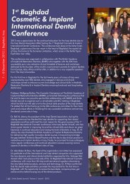
Download e-copy - Smile Dental Journal
Download e-copy - Smile Dental Journal
Download e-copy - Smile Dental Journal

Create successful ePaper yourself
Turn your PDF publications into a flip-book with our unique Google optimized e-Paper software.
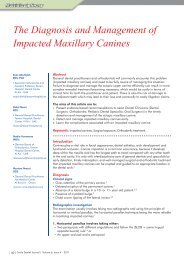
Flow chart of the sequence of management of impacted maxillary canines<br />
Clinical Examination at Age 10<br />
Absence of Buccal Bulge and Presence of Palatal Bulge<br />
Radiographic Localization<br />
Line of Arch<br />
Buccally Ectopic<br />
Palatally Ectopic<br />
Monitor Eruption<br />
of Canine / Space<br />
Creation<br />
Monitor Eruption of Canine<br />
Extract Deciduous Canines / Space Creation<br />
Canine not Erupting in 1 year<br />
Radiographic Localization: Beneficial Change in Position<br />
YES<br />
NO<br />
Canine Buccally or Palatally Impacted<br />
Surgical Exposure & Orthodontic Alignment<br />
NO<br />
Surgical Removal or Auto-Transplantation<br />
NO<br />
No Treatment and continuous Monitoring<br />
2. Vertical parallax involves taking either:<br />
• An upper occlusal (70–75°) and an<br />
orthopantomogram (OPG) or<br />
• A periapical and an orthopantomogram (OPG). 1,4<br />
3. Advanced three-dimensional (3D) imaging<br />
techniques: Cone-beam computed tomography<br />
(CBCT) 1-4<br />
Radiographic features<br />
• Either non-vertical or no resorption of the deciduous<br />
canine root. 3<br />
• Canine crown overlapping adjacent incisor roots. 3<br />
• Resorption of adjacent incisor roots. 2,3<br />
• Magnification of the permanent maxillary canine<br />
crown on a panoramic radiograph. 3<br />
Management<br />
Interceptive treatment by extraction of the<br />
deciduous canine<br />
• The patient should be aged between 10-13 years. 1,4<br />
• Better results are achieved in the absence of<br />
crowding. 1,4<br />
• Position of the canine in the dental arch and in<br />
its relationship to the adjacent lateral decides the<br />
outcome of the interceptive treatment. 1,4<br />
• The need to maintain space (or even create additional<br />
space) requires consideration. 1,4<br />
• If radiographic examination reveals no improvement<br />
in the impacted canine’s position 12 months after<br />
extraction of the deciduous canine, alternative<br />
treatment should be considered. 1,4<br />
Surgical exposure and orthodontic alignment<br />
• The patient should be well motivated and willing to<br />
wear fixed orthodontic appliances. 1,4<br />
• The patient should have good medical and oral health,<br />
and maintain proper oral hygiene. 1,4<br />
• The patient is considered to be unsuitable for<br />
interceptive treatment. 1,4<br />
• The degree of malposition of the impacted canine<br />
should not be too great to preclude orthodontic<br />
alignment. 1,4<br />
• Exposure and alignment of the impacted canine is<br />
indicated in cases when severe root resorption of an<br />
incisor tooth has occurred necessitating its extraction. 4<br />
• The optimal time for surgical exposure and orthodontic<br />
alignment is during adolescence. 4<br />
• Open communication between the orthodontist and<br />
oral surgeon is essential for the choice of appropriate<br />
surgical techniques.<br />
• Careful selection of surgical and orthodontic<br />
techniques is essential for the successful alignment of<br />
impacted maxillary canines.<br />
• Measured orthodontic forces in a favorable direction<br />
leads to successful alignment.<br />
Surgical removal of the palatally impacted<br />
permanent canine<br />
• This treatment option should be considered if the<br />
patient declines active treatment and/or is happy with<br />
their dental appearance. 1,4<br />
• Surgical removal of the impacted canine should be<br />
considered if there is radiographic evidence of early<br />
root resorption of the adjacent incisor. 1,4<br />
<strong>Smile</strong> <strong>Dental</strong> <strong>Journal</strong> | Volume 6, Issue 4 - 2011| 41 |