

PDF Download - Glidewell Dental Labs
PDF Download - Glidewell Dental Labs
PDF Download - Glidewell Dental Labs

Create successful ePaper yourself
Turn your PDF publications into a flip-book with our unique Google optimized e-Paper software.
Spectrophotometry<br />
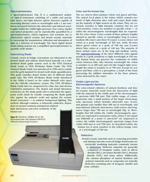
A spectrophotometer (Fig. 5) is a sophisticated analytical<br />
optical instrument consisting of a stable and precise<br />
light source and light detector (photo detector) capable of<br />
measuring discrete colors or wavelengths with a 1 nm resolution<br />
measured between the ultraviolet and infrared wavelengths<br />
within the visible light spectrum. True colors, shades<br />
and optical properties can be reproducibly quantified by a<br />
spectrophotometer, which engineers and scientists use to<br />
characterize optical ceramic and dental ceramic materials<br />
by measuring the transmission, reflection and absorption of<br />
light as a function of wavelength. The latest digital dental<br />
shade-taking systems use a simplified spectrophotometer to<br />
quantify tooth shades.<br />
Determining Shade<br />
Ceramic crown & bridge restorations are fabricated to the<br />
desired shade and esthetic detail based typically on a standardized<br />
shade guide system, such as the VITA Classical<br />
Shade Guide or VITA 3D-Master Shade Guide. The VITA<br />
Classical Shade Guide was introduced in 1956 and is considered<br />
the gold standard for dental tooth shade quantification.<br />
This guide classifies dental shades into 16 different shade<br />
guide tabs. The VITA 3D-Master Shade Guide introduced<br />
in the 1990s is based on the earlier Munsell color space<br />
and the CIELAB colorimetry system. The VITA 3D-Master<br />
Shade Guide tabs are categorized by value, hue and chroma<br />
colorimetry parameters. The dentist and dental laboratory<br />
technician use the shade guide tabs to determine the appropriate<br />
tooth shade by visually comparing the shade guide<br />
tabs against the patient’s tooth and against the ceramic<br />
dental restoration — in ambient background lighting. This<br />
method, although common, is inherently subjective, dependent<br />
on myriad variations arising from ambient<br />
light interactions and color sensitivity of the<br />
human eye.<br />
Figure 5: PerkinElmer Lambda 35 UV/VIS<br />
Spectrophotometer with <strong>Labs</strong>phere RSA-PE-20<br />
Reflectance Spectroscopy Accessory<br />
Color and the Human Eye<br />
The eye detects three primary colors: red, green and blue.<br />
The optical focal plane is the retina, which contains two<br />
kinds of light detection sites: rods and cones. Rods make<br />
up the majority of light sensors in the retina. They are extremely<br />
sensitive to light intensity variation, but are not<br />
color-sensitive. Cones are sensitive to specific colors — or<br />
rather the electromagnetic wavelengths that are responsible<br />
for color vision. Cones consist of three primary lengths<br />
attuned to specific colors or wavelengths: long (L-cones),<br />
medium (M-cones) and short (S-cones). L-cones detect red<br />
colors at a peak wavelength of 565 nm, while M-cones<br />
detect green colors at a peak of 540 nm and S-cones<br />
detect blue colors at a peak of 440 nm. The majority of<br />
these retinal cones are of the long and medium variety.<br />
The superpositioning of the three different color signals<br />
results in a peak sensitivity to light wavelength at 550 nm.<br />
The human brain can perceive the continuum of visible<br />
colors between blue (the shortest wavelength the retina<br />
is sensitive to at 400 nm) and red (the longest light wavelength<br />
the retina is sensitive to at 700 nm). Except for red,<br />
green and blue, color is a perception, a result of the brain<br />
processing the additive intensities of the three primary<br />
colors detected by the cones.<br />
Visible Light and the<br />
Electromagnetic Spectrum<br />
The color-related esthetics of natural dentition and dental<br />
ceramic materials result from the interaction of light<br />
with the material in the visible part of the electromagnetic<br />
spectrum (400–700 nm). This visible range, of course,<br />
represents only a small portion of the larger electromagnetic<br />
spectrum, which includes ultraviolet rays, X-rays,<br />
and gamma rays smaller than 400 nm in wavelength; and<br />
near-infrared, infrared, microwaves, radio waves, and longwaves<br />
larger than 700 nm in wavelength (Fig. 4). Wavelengths<br />
that fall within the visible electromagnetic spectrum<br />
are expressed in nanometer units, where 1 nm equals<br />
one billionth of a meter (1 nm=1x10 -9 m). Expressed in<br />
colors, the visible electromagnetic spectrum ranges from<br />
violet (400 nm) to red (700 nm). As described earlier, the<br />
human eye is most sensitive to the yellow-orange color<br />
at a wavelength of 550 nm.<br />
Dielectrics<br />
<strong>Dental</strong> ceramic materials such as veneering porcelains<br />
and alumina/zirconia crown & bridge frameworks are<br />
electrically insulating materials commonly known<br />
as dielectrics. Dielectric materials are generally<br />
comprised of inorganic oxides, such as silicon<br />
dioxide (SiO 2<br />
), zirconium dioxide (ZrO 2<br />
), titanium<br />
dioxide (TiO 2<br />
) and many others. Additionally,<br />
the calcium phosphate of natural dentition and<br />
polymer materials such as composites are typically<br />
dielectrics consisting also of inorganic nitrides and<br />
some carbides.<br />
52<br />
– www.inclusivemagazine.com –


