- Page 1 and 2:
Basel Seminars in Pathology Postgra
- Page 3 and 4:
UPDATE ON PATHOLOGY STANDARDS FOR B
- Page 5 and 6:
Molecular Pathways for Bladder Canc
- Page 7:
HISTOLOGIC GRADE UROTHELIAL NEOPLAS
- Page 10 and 11:
What’s New in the 2012 Consensus
- Page 12 and 13:
Reporting of Bladder Cancer • Gui
- Page 14 and 15:
• Seen with instrumentation with
- Page 16 and 17:
Squamous Metaplasia • Should be r
- Page 18 and 19:
GRADING OF UROTHELIAL LESIONS • F
- Page 20 and 21:
NORMAL
- Page 22 and 23:
Urothelial Dysplasia • Overall fe
- Page 24 and 25:
Urothelial Dysplasia • The diagno
- Page 28:
ATYPIA OF UNKNOWN SIGNIFICANCE (WHO
- Page 31 and 32:
Grading of Urothelial Carcinoma •
- Page 33 and 34:
Papilloma
- Page 35 and 36:
PUNLMP
- Page 37 and 38:
LOW GRADE
- Page 39 and 40:
HIGHGRADE
- Page 41 and 42:
P A P I L O M A L M P L O W G R A D
- Page 43 and 44:
Courtesy R. Montironi, Italy Exophy
- Page 45 and 46:
Inverted High Grade without invasio
- Page 47 and 48:
Inverted Papilloma
- Page 49 and 50:
Inverted PUNLMP
- Page 51 and 52:
Inverted LG
- Page 53 and 54:
Inverted HG
- Page 55 and 56:
Inverted HG, Non Inv
- Page 57 and 58:
WHO (2004) /ISUP : Prognostic Signi
- Page 59 and 60:
Contributions of WHO (2004) /ISUP
- Page 61 and 62:
Handling grade heterogeneity in bla
- Page 63 and 64:
Grading Papillary Urothelial Neopla
- Page 65:
Papillary Hyperplasia with Cytologi
- Page 68 and 69: Dysplasia with early papillary feat
- Page 70 and 71: Grading Invasive Cancer • Practic
- Page 74 and 75: • 2 types of muscle - awareness i
- Page 76 and 77: Muscularis Mucosae Muscle • Hyper
- Page 78 and 79: M. mucosae muscle patterns Typical
- Page 80 and 81: M. mucosae muscle pattern Hypertrop
- Page 82 and 83: Muscularis Propria • Several term
- Page 84 and 85: Muscle Involved by UCa, Indetermina
- Page 88 and 89: Assessment of pT2 vs. pT3 - cystect
- Page 91 and 92: Microinvasive Urothelial Carcinoma
- Page 93 and 94: Substratification or Substaging of
- Page 95 and 96: Extensive invasion
- Page 97 and 98: Retraction a mimic of vascular-lymp
- Page 99 and 100: Vascular Lymphatic Invasion (LVI)
- Page 101 and 102: 2013 International Society of Urolo
- Page 103 and 104: Lymph node involvement: • Approx.
- Page 105 and 106: THE BEST BLADDER CANCER PATHOLOGY S
- Page 107 and 108: PSEUDONEOPLASTIC MIMICS OF BLADDER
- Page 109 and 110: EMPHYSEMATOUS & BULLOUS CYSTITIS
- Page 113 and 114: AMYLOIDOSIS
- Page 115 and 116: MALAKOPLAKIA
- Page 117: NORMAL PARAGANGLIONIC TISSUE
- Page 124 and 125: CIS
- Page 129 and 130: FLAT LESIONS WITH ATYPIA PROBLEMS A
- Page 132: FLAT LESIONS WITH ATYPIA PROBLEMS A
- Page 135 and 136: RADIATION ATYPIA
- Page 138: Polyoma virus Isolated or clusters
- Page 144: ALL THAT IS PAPILLARY BLADDER IS NO
- Page 150 and 151: BEFORE YOU ASSIGN A WHO (2004)/ISUP
- Page 152 and 153: Broad-based stalk or core • Polyp
- Page 155: Papillary Polypoid cystitis
- Page 159: FIBROEPITHELIAL POLYP
- Page 162: Avulsion
- Page 166 and 167: Epstein, Reuter, Amin Bladder Biops
- Page 168 and 169:
TIGHTLY CLUSTERED UROTHELIUM
- Page 170 and 171:
APPROACH • Be very hesitant to ma
- Page 172 and 173:
NEPHROGENIC ADENOMA Pitfalls: • P
- Page 174 and 175:
NEPHROGENIC ADENOMA
- Page 177:
NEPHROGENIC ADENOMA
- Page 184 and 185:
Fibromyxoid variant
- Page 186 and 187:
FLORID REACTIVE PROLIFERATIONS •
- Page 188:
Nested Variant Von Brunn’s nests
- Page 213 and 214:
Microcystic variant Cystitis cystic
- Page 221 and 222:
FLORID CYSTITIS GLANDULARIS WITH MU
- Page 231:
Pseudosarcomatous stromal reaction
- Page 234 and 235:
PSEUDOSARCOMATOUS MYOFIBROBLASTIC P
- Page 245 and 246:
PSEUDOSARCOMATOUS MYOFIBROBLASTIC P
- Page 247 and 248:
IHC IN BLADDER PATHOLOGY • Provin
- Page 249 and 250:
CA in a cervical LN.
- Page 251 and 252:
UROTHELIAL CARCINOMA (Prim. or Meta
- Page 253:
URINARY BLADDER - IHC •Diagnosis
- Page 256 and 257:
Uroplakin 3
- Page 258 and 259:
Plasmacytoid U.Ca - CK20
- Page 260 and 261:
S100P(commercial) • Nuclear stain
- Page 262 and 263:
• Nuclear staining • lower sens
- Page 264 and 265:
UROTHELIAL CA
- Page 266 and 267:
PARAGANGLIOMA OF THE BLADDER - DIFF
- Page 269 and 270:
CIS REACTIVE ATYPIA
- Page 271 and 272:
IMMUNOHISTOCHEMISTRY IN FLAT LESION
- Page 273 and 274:
NORMAL p53
- Page 275 and 276:
CD-44
- Page 277 and 278:
p53
- Page 279 and 280:
CD-44
- Page 281 and 282:
Reactive- CK20 Reactive- p53 Fig 9C
- Page 283 and 284:
p53
- Page 285 and 286:
CD44
- Page 287 and 288:
p53 Regenerative basal cells vs. cl
- Page 289 and 290:
PAGETOID CIS
- Page 291 and 292:
CK20
- Page 293 and 294:
p53 CK20 CD44
- Page 295 and 296:
Post BCG Reactive Post BCG CIS
- Page 297 and 298:
• 2 types of muscle - awareness i
- Page 299 and 300:
Typical Hypertrophic-Haphazard Hype
- Page 301 and 302:
Muscularis mucosae or muscularis pr
- Page 303 and 304:
IMMUNOHISTOCHEMICAL MARKERS IN BLAD
- Page 306 and 307:
SMA hyperplastic
- Page 308 and 309:
SMA
- Page 310 and 311:
Hypertrophic muscularis mucosae
- Page 312 and 313:
Smoothelin - M. Mucosae
- Page 314 and 315:
SMOOTHELIN IN DESMOPLASIA: NEGATIVE
- Page 316:
TYPES OF MUSCULARIS PROPRIA INVASIO
- Page 319:
Smoothelin SPLAYING/FRACTURING OF M
- Page 322 and 323:
SMA
- Page 324 and 325:
METASTATIC ADENOCARCINOMA TO THE BL
- Page 328 and 329:
CK 7 CK20 CDX2 B-CATENIN
- Page 330 and 331:
New Prostate Lineage Associated Mar
- Page 332 and 333:
?Urothelial Carcinoma vs. ?Prostati
- Page 334 and 335:
UCa GATA3 CK5/6 S100P
- Page 336:
Prostatic adenocarcinoma ERG IHC Ur
- Page 341 and 342:
AMACR
- Page 343 and 344:
Clear cell Ca Neph. adenoma
- Page 345 and 346:
U Ca. with small tubules Nephrogeni
- Page 347 and 348:
NA Pax 2 S100A1 Ki67
- Page 349 and 350:
U Ca Pax 2 S100A1 Ki67
- Page 351 and 352:
Spindle cell lesions of bladder •
- Page 353 and 354:
PSFMT/ PMP Immunohistochemistry •
- Page 355 and 356:
Keratin-AE1/AE3 Desmin
- Page 360 and 361:
AE1/AE3
- Page 364 and 365:
SMA
- Page 366 and 367:
PMP ALK 1 KERATIN AE1/3 SMA
- Page 369 and 370:
Tubulointerstitial lesions Ian Robe
- Page 371 and 372:
Tubules - Normal
- Page 373 and 374:
Tubules - Atrophy Distribution give
- Page 375 and 376:
Interstitial inflammation - tubuloi
- Page 377 and 378:
Tubulointerstitial nephritis The na
- Page 379 and 380:
AKI, ANCA positive, cortex - TIN, g
- Page 381 and 382:
Beware TIN + red cell casts In rena
- Page 383 and 384:
Male 59 years. Acute rise in creati
- Page 385 and 386:
IgG4-related TIN
- Page 387 and 388:
Male 70 years AKI Rigors, breathles
- Page 389 and 390:
Male, 57 years. Acute renal failure
- Page 391 and 392:
Ascending infection (pyelonephritis
- Page 393 and 394:
Ascending infection (pyelonephritis
- Page 395 and 396:
PVN can be seen in native kidneys L
- Page 397 and 398:
Tubulointerstitial infiltrates in t
- Page 399 and 400:
Female, 57 years. Acute renal failu
- Page 401 and 402:
Haemoglobin Myoglobin Diagnosis?
- Page 403 and 404:
Diagnosis?
- Page 405 and 406:
Light chain tubulopathy Typically a
- Page 407 and 408:
Light chain tubulopathy kappa lambd
- Page 409 and 410:
Acute tubular injury in the post mo
- Page 411 and 412:
Acute tubular injury in the post mo
- Page 413 and 414:
Tacrolimus toxicity Diagnosis?
- Page 415 and 416:
Drug-associated tubular injury Mech
- Page 417 and 418:
The End
- Page 419 and 420:
Session plan Renal Pathology for th
- Page 421 and 422:
Tumour nephrectomy specimens Common
- Page 423 and 424:
Glomerular lesions - sclerosis Scle
- Page 425 and 426:
Diabetic nephropathy Earliest chang
- Page 427 and 428:
Diabetic nephropathy: Linear GBM po
- Page 429 and 430:
Diabetic nephropathy: Nodular scler
- Page 431 and 432:
Amyloidosis
- Page 433 and 434:
Amyloidosis
- Page 435 and 436:
Amyloidosis Beware! Congo red may b
- Page 437 and 438:
Immunotactoid Fibrillary GN
- Page 439 and 440:
Light chain deposition disease
- Page 441 and 442:
Light chain deposition disease
- Page 443 and 444:
Idiopathic FSGS Known cause of podo
- Page 445 and 446:
Focal segmental glomerulosclerosis
- Page 447 and 448:
Glomerular tip lesion A manifestati
- Page 449 and 450:
Glomerular lesions: proliferation M
- Page 451 and 452:
IgA nephropathy Commonest glomerulo
- Page 453 and 454:
Mesangial deposits have a character
- Page 455 and 456:
Classification of IgA nephropathy O
- Page 457 and 458:
MPGN, type I • Haematuria, protei
- Page 459 and 460:
C3 glomerulopathy = Isolated C3 dep
- Page 461 and 462:
Haematuria (90%), nephrotic syndrom
- Page 463 and 464:
Dense deposit disease
- Page 465 and 466:
Crescentic glomerulonephritis patte
- Page 467 and 468:
Arteritis (approx. 20%) Renal vascu
- Page 469 and 470:
Anti-GBM disease • Autoantibodies
- Page 471 and 472:
Anti-GBM disease Linear IgG in glom
- Page 473 and 474:
Male 46 years. Suffered from diabet
- Page 475 and 476:
Male 46 years. Suffered from diabet
- Page 477 and 478:
The End
- Page 479 and 480:
What to do? • Download Kidney Qui
- Page 481 and 482:
What to do? • Participants will s
- Page 483 and 484:
PathoPic A
- Page 485 and 486:
PathoPic C
- Page 489 and 490:
What is your main diagnosis? 1. chr
- Page 491 and 492:
What is your main diagnosis? 1. art
- Page 493 and 494:
What is your main diagnosis? 1. mal
- Page 495 and 496:
Solutions (I.S. Roberts)
- Page 497 and 498:
B PathoPic Dg: crescentic glomerulo
- Page 499 and 500:
D PathoPic Dg: no significant patho
- Page 501 and 502:
Dg: amyloidosis F
- Page 503 and 504:
Patterns of scarring Granular subca
- Page 505 and 506:
Patterns of scarring Granular subca
- Page 507 and 508:
Deep pitted cortical scars: Pattern
- Page 509 and 510:
Patterns of scarring Deep pitted co
- Page 511 and 512:
Patterns of scarring Segmental tran
- Page 513 and 514:
Malignant hypertension A cause of a
- Page 515 and 516:
Malignant hypertension A cause of a
- Page 517 and 518:
Malignant hypertension A cause of a
- Page 519 and 520:
Malignant hypertension A cause of a
- Page 521 and 522:
Set 2
- Page 523 and 524:
PathoPic H
- Page 527 and 528:
PathoPic L
- Page 529 and 530:
What is your main diagnosis? 1. col
- Page 531 and 532:
What is your main diagnosis? 1. nec
- Page 533 and 534:
What is your main diagnosis? 1. Ran
- Page 535 and 536:
Descripition Involvement of cortex
- Page 537 and 538:
Description enlargement of the kidn
- Page 539 and 540:
Differential diagnosis Acute uric a
- Page 541 and 542:
Differences between megalocytic IN
- Page 543 and 544:
Comments to papillary necrosis
- Page 545 and 546:
Prevalence of Lesions of the Renal
- Page 547 and 548:
Pathogenetic types of papillary nec
- Page 549 and 550:
Types of papillary necrosis: Extend
- Page 551 and 552:
Phenacetin kidney Keep in mind: bla
- Page 553 and 554:
Phenacetin kidney Note bone formati
- Page 555 and 556:
Phenacetin kidney Capillary scleros
- Page 557 and 558:
Yellow papillary necrosis Think of
- Page 559 and 560:
Histological examples of papillary
- Page 561 and 562:
Typical examples of papillary necro
- Page 563 and 564:
Typical examples of papillary necro
- Page 565 and 566:
Common Challenges in Kidney Cancer
- Page 567 and 568:
ISUP Consensus Conference Vancouver
- Page 570 and 571:
Image-guided biopsy. Shah et al.: H
- Page 572 and 573:
Can we confidently diagnose renal o
- Page 574 and 575:
„Translocation“ Type of Renal C
- Page 576:
Xp11 Translocation Carcinoma in Adu
- Page 579 and 580:
Clear cell-papillary RCC, sporadic
- Page 581 and 582:
Should CCPRCC/CCTPRCC be Recognized
- Page 583:
Do you think CCPRCC/CCTPRCC and RAT
- Page 586 and 587:
DIFFERENTIAL DIAGNOSIS OF EOSINOPHI
- Page 588 and 589:
What percent of a tumor needs to be
- Page 590 and 591:
Assess prognosis in epithelioid AML
- Page 593 and 594:
Mixed Epithelial and Stromal Tumor
- Page 595 and 596:
Are Cystic Nephroma and Mixed Epith
- Page 597 and 598:
Do you consider CN and MEST variati
- Page 599 and 600:
Tubulocystic RCC
- Page 601 and 602:
Challenges for Pathologists Diagnos
- Page 603 and 604:
Sporadic Renal Cell Carcinoma Moch
- Page 606:
Definition Adenoma • Tumours with
- Page 609 and 610:
Survival (%) Type and Prognosis of
- Page 611 and 612:
How do you subtype papillary renal
- Page 613 and 614:
Hereditary Leiomyomatosis and RCC S
- Page 617 and 618:
Should HLRCC be Recognized as a Dis
- Page 619 and 620:
Grading of Chromophobe RCC Amin MA,
- Page 621 and 622:
How should we grade papillary RCC?
- Page 624 and 625:
Unlike the Skywalker family, once t
- Page 626 and 627:
Survival (%) Prognostic Relevance o
- Page 628 and 629:
If a Tumor Shows Sarcomatoid Morpho
- Page 630 and 631:
Tumor Staging Bonsib SM. Renal lymp
- Page 632 and 633:
For staging purposes, do you consid
- Page 634 and 635:
Should the component of necrosis be
- Page 636 and 637:
Challenges for Pathologists Diagnos
- Page 638 and 639:
A proposed marker-based strategy de
- Page 640 and 641:
VHL and tumor development Frew and
- Page 643 and 644:
Randomised studies in the first lin
- Page 645 and 646:
J Urology 180, pp. 860-866, 2008
- Page 647 and 648:
Frequenz VHL Mutation in clear cell
- Page 649 and 650:
Thank you
- Page 652 and 653:
Potential of Needle Biopsy • Diag
- Page 657 and 658:
Hereditary Renal Cancer Syndromes
- Page 660 and 661:
How should HOCT be recognized? 1. S
- Page 662 and 663:
Novel renal tumor types with clear
- Page 664 and 665:
Renal Tumors with Clear Cytoplasm A
- Page 666 and 667:
CA IX and Tumor Development Mutatio
- Page 668 and 669:
Montani et al.: Am J Surg Path, May
- Page 670 and 671:
Is loss of VHL sufficient to cause
- Page 672 and 673:
Inside the primary cilium Receptors
- Page 674 and 675:
a b c d e f Montani et al.: Am J Su
- Page 676 and 677:
Additional mutation in VHL-/- cells
- Page 678 and 679:
Is it Acceptable for MC-RCC to have
- Page 680 and 681:
Renal Cyst in ADPKD Ki-67
- Page 682 and 683:
Novel renal tumor types with clear
- Page 684 and 685:
TFE3-Proteine-Expression
- Page 686 and 687:
• 14/28 pts: Stage 4 • 11/13 pt
- Page 689 and 690:
Translocation Carcinomas (MiTF/TFE
- Page 691:
When should TFE3 and TFEB analysis
- Page 694:
Clear cell-papillary RCC, sporadic
- Page 698 and 699:
Differential Diagnosis of Cystic Re
- Page 701 and 702:
Are Cystic Nephroma and Mixed Epith
- Page 703 and 704:
smaller cysts with phyllodes glands
- Page 705 and 706:
Immunohistochemistry MTSC (%) Papil
- Page 707 and 708:
Which IHC marker do you think is th
- Page 709 and 710:
PAX2 expression pattern in 16 spora
- Page 711 and 712:
PAX2 expression and overall surviva
- Page 715 and 716:
Tumors with Spindle Cell Morphology
- Page 717:
Small Blue Round Cell Tumors of Kid
- Page 720 and 721:
CD-99
- Page 724:
CD 99 Ewing-Sarcoma/PNET
- Page 728 and 729:
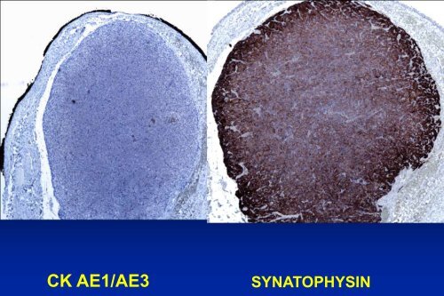
Synaptophysin
- Page 731:
The future has arrived 82
- Page 734:
VHL and Deep Sequencing • several
- Page 737 and 738:
Loss of PBRM1 expression is correla
- Page 739 and 740:
Cell Surface Capturing Proteomics:
- Page 741:
Conclusions •Renal mass biopsy -