Annual General Meeting of the Irish Thoracic Society - IJMS | Irish ...
Annual General Meeting of the Irish Thoracic Society - IJMS | Irish ...
Annual General Meeting of the Irish Thoracic Society - IJMS | Irish ...
Create successful ePaper yourself
Turn your PDF publications into a flip-book with our unique Google optimized e-Paper software.
31<br />
1<br />
SESSION THREE ONE<br />
ANNUAL MEETING OF THE IRISH THORACIC SOCIETY • 11 - 12 November 2005 • WESTWOOD HOUSE HOTEL, GALWAY<br />
ANNUAL MEETING OF THE IRISH THORACIC SOCIETY • 11 - 12 November 2005 • WESTWOOD HOUSE HOTEL, GALWAY<br />
3<br />
SESSION<br />
SESSION THREE ONE<br />
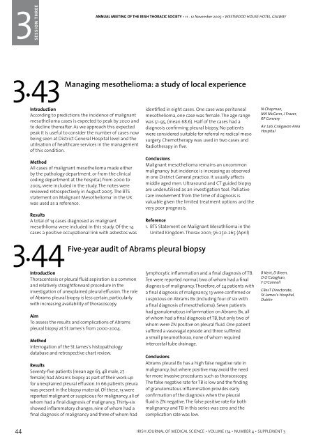
3.43<br />
Managing meso<strong>the</strong>lioma: a study <strong>of</strong> local experience<br />
3.45<br />
Single surgeons audit <strong>of</strong> carinal resections<br />
Introduction<br />
According to predictions <strong>the</strong> incidence <strong>of</strong> malignant<br />
meso<strong>the</strong>lioma cases is expected to peak by 2020 and<br />
to decline <strong>the</strong>reafter. As we approach this expected<br />
peak it is useful to consider <strong>the</strong> number <strong>of</strong> cases now<br />
being seen at District <strong>General</strong> Hospital level and <strong>the</strong><br />
utilisation <strong>of</strong> healthcare services in <strong>the</strong> management<br />
<strong>of</strong> this condition.<br />
Method<br />
All cases <strong>of</strong> malignant meso<strong>the</strong>lioma made ei<strong>the</strong>r<br />
by <strong>the</strong> pathology department, or from <strong>the</strong> clinical<br />
coding department at <strong>the</strong> hospital, from 2000 to<br />
2005, were included in <strong>the</strong> study. The notes were<br />
reviewed retrospectively in August 2005. The BTS<br />
statement on Malignant Meso<strong>the</strong>lioma 1 in <strong>the</strong> UK<br />
was used as a reference.<br />
Results<br />
A total <strong>of</strong> 14 cases diagnosed as malignant<br />
mesothlioma were included in this study. Of <strong>the</strong> 14<br />
cases a positive occupational link with asbestos was<br />
3.44<br />
identified in eight cases. One case was peritoneal<br />
meso<strong>the</strong>lioma, one case was female. The age range<br />
was 51-95, (mean 68.6). Half <strong>of</strong> <strong>the</strong> cases had a<br />
diagnosis confirming pleural biopsy. No patients<br />
were considered suitable for referral re radical meso<br />
surgery. Chemo<strong>the</strong>rapy was used in two cases and<br />
Radio<strong>the</strong>rapy in five.<br />
Conclusions<br />
Malignant meso<strong>the</strong>lioma remains an uncommon<br />
malignancy but incidence is increasing as observed<br />
in one District <strong>General</strong> practice. It usually affects<br />
middle aged men. Ultrasound and CT guided biopsy<br />
are underutilised as an investigation tool. Palliative<br />
care involvement from <strong>the</strong> time <strong>of</strong> diagnosis is<br />
valuable given <strong>the</strong> limited treatment options and <strong>the</strong><br />
very poor prognosis.<br />
Reference<br />
1. BTS Statement on Malignant Mesothlioma in <strong>the</strong><br />
United Kingdom. Thorax 2001; 56:250-265 (April)<br />
Five-year audit <strong>of</strong> Abrams pleural biopsy<br />
Introduction<br />
Thoracentesis or pleural fluid aspiration is a common<br />
and relatively straightforward procedure in <strong>the</strong><br />
investigation <strong>of</strong> unexplained pleural effusion. The role<br />
<strong>of</strong> Abrams pleural biopsy is less certain, particularly<br />
with increasing availability <strong>of</strong> thoracoscopy.<br />
Aim<br />
To assess <strong>the</strong> results and complications <strong>of</strong> Abrams<br />
pleural biopsy at St James’s from 2000-2004.<br />
Method<br />
Interrogation <strong>of</strong> <strong>the</strong> St James’s histopathology<br />
database and retrospective chart review.<br />
Results<br />
Seventy-five patients (mean age 63, 48 male, 27<br />
female) had Abrams biopsy as part <strong>of</strong> <strong>the</strong>ir work-up<br />
for unexplained pleural effusion. In 66 patients pleura<br />
was present in <strong>the</strong> biopsy material. Of <strong>the</strong>se, 13 were<br />
reported malignant or suspicious for malignancy, all <strong>of</strong><br />
whom had a final diagnosis <strong>of</strong> malignancy. Thirty-six<br />
showed inflammatory changes, nine <strong>of</strong> whom had a<br />
final diagnosis <strong>of</strong> malignancy and three <strong>of</strong> whom had<br />
lymphocytic inflammation and a final diagnosis <strong>of</strong> TB.<br />
Ten were reported normal, two <strong>of</strong> whom had a final<br />
diagnosis <strong>of</strong> malignancy. Therefore, <strong>of</strong> 24 patients with<br />
a final diagnosis <strong>of</strong> malignancy, 13 were confirmed or<br />
suspicious on Abrams Bx (including four <strong>of</strong> six with<br />
a final diagnosis <strong>of</strong> meso<strong>the</strong>lioma). Seven patients<br />
had granulomatous inflammation on Abrams Bx, all<br />
<strong>of</strong> whom had a final diagnosis <strong>of</strong> TB, but only two <strong>of</strong><br />
whom were ZN positive on pleural fluid. One patient<br />
suffered a vasovagal episode and three suffered<br />
a small pneumothorax, none <strong>of</strong> whom required<br />
intercostal tube drainage.<br />
Conclusions<br />
Abrams pleural Bx has a high false negative rate in<br />
malignancy, but where positive may avoid <strong>the</strong> need<br />
for more invasive procedures such as thoracoscopy.<br />
The false negative rate for TB is low and <strong>the</strong> finding<br />
<strong>of</strong> granulomatous inflammation provides early<br />
confirmation <strong>of</strong> <strong>the</strong> diagnosis when <strong>the</strong> pleural<br />
fluid is ZN negative. The false positive rate for both<br />
malignancy and TB in this series was zero and <strong>the</strong><br />
complication rate was low.<br />
N Chapman,<br />
MA McCann, J Frazer,<br />
RP Convery<br />
Air Lab, Craigavon Area<br />
Hospital<br />
B Kent, D Breen,<br />
D O’Calaghan,<br />
F O’Connell<br />
CResT Directorate,<br />
St James’s Hospital,<br />
Dublin<br />
Carinal resection is one <strong>of</strong> <strong>the</strong> most complicated<br />
procedures in tracheo-bronchial surgeries. We<br />
present our experience <strong>of</strong> carinal resections<br />
with histological analysis, operative techniques,<br />
complications and long-term survival.<br />
Since 2001 we have performed six carinal resections<br />
in St. James’s Hospital. Indications for surgery<br />
included Non Small Cell Carcinoma (NSCCa). The<br />
length extended from one to five tracheal rings.<br />
The operative approach was right postero-lateral<br />
thoracotomy in all <strong>the</strong> cases.<br />
3.46<br />
One out <strong>of</strong> <strong>the</strong> six patients died post-operatively due<br />
to infection. As quoted in <strong>the</strong> literature mortality and<br />
<strong>the</strong> incidence <strong>of</strong> complications were significantly<br />
correlated to length <strong>of</strong> resection and preoperative<br />
patients functional status.<br />
The feasibility <strong>of</strong> carinal resection is limited by<br />
<strong>the</strong> patient’s pulmonary function status and <strong>the</strong><br />
anatomical extension <strong>of</strong> <strong>the</strong> tumour growth.<br />
Thorough selection <strong>of</strong> patients may improve<br />
immediate and long-term results.<br />
Malignant chest wall tumours and tumours invading<br />
<strong>the</strong> chest wall: surgical techniques and results<br />
Primary chest wall tumours and lung cancer<br />
invading chest wall are <strong>the</strong> most common diseases<br />
indicating chest wall resection and reconstruction.<br />
We evaluated patients who underwent chest wall<br />
resection and reconstruction for primary chest wall<br />
tumours and lung cancer invading chest wall.<br />
Fourteen patients underwent chest wall resection<br />
and reconstruction for primary chest wall tumours<br />
and local invasion <strong>of</strong> lung cancer year between<br />
February 2001 to December 2004. Wide chest wall<br />
resection was performed and reconstruction was<br />
done by sandwich method with Composite Marlex<br />
mesh and Methyl-Metacryalate. In <strong>the</strong> majority <strong>of</strong><br />
3.47<br />
cases it was possible to approximate <strong>the</strong> s<strong>of</strong>t tissues<br />
over <strong>the</strong> reconstruction. However in a small number<br />
<strong>of</strong> cases Plastic surgical colleagues helped in closing<br />
<strong>the</strong> defect.<br />
There was no operative or postoperative mortality.<br />
We looked at <strong>the</strong> mean length <strong>of</strong> in-hospital stay,<br />
histological types, Morbidity and Long-term survival.<br />
Wide excision reduces <strong>the</strong> chances <strong>of</strong> local<br />
recurrence. Simultaneous skeletal reconstruction <strong>of</strong><br />
<strong>the</strong> chest wall prevents postoperative complications<br />
and restores <strong>the</strong> respiratory dynamics by avoiding<br />
paradoxical or harmful movements.<br />
Role <strong>of</strong> vascular reconstruction in <strong>the</strong> resection <strong>of</strong><br />
thoracic malignancy<br />
Since <strong>the</strong> revised staging <strong>of</strong> lung cancer in 1997 1 , local<br />
invasion <strong>of</strong> chest wall or vascular structures does not<br />
preclude curative lung resection.<br />
Vascular reconstruction is an important adjunct to<br />
resection <strong>of</strong> intra thoracic malignancy This includes<br />
primary lung tumours invading major vessels in<br />
<strong>the</strong> chest such as Superior Venacava and head and<br />
neck vessels as well as invasive thymomas which<br />
normally involves <strong>the</strong> superior venacava and <strong>the</strong><br />
brachiocephalic vessels, in addition a small group <strong>of</strong><br />
lung cancers are respectable with <strong>the</strong> exception <strong>of</strong><br />
involvement <strong>of</strong> <strong>the</strong> left atrium and again a portion <strong>of</strong><br />
left atrium may be excised to give a clear margin.<br />
We present a group <strong>of</strong> patients who had resection<br />
and reconstruction <strong>of</strong> a variety <strong>of</strong> vascular structures,<br />
including Superior Venacava, Brachiocephalic vein,<br />
Subclavian artery, Left Atrium and Pulmonary artery.<br />
Despite <strong>the</strong> technically complex nature <strong>of</strong> <strong>the</strong>se<br />
operations we believe that <strong>the</strong>y are justified if <strong>the</strong><br />
thoracic malignancy is totally resectable o<strong>the</strong>rwise.<br />
Reference<br />
1. Adapted from Mountain, CF, Chest 1997; 111:1710<br />
K Doddakula, T Akbar,<br />
M El Siddig, A Raza,<br />
V Young<br />
Dept <strong>of</strong> Cardiothoracic<br />
Surgery, St James’s<br />
Hospital, Dublin<br />
K Doddakula,<br />
W Ahmed, T Akbar,<br />
I Chong, V Young<br />
Dept <strong>of</strong> Cardiothoracic<br />
Surgery, St James’s<br />
Hospital, Dublin<br />
K Doddakula,<br />
W Ahmed, T Akbar,<br />
I Chong, V Young<br />
Dept <strong>of</strong> Cardiothoracic<br />
Surgery, St James’s<br />
Hospital, Dublin<br />
44 IRISH JOURNAL OF MEDICAL SCIENCE • VOLUME 174 • NUMBER 4 • SUPPLEMENT 3<br />
IRISH JOURNAL OF MEDICAL SCIENCE • VOLUME 174 • NUMBER 4 • SUPPLEMENT 3 45


