Annual General Meeting of the Irish Thoracic Society - IJMS | Irish ...
Annual General Meeting of the Irish Thoracic Society - IJMS | Irish ...
Annual General Meeting of the Irish Thoracic Society - IJMS | Irish ...
Create successful ePaper yourself
Turn your PDF publications into a flip-book with our unique Google optimized e-Paper software.
41<br />
1<br />
SESSION FOUR ONE<br />
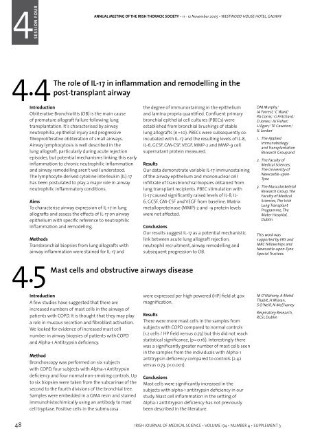
ANNUAL MEETING OF THE IRISH THORACIC SOCIETY • 11 - 12 November 2005 • WESTWOOD HOUSE HOTEL, GALWAY<br />
ANNUAL MEETING OF THE IRISH THORACIC SOCIETY • 11 - 12 November 2005 • WESTWOOD HOUSE HOTEL, GALWAY<br />
SESSION<br />
4SESSION FOUR ONE<br />
4.4 4.6 The role <strong>of</strong> IL-17 in inflammation and remodelling in <strong>the</strong><br />
post-transplant airway<br />
4.5 Mast cells and obstructive airways disease 4.7<br />
Introduction<br />
Obliterative Bronchiolitis (OB) is <strong>the</strong> main cause<br />
<strong>of</strong> premature allograft failure following lung<br />
transplantation. It’s characterised by airway<br />
neutrophilia, epi<strong>the</strong>lial injury and progressive<br />
fibroproliferative obliteration <strong>of</strong> small airways.<br />
Airway lymphocytosis is well described in <strong>the</strong><br />
lung allograft, particularly during acute rejection<br />
episodes, but potential mechanisms linking this early<br />
inflammation to chronic neutrophilic inflammation<br />
and airway remodelling aren’t well understood.<br />
The lymphocyte-derived cytokine interleukin (IL)-17<br />
has been postulated to play a major role in airway<br />
neutrophilic inflammatory conditions.<br />
Aims<br />
To characterise airway expression <strong>of</strong> IL-17 in lung<br />
allografts and assess <strong>the</strong> effects <strong>of</strong> IL-17 on airway<br />
epi<strong>the</strong>lium with specific reference to neutrophilic<br />
inflammation and remodelling.<br />
Methods<br />
Transbronchial biopsies from lung allografts with<br />
airway inflammation were stained for IL-17 and<br />
<strong>the</strong> degree <strong>of</strong> immunostaining in <strong>the</strong> epi<strong>the</strong>lium<br />
and lamina propria quantified. Confluent primary<br />
bronchial epi<strong>the</strong>lial cell cultures (PBECs) were<br />
established from bronchial brushings <strong>of</strong> stable<br />
lung allografts (n=10). PBECs were subsequently coincubated<br />
with IL-17 and <strong>the</strong> resulting levels <strong>of</strong> IL-8,<br />
IL-6, GCSF, GM-CSF, VEGF, MMP-2 and MMP-9 cell<br />
supernatant protein measured.<br />
Conclusions<br />
Our results suggest IL-17 as a potential mechanistic<br />
link between acute lung allograft rejection,<br />
neutrophil recruitment, airway remodelling and<br />
subsequent progression to OB.<br />
DM Murphy, 1<br />
IA Forrest, 1 C Ward, 1<br />
PA Corris, 1 G Pritchard, 1<br />
D Jones, 2 AJ Fisher, 1<br />
JJ Egan, 3 TE Cawston, 2<br />
JL Lordan 1<br />
1. The Applied<br />
Immunobiology<br />
and Transplantation<br />
Research Group and<br />
Results<br />
Our data demonstrate variable IL-17 immunostaining<br />
<strong>of</strong> <strong>the</strong> airway epi<strong>the</strong>lium and mononuclear cell<br />
infiltrate <strong>of</strong> transbronchial biopsies obtained from<br />
lung transplant recipients. PBEC stimulation with<br />
IL-17 caused significantly raised levels <strong>of</strong> IL-8, IL-<br />
6, GCSF, GM-CSF and VEGF from baseline. Matrix<br />
metalloproteinase (MMP)-2 and -9 protein levels<br />
were not affected.<br />
2. The Faculty <strong>of</strong><br />
Medical Sciences,<br />
The University <strong>of</strong><br />
Newcastle-upon-<br />
Tyne<br />
3. The Musculoskeletal<br />
Research Group, The<br />
Faculty <strong>of</strong> Medical<br />
Sciences, The <strong>Irish</strong><br />
Lung Transplant<br />
Programme, The<br />
Mater Hospital,<br />
Dublin<br />
This work was<br />
supported by ERS and<br />
MRC fellowships and<br />
Newcastle-upon-Tyne<br />
Special Trustees.<br />
Viral infection and cytokine responses in exacerbations<br />
<strong>of</strong> COPD<br />
Introduction<br />
In COPD exacerbations can be triggered by viral<br />
and bacterial infections. Real-time PCR allows<br />
faster and more accurate detection <strong>of</strong> respiratory<br />
viral infections and assessment <strong>of</strong> cytokine mRNA<br />
responses. We studied patients with COPD when<br />
stable and during exacerbations to determine <strong>the</strong><br />
role <strong>of</strong> viral infection and airway inflammation.<br />
Method<br />
Patients were recruited with 24 hours <strong>of</strong> hospital<br />
admission for a COPD exacerbation (AECOPD).<br />
Patients with stable COPD who had no change in<br />
treatment or symptoms over <strong>the</strong> previous eight<br />
weeks were also recruited (SCOPD). Sputum, nasal<br />
and throat swab specimens were obtained and<br />
screened for respiratory viruses using nested PCR.<br />
Real-time PCR was used to measure mRNA cytokine<br />
responses. Ribosomal RNA (18s rRNA) was employed<br />
as a housekeeping gene. Supernatant from sputum<br />
specimens was analysed for corresponding protein<br />
concentrations <strong>of</strong> cytokines using <strong>the</strong> BioPlex system.<br />
All mRNA levels are adjusted for 18s rRNA and<br />
expressed as copies /ml <strong>of</strong> sputum (c/ml).<br />
Results<br />
One hundred and thirty six patients were recruited<br />
during an acute exacerbation and 68 when stable.<br />
Mean (±SD) age <strong>of</strong> each group was 70 yrs (± 9) and<br />
66 yrs (± 9) respectively. FEV 1<br />
(% predicted) was as<br />
follows; AECOPD 0.84 ± 0.5 (39%), SCOPD 1.00 ± 0.5<br />
(48%). Smoking history AECOPD 48 ± 39 and SCOPD<br />
42 ± 26 pack years. A respiratory virus was detected<br />
in 50 (37%) AECOPD and in 8 (12%) SCOPD patients (p<br />
< 0.001). TNF-α mRNA levels were higher in AECOPD<br />
(1392 c/ml) than SCOPD (139 c/ml) patients. IL-6 and<br />
IL-8 mRNA was also significantly increased during<br />
AECOPD (219c/ml, 38429 c/ml,) in comparison to<br />
stable patients (27 c/ml, 4071 c/ml p < 0.005). GRO-α<br />
and GM-CSF mRNA levels were increased during<br />
exacerbations (p < 0.005). Expression <strong>of</strong> growth<br />
factors TGF-β1 and TGF-β2 were also increased in<br />
AECOPD. Corresponding protein concentrations <strong>of</strong><br />
IL-6, TNF-α, Interferon-γ and IL-4 were raised during<br />
exacerbations (p < 0.005). TGF-β1 and TNF-α mRNA<br />
levels were higher during those exacerbations in<br />
which a virus was not isolated.<br />
Conclusions<br />
Patients with AECOPD have an increased airway<br />
inflammatory response when compared to <strong>the</strong> stable<br />
patients. The expression <strong>of</strong> some cytokine targets are<br />
modulated by respiratory viral infection.<br />
Increased levels <strong>of</strong> cysteinyl and metalloproteases<br />
mediated by a TH 1 response in COPD patients<br />
TE McManus, 1,2<br />
AM Marley, 1<br />
F De Courcey, 3<br />
N Baxter, 1 SN Christie, 2<br />
HJ O’Neill, 2 JS Elborn, 3,4<br />
PV Coyle, 2 JC Kidney 1<br />
1. Dept <strong>of</strong> Respiratory<br />
Medicine, Mater<br />
Hospital<br />
2. Regional Virus<br />
Laboratory, Royal<br />
Victoria Hospital<br />
3. Respiratory Research<br />
Group, Institute <strong>of</strong><br />
Clinical Science<br />
4. Dept <strong>of</strong> Respiratory<br />
Medicine, Belfast<br />
City Hospital<br />
Introduction<br />
A few studies have suggested that <strong>the</strong>re are<br />
increased numbers <strong>of</strong> mast cells in <strong>the</strong> airways <strong>of</strong><br />
patients with COPD. It is thought that <strong>the</strong>y may play<br />
a role in mucous secretion and fibroblast activation.<br />
We looked for evidence <strong>of</strong> increased mast cell<br />
number in airway biopsies <strong>of</strong> patients with COPD<br />
and Alpha-1 Antitrypsin deficiency.<br />
Method<br />
Bronchoscopy was performed on six subjects<br />
with COPD, four subjects with Alpha-1 Antitrypsin<br />
deficiency and four normal non-smoking controls. Up<br />
to six biopsies were taken from <strong>the</strong> subcarinae <strong>of</strong> <strong>the</strong><br />
second to <strong>the</strong> fourth divisions <strong>of</strong> <strong>the</strong> bronchial tree.<br />
Samples were embedded in a GMA resin and stained<br />
immunohistochimically using an antibody to mast<br />
cell tryptase. Positive cells in <strong>the</strong> submucosa<br />
were expressed per high powered (HP) field at 40x<br />
magnification.<br />
Results<br />
There were more mast cells in <strong>the</strong> samples from<br />
subjects with COPD compared to normal controls<br />
(1.2 cells / HP field versus 0.73) but this did not reach<br />
statistical significance, (p=0.16). Interestingly <strong>the</strong>re<br />
was a significantly greater number <strong>of</strong> mast cells seen<br />
in <strong>the</strong> samples from <strong>the</strong> individuals with Alpha-1<br />
antitrypsin deficiency compared to controls (2.42<br />
versus 0.73, p


