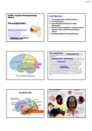
The Frontal lobes - Mahidol University
The Frontal lobes - Mahidol University
The Frontal lobes - Mahidol University

Create successful ePaper yourself
Turn your PDF publications into a flip-book with our unique Google optimized e-Paper software.
19/07/54<br />
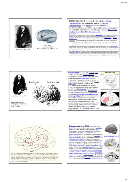
Paul Broca (1824-1880)<br />
Tan’s Brain<br />
(removed in 1861)<br />
Now in the Museum of Man, Paris<br />
Expressive aphasia, known as Broca's aphasia in clinical<br />
neuropsychology and agrammatic aphasia in cognitive<br />
neuropsychology, is an aphasia caused by damage to or<br />
developmental issues in anterior regions of the brain, including (but<br />
not limited to) the left inferior frontal region known as Broca's area<br />
(Brodmann area 44 and Brodmann area 45)<br />
Presentation<br />
Sufferers of this form of aphasia exhibit the common problem of agrammatism. For them, speech is difficult to initiate,<br />
non‐fluent, labored, and halting. Similarly, writing is difficult as well. Intonation and stress patterns are deficient.<br />
Language g is reduced to disjointed words and sentence construction is poor, omitting function words and inflections<br />
(bound morphemes). A person with expressive aphasia might say "Son ... <strong>University</strong> ... Smart ... Boy ... Good ... Good ... "<br />
For example, in the following passage, a Broca's aphasic patient is trying to explain how he came to the hospital for<br />
dental surgery:<br />
Yes... ah... Monday... er... Dad and Peter H... (his own name), and Dad.... er... hospital... and ah... Wednesday...<br />
Wednesday, nine o'clock... and oh... Thursday... ten o'clock, ah doctors... two... an' doctors... and er... teeth...<br />
yah.[1]<br />
In extreme cases, patients may be only able to produce a single word. <strong>The</strong> most famous case of this was Paul Broca's<br />
patient Leborgne, nicknamed "Tan", after the only syllable he could say. Even in such cases, over-learned and rote-learned<br />
speech patterns may be retained—for instance, some patients can count from one to ten, but cannot produce the same<br />
numbers in ordinary conversation.<br />
While word comprehension is generally preserved, meaning interpretation dependent on syntax and phrase structure is<br />
substantially impaired. This can be demonstrated by using phrases with unusual structures. A typical Broca's aphasic<br />
patient will misinterpret "the dog is bitten by the man" by switching the subject and object. Patients who recover go on to<br />
say that they knew what they wanted to say but could not express themselves. Residual deficits will often be seen.<br />
Broca's area is a region of the hominid brain<br />
with functions linked to speech production.<br />
<strong>The</strong> production of language has been linked to the<br />
Broca’s area since Pierre Paul Broca reported<br />
impairments in two patients. <strong>The</strong>y had lost the<br />
ability to speak after injury to the posterior inferior<br />
frontal gyrus of the brain. Since then, the<br />
approximate region he identified has become<br />
known as Broca’s area, and the deficit in language<br />
production as Broca’s aphasia. Broca’s area is now<br />
typically defined in terms of the pars opercularis<br />
and pars triangularis of the inferior frontal gyrus,<br />
represented in Brodmann’s<br />
cytoarchitectonic map<br />
as areas 44 and 45. Studies of chronic aphasia have<br />
implicated an essential role of Broca’s area in<br />
various speech and language functions. Further,<br />
functional MRI studies have also identified<br />
activation patterns in Broca’s area associated with<br />
various language tasks. However, slow destruction<br />
of the Broca's area by brain tumors can leave<br />
speech relatively intact suggesting its functions can<br />
shift to nearby areas in the brain.<br />
Brodmann area 44, , or BA44<br />
44, is part of the frontal<br />
cortex in the human brain. Situated just anterior to premotor<br />
cortex (BA6) and on the lateral surface, inferior to BA9.<br />
This area is also known as pars opercularis (of the inferior<br />
frontal gyrus), and it refers to a subdivision of the<br />
cytoarchitecturally defined frontal region of cerebral cortex. In<br />
the human it corresponds approximately to the opercular part<br />
of inferior frontal gyrus (H). Thus, it is bounded caudally by the<br />
inferior precentral sulcus (H) and rostrally by the anterior<br />
ascending limb of lateral sulcus (H). It surrounds the diagonal<br />
sulcus (H). In the depth of the lateral sulcus it borders on the<br />
insula. Cytoarchitectonically it is bounded caudally and dorsally<br />
by the agranular frontal area 6, dorsally by the granular frontal<br />
area 9 and rostrally by the triangular area 45 (Brodmann‐1909).<br />
Together with left‐hemisphere BA 45, the left hemisphere . BA<br />
44 comprises Broca's area a region involved in semantic tasks.<br />
Some data suggest that BA44 is more involved in phonological<br />
and syntactic processing. Some recent findings also suggest the<br />
implication of this region in music perception. In 95.5% of righthanders<br />
and 61.4% of left‐handers, therefore about 90% of the<br />
clinical population, speech is lateralised in the left hemisphere.<br />
Brain: Brodmann area 44<br />
7/19/2011 NEUROPSYCHIATRY 143<br />
24