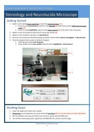
QBI HISTOLOGY AND MICROSCOPY GUIDE
QBI HISTOLOGY AND MICROSCOPY GUIDE
QBI HISTOLOGY AND MICROSCOPY GUIDE

Create successful ePaper yourself
Turn your PDF publications into a flip-book with our unique Google optimized e-Paper software.
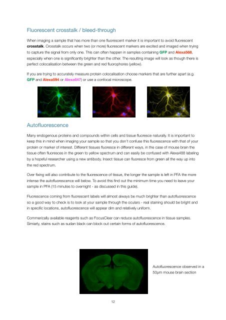
Fluorescent crosstalk / bleed-through<br />
When imaging a sample that has more than one fluorescent marker it is important to avoid fluorescent<br />
crosstalk. Crosstalk occurs when two (or more) fluorescent markers are excited and imaged when trying<br />
to capture the signal from only one. This can often happen in samples containing GFP and Alexa568,<br />
especially when one is significantly brighter than the other. The resulting image will look as though there is<br />
perfect colocalisation between the green and red fluorophores (yellow).<br />
If you are trying to accurately measure protein colocalisation choose markers that are further apart (e.g.<br />
GFP and Alexa594 or Alexa647) or use a confocal microscope.<br />
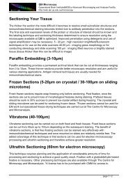
Autofluorescence<br />
Many endogenous proteins and compounds within cells and tissue fluoresce naturally. It is important to<br />
keep this in mind when imaging your sample so that you don’t confuse this fluorescence with that of your<br />
protein or marker of interest. Different tissues fluoresce in different ways, in the case of mouse brain the<br />
tissue often fluoresces in the green to yellow spectrum and can easily be confused with Alexa488 labeling<br />
by a hopeful researcher using a new antibody. Insect tissue can fluoresce from green all the way up into<br />
the red spectrum.<br />
Over fixing will also contribute to the fluorescence of tissue, the longer the sample is left in PFA the more<br />
intense the autofluorescence will below. To avoid this find out the minimum time you need to leave your<br />
sample in PFA (15 minutes to overnight - as discussed in this guide).<br />
Fluorescence coming from fluorescent labels will almost always be much brighter than autofluorescence<br />
so a good way to check is to look at your sample through the oculars - real staining should be bright and<br />
in specific locations, autofluorescence will appear dim and relatively uniform.<br />
Commerically available reagents such as FocusClear can reduce autofluorescence in tissue samples.<br />
Simiarly, stains such as sudan black can block out certain forms of autofluorescence.<br />
Autofluorescence observed in a<br />
50µm mouse brain section<br />
12