QBI HISTOLOGY AND MICROSCOPY GUIDE
QBI HISTOLOGY AND MICROSCOPY GUIDE
QBI HISTOLOGY AND MICROSCOPY GUIDE

You also want an ePaper? Increase the reach of your titles
YUMPU automatically turns print PDFs into web optimized ePapers that Google loves.
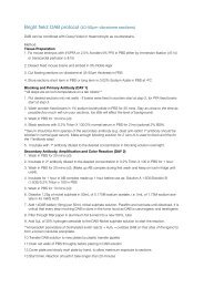
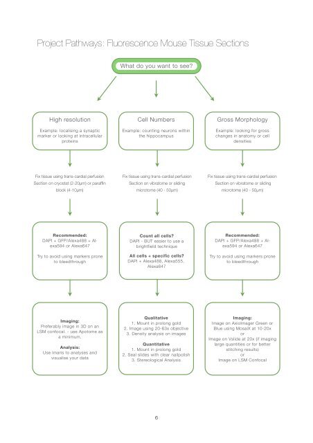
Project Pathways: Fluorescence Mouse Tissue Sections<br />
What do you want to see?<br />
High resolution<br />
Example: localising a synaptic<br />
marker or looking at intracellular<br />
proteins<br />
Cell Numbers<br />
Example: counting neurons within<br />
the hippocampus<br />
Gross Morphology<br />
Example: looking for gross<br />
changes in anatomy or cell<br />
densities<br />
Fix tissue using trans-cardial perfusion<br />
Section on cryostat (2-20µm) or paraffin<br />
block (4-10µm)<br />
Fix tissue using trans-cardial perfusion<br />
Section on vibratome or sliding<br />
microtome (40 - 50µm)<br />
Fix tissue using trans-cardial perfusion<br />
Section on vibratome or sliding<br />
microtome (40 - 50µm)<br />
Recommended:<br />
DAPI + GFP/Alexa488 + Alexa594<br />
or Alexa647<br />
Try to avoid using markers prone<br />
to bleedthrough<br />
Count all cells?<br />
DAPI - BUT easier to use a<br />
brightfield technique<br />
All cells + specific cells?<br />
DAPI + Alexa488, Alexa555,<br />
Alexa647<br />
Recommended:<br />
DAPI + GFP/Alexa488 + Alexa594<br />
or Alexa647<br />
Try to avoid using markers prone<br />
to bleedthrough<br />
Imaging:<br />
Preferably image in 3D on an<br />
LSM confocal. - use Apotome as<br />
a minimum.<br />
Analysis:<br />
Use Imaris to analyses and<br />
visualise your data<br />
Qualitative<br />
1. Mount in prolong gold<br />
2. Image using 20-63x objective<br />
3. Density analysis on images<br />
Quantitative<br />
1. Mount in prolong gold<br />
2. Seal slides with clear nailpolish<br />
3. Stereological Analysis<br />
Imaging:<br />
Image on AxioImager Green or<br />
Blue using MosaiX at 10-20x<br />
or<br />
Image on Vslide at 20x (if imaging<br />
large quantities or for better<br />
stitching results)<br />
or<br />
Image on LSM Confocal<br />
6