QBI HISTOLOGY AND MICROSCOPY GUIDE
QBI HISTOLOGY AND MICROSCOPY GUIDE
QBI HISTOLOGY AND MICROSCOPY GUIDE

You also want an ePaper? Increase the reach of your titles
YUMPU automatically turns print PDFs into web optimized ePapers that Google loves.
<strong>QBI</strong> Slide Scanners<br />
What can they be used for?<br />
Digital archiving and high through-put imaging - the slide scanners can be used in an automated way to<br />
digitally archive slides at full resolution or scan large quantities of tissue sections for further analysis. Each system<br />
can handle 80 slides per run.<br />
High-resolution scanning - the slide scanners can be controlled manually to image one, or a few tissue sections<br />
at high resolution very quickly - this is an alternative to using MosaiX on the standard <strong>QBI</strong> microscopes and allows<br />
much higher magnification in a fraction of the time.<br />
Fluorescence imaging - fluorescence scanning from 10x to 63x magnification is also available. This can be used<br />
for imaging cells, neurospheres or tissue sections and can allow 3-dimensional reconstructions and advanced<br />
analysis using Imaris. <strong>QBI</strong>’s fluorescence scanning capabilities are state-of-the-art and capable of undertaking high<br />
through-put and high resolution analysis projects yet to be developed in other institutes around the world.<br />
Brightfield Scanning:<br />
Scanning individual sections usually takes under a<br />
minute. Details for two of the most common high<br />
through-put scanning options are below:<br />
20x Standard Colour Scan (full slide)<br />
10 - 25 minutes per slide<br />
~8-16GB full resolution image (1-4GB compressed)<br />
10x High-Resolution Colour Scan (full slide)<br />
3 - 10 minutes per slide<br />
~1-3GB full resolution image<br />
(high-resolution scanning uses a monochrome camera and<br />
takes 3 seperate images using red, green and blue lights)<br />
Slides are typically saved in *.IMS format and can be<br />
opened and cropped in Imaris or MetaViewer. However<br />
they can be saved in TIF or JPG format.<br />
Fluorescence Scanning:<br />
Scan times vary considerably depending on the type<br />
of project. Slides can be scanned as a single plane or<br />
complete Z-stacks can be captured:<br />
20x Single Channel Scan (full slide)<br />
10 - 25 minutes per slide<br />
~1-4GB full resolution image<br />
20 x Three Channel Scan (full slide)<br />
1 - 2 hours per slide<br />
~8-16GB full resolution image<br />
40 x Three Channel Scan / 60 slice z-stack<br />
1 - 2 hours per section<br />
~8-10GB full resolution image<br />
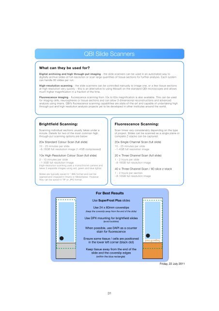
For Best Results<br />
Use SuperFrost Plus slides<br />
Use 24 x 60mm coverslips<br />
(keep the coverslip away from the end of the slide)<br />
Use DPX mounting for brightfield slides<br />
(avoid bubbles)<br />
When possible, use DAPI as a counter<br />
stain for fluorescence<br />
Ensure some tissue / cells are positioned<br />
in the lower left corner (black dot)<br />
SuperFrost<br />
Keep tissue away from the end of the<br />
slide and the coverslip edges<br />
(within the blue rectangle)<br />
Friday, 22 July 2011<br />
31