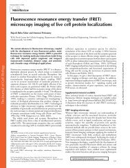
QBI HISTOLOGY AND MICROSCOPY GUIDE
QBI HISTOLOGY AND MICROSCOPY GUIDE
QBI HISTOLOGY AND MICROSCOPY GUIDE

Create successful ePaper yourself
Turn your PDF publications into a flip-book with our unique Google optimized e-Paper software.
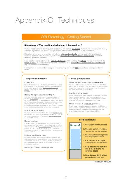
Appendix C: Techniques<br />
<strong>QBI</strong> Stereology : Getting Started<br />
Stereology - Why use it and what can it be used for?<br />
Traditional approaches to counting, such as counting cell profiles, are biased. Furthermore, calculating cell density<br />
in sample regions may not reflect a change in cell number or be representative of the whole tissue.<br />
Stereology can be used to accurately estimate the total number of cells within a region of interest by only<br />
counting ~200-800 cells. This allows you to make statistically valid quantitative comparisons instead of<br />
observing qualitative differences.<br />
It can also be used to determine the size of cells/nuclei whilst counting, the volume of a region of interest, the<br />
length of fibres (e.g. vasculature or neuronal projections), or to analyse three-dimensional cytoarchitecture<br />
(e.g. cell-cell spatial relationships).<br />
It is important to understand stereology is time consuming and should NOT be used as a technique to fish for<br />
results.<br />
Things to remember:<br />
It takes time.<br />
For each sample group a Pilot Study should be conducted<br />
on one animal to determine the best sampling parameters.<br />
A pilot study generally takes 1 whole day / region of<br />
interest. Following on, additional animals should take ~1/2<br />
a day each.<br />
Identify the region you are counting in.<br />
You need to be able to identify the region you want to<br />
count in at all depths throughout the brain. You can use<br />
the mouse brain atlas, or sometimes the cells of that region<br />
will be clustered and drawing a border around them will be<br />
easy. If you can’t accurately identify where you are<br />
counting you may need to reconsider your project - varying<br />
region outlines can cause big variations in cell counts.<br />
Sample the whole region.<br />
You need to have set of sections that spans right from the<br />
beginning of your region of interest to the very end. You do<br />
not need every section - often people use serial sections<br />
e.g. every 6th section.<br />
Missing sections.<br />
It is OK if a section gets damaged and can’t be plated, or<br />
folds up and can’t be imaged - just make a note of it. The<br />
important thing is that you need to know that the section is<br />
missing so that it can be accounted for in the analysis.<br />
Proper stereology will be impossible if there is no record of<br />
missing sections.<br />
Sections need to stay thick.<br />
For stereology it is important to have thick sections, for<br />
this reason we often cut at 40µm or more. These sections<br />
also need to stay thick - if a section shrinks below 20µm<br />
thick then it becomes difficult or impossible to carry out<br />
stereological analysis.<br />
Discuss your project before you start.<br />
Tissue preparation:<br />
Tissue sections should be cut at 40-50µm.<br />
You can cut thicker but you need to make sure the whole<br />
section will be labelled during your staining protocol - often<br />
times if the tissue is too thick the stain or antibodies will<br />
not reach the middle of the section.<br />
Avoid drying the tissue.<br />
Drying the tissue will make it thinner. Ensure you keep your<br />
tissue sections hydrated, avoid processing steps that<br />
involve drying, ethanol or xylene, If a section needs to be<br />
dried you can rehydrate overnight in PBS+azide.<br />
Mount sections in an aqueous solution.<br />
This is usually done for fluorescent samples but DPX is<br />
often used for brightfield. DPX can make sections very thin,<br />
especially if xylene has been used in the protocol. For<br />
brightfield sections use Mowiol instead of DPX.<br />
For Best Results<br />
1. Use SuperFrost Plus slides<br />
2. Use 22 x 50mm coverslips<br />
seal onto slide with clear nailpolish<br />
3. Use mowiol mounting media<br />
if mounting brightfield sections<br />
4. Cut sections at 40-50µm<br />
avoid drying out and dehydration<br />
4. Keep tissue away from the<br />
end of the slide and the<br />
coverslip edges<br />
5. Keep tissue within the blue<br />
rectangle (brightfield only)<br />
Thursday, 21 July 2011<br />
30