QBI HISTOLOGY AND MICROSCOPY GUIDE
QBI HISTOLOGY AND MICROSCOPY GUIDE
QBI HISTOLOGY AND MICROSCOPY GUIDE
Create successful ePaper yourself
Turn your PDF publications into a flip-book with our unique Google optimized e-Paper software.
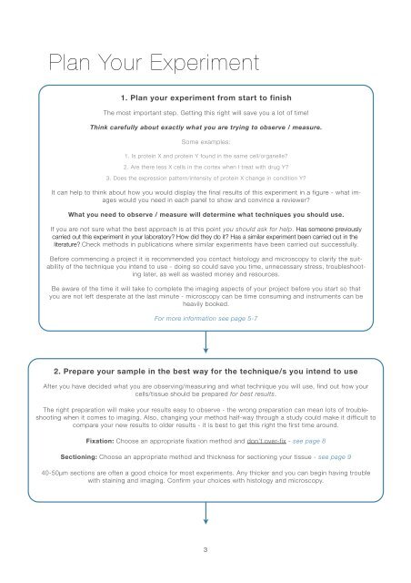
Plan Your Experiment<br />
1. Plan your experiment from start to finish<br />
The most important step. Getting this right will save you a lot of time!<br />
Think carefully about exactly what you are trying to observe / measure.<br />
Some examples:<br />
1. Is protein X and protein Y found in the same cell/organelle?<br />
2. Are there less X cells in the cortex when I treat with drug Y?<br />
3. Does the expression pattern/intensity of protein X change in condition Y?<br />
It can help to think about how you would display the final results of this experiment in a figure - what images<br />
would you need in each panel to show and convince a reviewer?<br />
What you need to observe / measure will determine what techniques you should use.<br />
If you are not sure what the best approach is at this point you should ask for help. Has someone previously<br />
carried out this experiment in your laboratory? How did they do it? Has a similar experiment been carried out in the<br />
literature? Check methods in publications where similar experiments have been carried out successfully.<br />
Before commencing a project it is recommended you contact histology and microscopy to clarify the suitability<br />
of the technique you intend to use - doing so could save you time, unnecessary stress, troubleshooting<br />
later, as well as wasted money and resources.<br />
Be aware of the time it will take to complete the imaging aspects of your project before you start so that<br />
you are not left desperate at the last minute - microscopy can be time consuming and instruments can be<br />
heavily booked.<br />
For more information see page 5-7<br />
2. Prepare your sample in the best way for the technique/s you intend to use<br />
After you have decided what you are observing/measuring and what technique you will use, find out how your<br />
cells/tissue should be prepared for best results.<br />
The right preparation will make your results easy to observe - the wrong preparation can mean lots of troubleshooting<br />
when it comes to imaging. Also, changing your method half-way through a study could make it difficult to<br />
compare your new results to older results - it is best to get this right the first time around.<br />
Fixation: Choose an appropriate fixation method and don’t over-fix - see page 8<br />
Sectioning: Choose an appropriate method and thickness for sectioning your tissue - see page 9<br />
40-50µm sections are often a good choice for most experiments. Any thicker and you can begin having trouble<br />
with staining and imaging. Confirm your choices with histology and microscopy.<br />
3


