

Dental Press
Dental Press
Dental Press

Create successful ePaper yourself
Turn your PDF publications into a flip-book with our unique Google optimized e-Paper software.
Consolaro A<br />
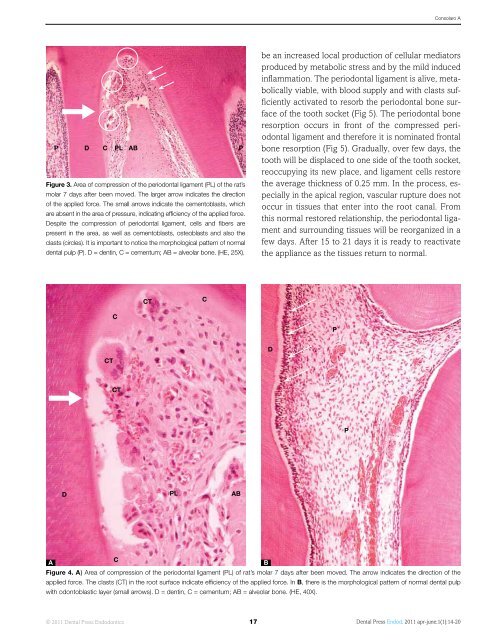
P D C PL AB<br />
Figure 3. Area of compression of the periodontal ligament (PL) of the rat’s<br />
molar 7 days after been moved. The larger arrow indicates the direction<br />
of the applied force. The small arrows indicate the cementoblasts, which<br />
are absent in the area of pressure, indicating efficiency of the applied force.<br />
Despite the compression of periodontal ligament, cells and fibers are<br />
present in the area, as well as cementoblasts, osteoblasts and also the<br />
clasts (circles). It is important to notice the morphological pattern of normal<br />
dental pulp (P). D = dentin, C = cementum; AB = alveolar bone. (HE, 25X).<br />
P<br />
be an increased local production of cellular mediators<br />
produced by metabolic stress and by the mild induced<br />
inflammation. The periodontal ligament is alive, metabolically<br />
viable, with blood supply and with clasts sufficiently<br />
activated to resorb the periodontal bone surface<br />
of the tooth socket (Fig 5). The periodontal bone<br />
resorption occurs in front of the compressed periodontal<br />
ligament and therefore it is nominated frontal<br />
bone resorption (Fig 5). Gradually, over few days, the<br />
tooth will be displaced to one side of the tooth socket,<br />
reoccupying its new place, and ligament cells restore<br />
the average thickness of 0.25 mm. In the process, especially<br />
in the apical region, vascular rupture does not<br />
occur in tissues that enter into the root canal. From<br />
this normal restored relationship, the periodontal ligament<br />
and surrounding tissues will be reorganized in a<br />
few days. After 15 to 21 days it is ready to reactivate<br />
the appliance as the tissues return to normal.<br />
CT<br />
C<br />
C<br />
P<br />
D<br />
CT<br />
CT<br />
P<br />
D<br />
PL<br />
AB<br />
A<br />
C<br />
B<br />
Figure 4. A) Area of compression of the periodontal ligament (PL) of rat’s molar 7 days after been moved. The arrow indicates the direction of the<br />
applied force. The clasts (CT) in the root surface indicate efficiency of the applied force. In B, there is the morphological pattern of normal dental pulp<br />
with odontoblastic layer (small arrows). D = dentin, C = cementum; AB = alveolar bone. (HE, 40X).<br />
© 2011 <strong>Dental</strong> <strong>Press</strong> Endodontics 17<br />
<strong>Dental</strong> <strong>Press</strong> Endod. 2011 apr-june;1(1):14-20


