

Dental Press
Dental Press
Dental Press

Create successful ePaper yourself
Turn your PDF publications into a flip-book with our unique Google optimized e-Paper software.
[ original article ] In vitro evaluation of shape changes in curved artificial root canals prepared with two rotary systems<br />
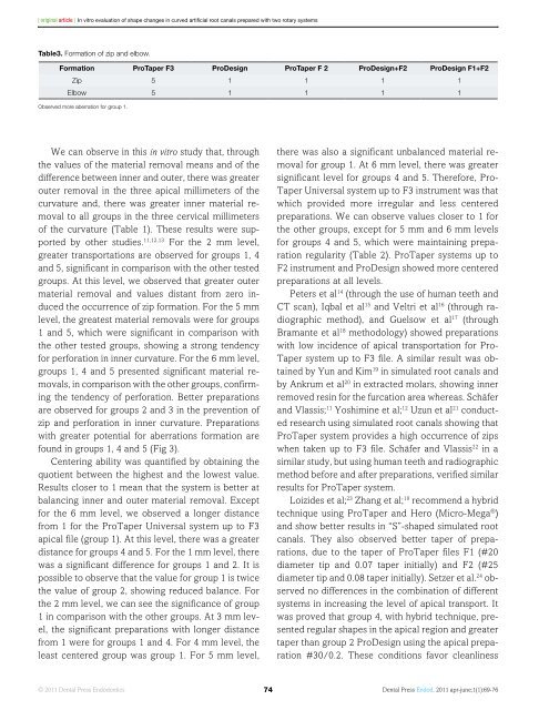
Table3. Formation of zip and elbow.<br />
Formation ProTaper F3 ProDesign ProTaper F 2 ProDesign+F2 ProDesign F1+F2<br />
Zip 5 1 1 1 1<br />
Elbow 5 1 1 1 1<br />
Observed more aberration for group 1.<br />
We can observe in this in vitro study that, through<br />
the values of the material removal means and of the<br />
difference between inner and outer, there was greater<br />
outer removal in the three apical millimeters of the<br />
curvature and, there was greater inner material removal<br />
to all groups in the three cervical millimeters<br />
of the curvature (Table 1). These results were supported<br />
by other studies. 11,12,13 For the 2 mm level,<br />
greater transportations are observed for groups 1, 4<br />
and 5, significant in comparison with the other tested<br />
groups. At this level, we observed that greater outer<br />
material removal and values distant from zero induced<br />
the occurrence of zip formation. For the 5 mm<br />
level, the greatest material removals were for groups<br />
1 and 5, which were significant in comparison with<br />
the other tested groups, showing a strong tendency<br />
for perforation in inner curvature. For the 6 mm level,<br />
groups 1, 4 and 5 presented significant material removals,<br />
in comparison with the other groups, confirming<br />
the tendency of perforation. Better preparations<br />
are observed for groups 2 and 3 in the prevention of<br />
zip and perforation in inner curvature. Preparations<br />
with greater potential for aberrations formation are<br />
found in groups 1, 4 and 5 (Fig 3).<br />
Centering ability was quantified by obtaining the<br />
quotient between the highest and the lowest value.<br />
Results closer to 1 mean that the system is better at<br />
balancing inner and outer material removal. Except<br />
for the 6 mm level, we observed a longer distance<br />
from 1 for the ProTaper Universal system up to F3<br />
apical file (group 1). At this level, there was a greater<br />
distance for groups 4 and 5. For the 1 mm level, there<br />
was a significant difference for groups 1 and 2. It is<br />
possible to observe that the value for group 1 is twice<br />
the value of group 2, showing reduced balance. For<br />
the 2 mm level, we can see the significance of group<br />
1 in comparison with the other groups. At 3 mm level,<br />
the significant preparations with longer distance<br />
from 1 were for groups 1 and 4. For 4 mm level, the<br />
least centered group was group 1. For 5 mm level,<br />
there was also a significant unbalanced material removal<br />
for group 1. At 6 mm level, there was greater<br />
significant level for groups 4 and 5. Therefore, Pro-<br />
Taper Universal system up to F3 instrument was that<br />
which provided more irregular and less centered<br />
preparations. We can observe values closer to 1 for<br />
the other groups, except for 5 mm and 6 mm levels<br />
for groups 4 and 5, which were maintaining preparation<br />
regularity (Table 2). ProTaper systems up to<br />
F2 instrument and ProDesign showed more centered<br />
preparations at all levels.<br />
Peters et al 14 (through the use of human teeth and<br />
CT scan), Iqbal et al 15 and Veltri et al 16 (through radiographic<br />
method), and Guelsow et al 17 (through<br />
Bramante et al 18 methodology) showed preparations<br />
with low incidence of apical transportation for Pro-<br />
Taper system up to F3 file. A similar result was obtained<br />
by Yun and Kim 19 in simulated root canals and<br />
by Ankrum et al 20 in extracted molars, showing inner<br />
removed resin for the furcation area whereas. Schäfer<br />
and Vlassis; 11 Yoshimine et al; 12 Uzun et al 21 conducted<br />
research using simulated root canals showing that<br />
ProTaper system provides a high occurrence of zips<br />
when taken up to F3 file. Schäfer and Vlassis 22 in a<br />
similar study, but using human teeth and radiographic<br />
method before and after preparations, verified similar<br />
results for ProTaper system.<br />
Loizides et al; 23 Zhang et al; 10 recommend a hybrid<br />
technique using ProTaper and Hero (Micro-Mega ® )<br />
and show better results in “S”-shaped simulated root<br />
canals. They also observed better taper of preparations,<br />
due to the taper of ProTaper files F1 (#20<br />
diameter tip and 0.07 taper initially) and F2 (#25<br />
diameter tip and 0.08 taper initially). Setzer et al. 24 observed<br />
no differences in the combination of different<br />
systems in increasing the level of apical transport. It<br />
was proved that group 4, with hybrid technique, presented<br />
regular shapes in the apical region and greater<br />
taper than group 2 ProDesign using the apical preparation<br />
#30/0.2. These conditions favor cleanliness<br />
© 2011 <strong>Dental</strong> <strong>Press</strong> Endodontics 74<br />
<strong>Dental</strong> <strong>Press</strong> Endod. 2011 apr-june;1(1):69-76


