

Dental Press
Dental Press
Dental Press

You also want an ePaper? Increase the reach of your titles
YUMPU automatically turns print PDFs into web optimized ePapers that Google loves.
[ original article ] Effect of intracanal posts on dimensions of cone beam computed tomography images of endodontically treated teeth<br />
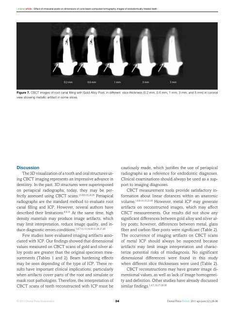
0.2 mm 0.6 mm 1 mm 3 mm 5 mm<br />
Figure 7. CBCT images of root canal filling with Gold Alloy Post, in different slice thickness (0.2 mm, 0.6 mm, 1 mm, 3 mm, and 5 mm) in coronal<br />
view showing metallic artifact in some slices.<br />
Discussion<br />
The 3D visualization of a tooth and oral structures using<br />
CBCT imaging represents an impressive advance in<br />
dentistry. In the past, 3D structures were superimposed<br />
on periapical radiographs; today, they may be perfectly<br />
assessed using CBCT scans. 2,5,8,9,10,24,29 Periapical<br />
radiographs are the standard method to evaluate root<br />
canal filling and ICP. However, several authors have<br />
described their limitations. 8,9,10 At the same time, high<br />
density materials may produce image artifacts, which<br />
may limit interpretation, reduce image quality, and induce<br />
diagnostic errors conditions. 3,4,7,12,13,14,16-21,26,27,28<br />
Few studies have evaluated imaging artifacts associated<br />
with ICP. Our findings showed that dimensional<br />
values measured on CBCT scans of gold and silver alloy<br />
posts are greater than the original specimen measurements<br />
(Tables 1 and 2). Beam hardening effects<br />
may be seen depending of the type of ICP. These results<br />
have important clinical implications, particularly<br />
when artifacts cover parts of the root and simulate or<br />
mask root pathologies. Therefore, the interpretation of<br />
CBCT scans of teeth reconstructed with ICP must be<br />
cautiously made, which justifies the use of periapical<br />
radiographs as a reference for endodontic diagnoses.<br />
Clinical examinations should always be used as a support<br />
to imaging diagnoses.<br />
CBCT measurement tools provide satisfactory information<br />
about linear distances within an anatomic<br />
volume. 1,8,9,10,15,23,30 However, metal ICP may generate<br />
artifacts on reconstructed images, which may affect<br />
CBCT measurements. Our results did not show any<br />
significant differences between gold alloy and silver alloy<br />
posts; however, differences between metal, glass<br />
fiber and carbon fiber posts were significant (Table 2).<br />
The occurrence of imaging artifacts on CBCT scans<br />
of metal ICP should always be suspected because<br />
artifacts may limit image interpretation and characterize<br />
potential risks of misdiagnosis. No significant<br />
dimensional differences were found in this study<br />
when different slice thicknesses were used (Table 2).<br />
CBCT reconstructions may have greater image dimensional<br />
values, as well as lack of image homogeneity<br />
and definition. Other studies have already discussed<br />
similar findings. 1,4,7,15,17-20,30<br />
© 2011 <strong>Dental</strong> <strong>Press</strong> Endodontics 34<br />
<strong>Dental</strong> <strong>Press</strong> Endod. 2011 apr-june;1(1):28-36


