

Dental Press
Dental Press
Dental Press

Create successful ePaper yourself
Turn your PDF publications into a flip-book with our unique Google optimized e-Paper software.
Deonízio MD, Sydney GB, Batista A, Estrela C<br />
The calcium hydroxide paste was prepared with a<br />
distilled water base, because it is a hydrosoluble vehicle,<br />
which increases the effectiveness of calcium<br />
hydroxide. 3,24,25 Barium sulphate was used as a radiopaque<br />
substance to differentiate the optical density<br />
of the calcium hydroxide from the dentine. The ratio of<br />
barium sulphate used to calcium hydroxide was 1:2. 13,17<br />
The insertion of the paste was performed using small<br />
quantities at a time. When activated, the Lentulo spiral<br />
launched the paste against the canal walls, and the use<br />
of a plugger allowed its condensation in all thirds.<br />
The speeds used were determined based on the<br />
maximum speeds possible in dental equipment<br />
(around 20,000 rpm). The higher the speed and the<br />
quantity of paste in the Lentulo, the greater the quantity<br />
of air that ends up being retained inside of the<br />
root canal, generating air bubbles formation that do<br />
not allow the complete filling and, consequently, the<br />
desired action. Thus, the speeds used in the study<br />
were 15,000 rpm, 10,000 rpm, and 5,000 rpm, which<br />
were maintained constant through an electric motor<br />
(Driller – São Paulo, Brazil).<br />
Digital radiography today represents one of the<br />
great advances in imaging, allowing speed and simplicity<br />
in the capture of images with a significant reduction<br />
in exposure time and allowing standardization,<br />
high-quality analysis, besides becoming a viable<br />
and safe alternative for the results interpretation, conferring<br />
greater diagnostic precision. The use of digital<br />
technology besides being reproducible is a system<br />
that allows almost instant images of the structures to<br />
be observed, without the need for chemical processing<br />
and with a reduced exposure time. 26<br />
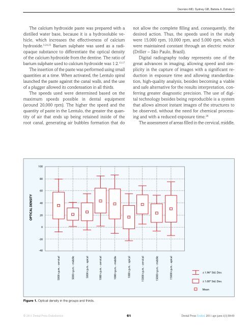
The assessment of areas filled in the cervical, middle,<br />
100<br />
80<br />
60<br />
OPTICAL DENSITY<br />
40<br />
20<br />
0<br />
-20<br />
-40<br />
5000 r.p.m. - cervical<br />
5000 r.p.m. - middle<br />
5000 r.p.m. - apical<br />
1080 r.p.m. - cervical<br />
1080 r.p.m. - middle<br />
1080 r.p.m. - apical<br />
15000 r.p.m. - cervical<br />
15000 r.p.m. - middle<br />
15000 r.p.m. - apical<br />
± 1.96* Std. Dev.<br />
± 1.00* Std. Dev.<br />
Mean<br />
Figure 1. Optical density in the groups and thirds.<br />
© 2011 <strong>Dental</strong> <strong>Press</strong> Endodontics 61<br />
<strong>Dental</strong> <strong>Press</strong> Endod. 2011 apr-june;1(1):58-63


