

Dental Press
Dental Press
Dental Press

You also want an ePaper? Increase the reach of your titles
YUMPU automatically turns print PDFs into web optimized ePapers that Google loves.
Souza MA, Menin MLF, Montagner F, Cecchin D, Farina AP<br />
Introduction<br />
The success of endodontic therapy is grounded<br />
not only in the correct diagnosis, but also the proper<br />
planning and technical implementation, and especially<br />
in caring for the maintenance of the aseptic chain<br />
during the patients care.<br />
The endodontic instruments are used to remove<br />
the remnants of pulp tissue during the procedures of<br />
cleaning and shaping of the root canal system. These<br />
instruments can be recycled for reuse after its first<br />
use. In a large study was reported that 88% of general<br />
practitioners dentists in the UK re-process the endodontic<br />
files after use 1 .<br />
The mandatory conduct of biosecurity recognizes<br />
that the endodontic instruments, to be reused, they<br />
must go through a cleaning process before sterilization<br />
2 , since the presence of organic matter and/or<br />
debris on the instruments may interfere with the sterilization<br />
process. These organic compounds creates<br />
barriers to protect the microorganisms, which may<br />
prevent the penetration of the sterilizing agent 3 .<br />
The procedures of pre-cleaning and autoclave can<br />
be used to sterilize endodontic instruments 4,5 . However,<br />
the complex architecture of endodontic files<br />
could difficult these procedures 5 . <strong>Dental</strong> structures<br />
and organic debris have been observed on the surface<br />
of rotary instruments, especially in the cracks 6 .<br />
According to a previous study, 66% of endodontic<br />
files retrieved dental general practitioners remained<br />
visibly contaminated 7 . Thus, there is the possibility of<br />
cross contamination associated with the inability to<br />
properly clean and sterilize them, and suggested that<br />
these instruments should be single-use devices.<br />
Therefore, the aim of this study was to determine<br />
the presence of debris left on the surface of endodontic<br />
files after performing a cleaning process and analyze<br />
its influence on the sterilization process.<br />
Materials and methods<br />
Fifty endodontic files K #25 were selected for this<br />
study, regardless of their trademark. The samples<br />
were divided into two groups (n = 25) according to<br />
the method of analysis, prior use and performance of<br />
disinfection protocol, according to Table 1.<br />
The endodontic instruments were obtained directly<br />
from students of the School of Dentistry of<br />
Pontifical Catholic University of Rio Grande do Sul<br />
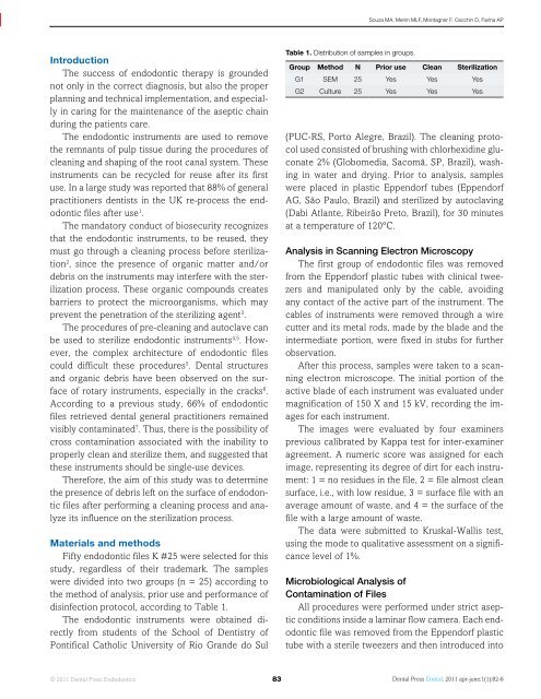
Table 1. Distribution of samples in groups.<br />
Group Method N Prior use Clean Sterilization<br />
G1 SEM 25 Yes Yes Yes<br />
G2 Culture 25 Yes Yes Yes<br />
(PUC-RS, Porto Alegre, Brazil). The cleaning protocol<br />
used consisted of brushing with chlorhexidine gluconate<br />
2% (Globomedia, Sacomã, SP, Brazil), washing<br />
in water and drying. Prior to analysis, samples<br />
were placed in plastic Eppendorf tubes (Eppendorf<br />
AG, São Paulo, Brazil) and sterilized by autoclaving<br />
(Dabi Atlante, Ribeirão Preto, Brazil), for 30 minutes<br />
at a temperature of 120ºC.<br />
Analysis in Scanning Electron Microscopy<br />
The first group of endodontic files was removed<br />
from the Eppendorf plastic tubes with clinical tweezers<br />
and manipulated only by the cable, avoiding<br />
any contact of the active part of the instrument. The<br />
cables of instruments were removed through a wire<br />
cutter and its metal rods, made by the blade and the<br />
intermediate portion, were fixed in stubs for further<br />
observation.<br />
After this process, samples were taken to a scanning<br />
electron microscope. The initial portion of the<br />
active blade of each instrument was evaluated under<br />
magnification of 150 X and 15 kV, recording the images<br />
for each instrument.<br />
The images were evaluated by four examiners<br />
previous calibrated by Kappa test for inter-examiner<br />
agreement. A numeric score was assigned for each<br />
image, representing its degree of dirt for each instrument:<br />
1 = no residues in the file, 2 = file almost clean<br />
surface, i.e., with low residue, 3 = surface file with an<br />
average amount of waste, and 4 = the surface of the<br />
file with a large amount of waste.<br />
The data were submitted to Kruskal-Wallis test,<br />
using the mode to qualitative assessment on a significance<br />
level of 1%.<br />
Microbiological Analysis of<br />
Contamination of Files<br />
All procedures were performed under strict aseptic<br />
conditions inside a laminar flow camera. Each endodontic<br />
file was removed from the Eppendorf plastic<br />
tube with a sterile tweezers and then introduced into<br />
© 2011 <strong>Dental</strong> <strong>Press</strong> Endodontics 83<br />
<strong>Dental</strong> <strong>Press</strong> Endod. 2011 apr-june;1(1):82-6


