Biennial Report 2011â2012
Biennial Report 2011â2012
Biennial Report 2011â2012

Create successful ePaper yourself
Turn your PDF publications into a flip-book with our unique Google optimized e-Paper software.
Department of Protein-Nucleic Acids Interactions<br />
Structure and function of restriction<br />
endonucleases: projects overview<br />
The recognition domain of the methyl-specific<br />
endonuclease McrBC flips out 5-methylcytosine<br />
DNA cytosine methylation is a widespread epigenetic<br />
mark. Biological effects of DNA methylation are mediated<br />
by the proteins that preferentially bind to 5-methylcytosine<br />
(5mC) in different sequence contexts. Until now two<br />
different structural mechanisms have been established for<br />
5mC recognition in eukaryotes; however, it is still unknown<br />
how discrimination of the 5mC modification is achieved<br />
in prokaryotes. To address this question we have solved the<br />
crystal structure of the N-terminal DNA-binding domain<br />
(McrB-N) of the methyl-specific endonuclease McrBC from<br />
Escherichia coli.<br />
The McrB-N protein shows a novel DNA-binding fold<br />
adapted for 5mC-recognition. In the McrB-N structure in<br />
complex with methylated DNA, the 5mC base is flipped<br />
out from the DNA duplex and positioned within a binding<br />
pocket. Base flipping elegantly explains why McrBC<br />
system restricts only T4-even phages impaired in glycosylation<br />
[Luria, S.E. and Human, M.L. (1952) A nonhereditary,<br />
host-induced variation of bacterial viruses. J. Bacteriol., 64,<br />
557-569]: flipped out 5-hydroxymethylcytosine is accommodated<br />
in the binding pocket but there is no room for the<br />
glycosylated base. The mechanism for 5mC recognition employed<br />
by McrB-N is highly reminiscent of that for eukaryotic<br />
SRA domains, despite the differences in their protein<br />
folds.<br />
Structural and functional studies of the<br />
Bse634I specificity<br />
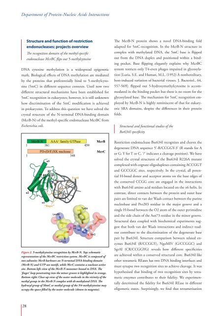
Figure 2. 5-methylcytosine recognition by McrB-N. Top: schematic<br />
representation of the McrBC restriction system. McrBC is composed of<br />
two subunits: McrB harbours an N-terminal DNA-binding domain<br />
(McrB-N) and GTP-ase motifs, while McrC contains a nuclease active<br />
site. Bottom left: view of the McrB-N monomer bound to DNA. The<br />
‘finger’ loop penetrating into the minor groove is highlighted in orange.<br />
Bottom right: Close-up view of the water molecule in the vicinity of the<br />
methyl group in the McrB-N complex with di-methylated DNA. The<br />
hydroxyl group of 5hmC or methyl group of the N4-methylcytosine may<br />
occupy the space filled by the water molecule (shown in magenta).<br />
Restriction endonuclease Bse634I recognizes and cleaves the<br />
degenerate DNA sequence 5’-R/CCGGY-3’ (R stands for A<br />
or G; Y for T or C, ‘/’ indicates a cleavage position). We have<br />
solved the crystal structures of the Bse634I R226A mutant<br />
complexed with cognate oligoduplexes containing ACCGGT<br />
and GCCGGC sites, respectively. In the crystal, all potential<br />
H-bond donor and acceptor atoms on the base edges of<br />
the conserved CCGG core are engaged in the interactions<br />
with Bse634I amino acid residues located on the α6 helix. In<br />
contrast, direct contacts between the protein and outer base<br />
pairs are limited to van der Waals contact between the purine<br />
nucleobase and Pro203 residue in the major groove and a<br />
single H-bond between the O2 atom of the outer pyrimidine<br />
and the side chain of the Asn73 residue in the minor groove.<br />
Structural data coupled with biochemical experiments suggest<br />
that both van der Waals interactions and indirect readout<br />
contribute to the discrimination of the degenerate base<br />
pair by Bse634I. Structure comparison between related enzymes<br />
Bse634I (R/CCGGY), NgoMIV (G/CCGGC) and<br />
SgrAI (CR/CCGGYG) reveals how different specificities<br />
are achieved within a conserved structural core. Bse634I like<br />
other tetrameric REases has two DNA binding interfaces and<br />
must synapse two recognition sites to achieve cleavage. It was<br />
hypothesised that binding of two recognition sites by tetrameric<br />
enzymes contributes to their fidelity. We experimentally<br />
determined the fidelity for Bse634I REase in different<br />
oligomeric states. Surprisingly, we find that tetramerisation<br />
28