DC beads loaded with Irinotecan : precilinical and early clinical ...
DC beads loaded with Irinotecan : precilinical and early clinical ...
DC beads loaded with Irinotecan : precilinical and early clinical ...

You also want an ePaper? Increase the reach of your titles
YUMPU automatically turns print PDFs into web optimized ePapers that Google loves.
<strong>DC</strong> <strong>beads</strong> <strong>loaded</strong> <strong>with</strong> <strong>Irinotecan</strong> :<br />
<strong>precilinical</strong> <strong>and</strong> <strong>early</strong> <strong>clinical</strong> datas in<br />
CRC liver metastases<br />
T de Baere - Villejuif - France
Colorectal cancer<br />
•World wide incidence: 400,000 patients<br />
•60% patients will develop liver metastases<br />
•Surgery is feasible in only a minority of<br />
patients <strong>and</strong> most patients are treated <strong>with</strong><br />
systemic chemotherapy.<br />
•Patients have a poor prognosis <strong>with</strong><br />
reported 1- <strong>and</strong> 3-year survival rates of<br />
31% <strong>and</strong> 2.6%, respectively .
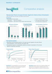
Colorectal cancer chemotherapy<br />
% response rate<br />
FOLFIRI<br />
FOLFOX<br />
5 FU<br />
10 %<br />
5FU-FA<br />
FA<br />
20 %<br />
LV5FU2<br />
25 %<br />
55 % 55 %<br />
Sytemic therapy<br />
• Improvement in response rate, namely due to<br />
Oxaliplatinum <strong>and</strong> <strong>Irinotecan</strong>
Colorectal cancer chemotherapy<br />
% response rate<br />
FUDR<br />
FOLFIRI<br />
FOLFOX<br />
Iri.<br />
Oxal.<br />
5 FU<br />
10 %<br />
5FU-FA<br />
FA<br />
20 %<br />
45 %<br />
LV5FU2<br />
25 %<br />
55 % 55 %<br />
67 %<br />
64 %<br />
Sytemic therapy<br />
Intra-arterial therapy<br />
• Improvement in response rate, namely due to<br />
Oxaliplatinum <strong>and</strong> <strong>Irinotecan</strong><br />
• Intra-arterial delivery provide higher response rate<br />
for a given drug
Rationale for intra-arterial route<br />
Drug<br />
% Liver<br />
extraction<br />
Clearance<br />
TB (l/min)<br />
5-FU 22-45 2-5<br />
FUDR 69-92 5-15<br />
IRINOTECAN 38-72 9-25<br />
MITO-C 7-18 3-5<br />
CDDP 8-50 0.3-0.5<br />
DOXO 45-50 -<br />
R art =<br />
CL<br />
Q A (1 - E r )<br />
- Rart : advantage of IA vs IV<br />
- Er :extraction ratio at 1st pass<br />
- QA : arterial flow - CL : Body clearance of the drug
Rationale for intra-arterial route<br />
101<br />
R art<br />
81<br />
61<br />
41<br />
21<br />
1 0.9<br />
0.5<br />
0.1<br />
E r<br />
R art =<br />
CL<br />
Q A (1 - E r )<br />
24 18 12 6<br />
Q A (L/h)<br />
• Drug eluting embols<br />
Complete embolization (Q A =0, R art ≈)
In vitro<br />
Loading/elution of embolics <strong>with</strong> <strong>Irinotecan</strong><br />
Hepaspheres<br />
• 100-150 μm dry/ 400-600 μm hydrated<br />
• Poly (vinyl alcohol-co-sodium acrylate)<br />
<strong>DC</strong> <strong>beads</strong><br />
• 500-700 μm hydrated<br />
PVA (<strong>with</strong> N-acryloyl-aminoacetaldehyde dimethylacetal)<br />
copolymerized <strong>with</strong> 2-acrylamido-2 methylpropanesulfonate<br />
sodium salt*.<br />
* Lewis et al J Mater Sci: Mater Med (2007) 18:1691–1699
<strong>Irinotecan</strong> loading<br />
<strong>DC</strong> <strong>beads</strong><br />
- <strong>loaded</strong> <strong>with</strong>in 1 hour<br />
- 92% uptake (vs 95% Taylor & al EJPS 2007)<br />
Hepasheres<br />
-uptake at 1hr / 86% uptake<br />
-Heterogenous loading
<strong>DC</strong> <strong>beads</strong> size<br />
700<br />
600<br />
Mean diameter (microns)<br />
500<br />
400<br />
300<br />
200<br />
100<br />
0<br />
<strong>DC</strong> <strong>beads</strong>/H2O <strong>DC</strong> <strong>beads</strong>/NaCl <strong>DC</strong> <strong>beads</strong>/50%<br />
Visipaque<br />
<strong>DC</strong> <strong>beads</strong>/after<br />
catheter<br />
<strong>DC</strong> <strong>beads</strong> in vial <strong>DC</strong> <strong>beads</strong>/Irino <strong>DC</strong> <strong>beads</strong>/Irino<br />
apr¸ slibˇ ration<br />
• Shrinking under irinotecan loading <strong>with</strong> return to baseline after release
Hepasphere size<br />
700<br />
600<br />
Mean diameter (microns)<br />
500<br />
400<br />
300<br />
200<br />
100<br />
0<br />
NaCl 0.9%<br />
NaCl:<br />
Visipaque 1:1<br />
after cat<br />
NaCl:Visipaque<br />
1:1 cat+ comp<br />
NaCl after Cat <strong>Irinotecan</strong> <strong>Irinotecan</strong><br />
coloured only<br />
<strong>Irinotecan</strong> after<br />
lib<br />
• Moderate shrinkage under irinotecan loading <strong>with</strong> return to baseline<br />
after release
Beads Elasticity<br />
20<br />
Elastic modulus 20% compr (kPa)<br />
15<br />
10<br />
5<br />
0<br />
<strong>DC</strong> Beads<br />
<strong>Irinotecan</strong><br />
Doxorubicin<br />
Hepaspheres<br />
Compression of a microsphere monolayer<br />
to determine elastic modulus
Release measurement<br />
Flow<br />
<br />
<br />
Sotax CE 6 apparatus (37C) <strong>with</strong> n=6 implant cells, constant-flow<br />
CY7 pump (NaCL 0.9%, 5 ml/min)<br />
Spectrophotometric determination at 359 nm (irinotecan)
<strong>Irinotecan</strong> before/after release<br />
<strong>DC</strong> <strong>beads</strong> before release<br />
Hepaspheres before release<br />
<strong>DC</strong> <strong>beads</strong> after release<br />
Hepaspheres after release
<strong>Irinotecan</strong> release<br />
<strong>DC</strong> Beads<br />
Drug load = 92%<br />
Drug release = 62%<br />
t 1/2 = 1 hr<br />
Hepasheres<br />
Drug load = 86%<br />
Drug release = 42%<br />
t 1/2 = 5 min
Tumor necrosis<br />
-Diffuse liver metastasis obtained through intra-portal vein injection of CC531-lac-Z<br />
rat colorectal carcinoma cells<br />
- Surgical access to GDA : occluded flow <strong>and</strong> blind delivery of embolics 7525 microns<br />
‣Bl<strong>and</strong> embolization<br />
‣ DEBIRI 20mg/kg<br />
‣ DEBIRI 30mg/kg<br />
Eyol E, Clin Exp Metastasi 2008.
Tumor necrosis<br />
-Diffuse liver metastasis obtained through intra-portal vein injection of CC531-lac-Z<br />
rat colorectal carcinoma cells<br />
140<br />
120<br />
Tumour cell number<br />
Liver weight<br />
100<br />
% treatment/control<br />
80<br />
60<br />
40<br />
p=0.05<br />
p=0.07<br />
p=0.04<br />
p=0.02<br />
20<br />
IRI-Beads animal<br />
Bl<strong>and</strong> embol animal<br />
0<br />
20 mg/kg 30 mg/kg Control<br />
<strong>Irinotecan</strong>-Beads<br />
Bl<strong>and</strong> Embo<br />
Significant reduction in tumour burden <strong>and</strong> liver weight of IRI-Beads vs<br />
bl<strong>and</strong> embolization<br />
Eyol E, Clin Exp Metastasi 2008.
Tumor necrosis<br />
-Diffuse liver metastasis obtained through intra-portal vein injection of CC531-lac-Z<br />
rat colorectal carcinoma cells<br />
In vitro 50% cell survival @ 50 μg at 48 h, 25 μg at 72 h<br />
No group <strong>with</strong> IA or IV <strong>Irinotecan</strong><br />
: 48 hours<br />
: 72 hours<br />
Eyol E, Clin Exp Metastasi 2008.
In vivo Pharmakokinetic<br />
VX2 liver tumor<br />
• IV : 12 mg<br />
• IA : 12 mg<br />
• TACE : 6-16.5 mg (m=9.35) / 100-300 microns<br />
– Complete stasis<br />
<br />
PharmacoKinetic CPT11 & SN38*<br />
• Venous sampling (10min - 24H)<br />
• Tissue sampling (1, 6, 24H)<br />
– Tumor<br />
– Liver<br />
<br />
Quantification of tumor necrosis<br />
* SN38 is the most active metabolite of <strong>Irinotecan</strong>
Peripheral venous sampling<br />
4500<br />
40<br />
4000<br />
Serum CPT11<br />
35<br />
Serum SN38<br />
3500<br />
3000<br />
2500<br />
30<br />
25<br />
IV<br />
IA<br />
TACE<br />
2000<br />
20<br />
1500<br />
15<br />
1000<br />
IV<br />
10<br />
500<br />
IA<br />
TACE<br />
5<br />
0<br />
10 20 40 60 2H 4H 6H 12H 24H<br />
0<br />
10 20 40 60 2H 4H 6H 12H 24H<br />
• Reduced systemic peak levels by more than 50%<br />
Lower systemic toxicity expected for a given dose
Peripheral sampling<br />
4500<br />
4000<br />
Serum CPT11<br />
3500<br />
3000<br />
2500<br />
2000<br />
1500<br />
1000<br />
500<br />
0<br />
IV<br />
IA<br />
TACE<br />
10 20 40 60 2H 4H 6H 12H 24H<br />
Taylor RR, Eur J Pharmaco Sciences 2007<br />
• Reduced systemic peak levels by more than 50%<br />
Lower systemic toxicity expected for a given dose
Tissue sampling<br />
10000<br />
9000<br />
CPT11 in tumor tis<br />
800<br />
700<br />
SN38 in tumor tissue<br />
8000<br />
7000<br />
6000<br />
IV<br />
IA<br />
TACE<br />
600<br />
500<br />
IV<br />
IA<br />
TACE<br />
5000<br />
400<br />
4000<br />
300<br />
3000<br />
200<br />
2000<br />
1000<br />
100<br />
0<br />
1H 6H 24H<br />
0<br />
1H 6H 24H<br />
• High <strong>and</strong> sustained levels of drug <strong>and</strong> SN38 in the<br />
tumor tissue: Higher exposure (AUC)<br />
• Too short follow-up to evaluate extend of dug exposure
Tissue sampling<br />
10000<br />
9000<br />
CPT11 in tumor tis<br />
CP11 in peripheral liver<br />
8000<br />
7000<br />
IV<br />
IA<br />
TACE<br />
6000<br />
5000<br />
4000<br />
3000<br />
2000<br />
1000<br />
0<br />
1H 6H 24H<br />
• No differences in the level of drug in the peripheral<br />
non-tumoral liver, excepted lower initial peak for DEB
Tumor necrosis<br />
110%<br />
Tumor necrosis<br />
100<br />
90%<br />
70%<br />
50%<br />
30%<br />
IV IA TACE<br />
Necrosis (%)<br />
80<br />
60<br />
40<br />
20<br />
Damaged<br />
Necrosis<br />
10%<br />
-10%<br />
1H 6H 24H<br />
0<br />
DEB1h DEB12h DEB24h DEB3d DEB7d DEB14d<br />
(Hong K, et al. Clin Cancer Res 2006;12:2563–7)<br />
• Baseline necrosis of 30%, usual in VX2 tumor<br />
• Equivalent percent of onecrosis for IA & TACE @ 6 h<br />
• Significantly higher necrosis for TACE @ 24 h
Tumor necrosis<br />
1000<br />
900<br />
800<br />
700<br />
Serum ASAT<br />
IV IA TACE<br />
600<br />
500<br />
400<br />
300<br />
200<br />
100<br />
0<br />
0H 1H 6H 24<br />
• ≈ 10 fold increase in transaminases<br />
• Start to decrease as <strong>early</strong> as 24 hours
Conclusion<br />
• <strong>DC</strong> Beads can load rapidly 96% of <strong>Irinotecan</strong><br />
• <strong>DC</strong> Beads provide much slower <strong>and</strong> more<br />
complete release of <strong>Irinotecan</strong> than Hepaspheres<br />
• <strong>DC</strong> IRI-Beads produce lower systemic exposure<br />
to <strong>Irinotecan</strong> when compare to IV or IAH<br />
injection<br />
• <strong>DC</strong> IRI-Beads provides higher degree of tumor<br />
necrosis than IV or IAH injection of <strong>Irinotecan</strong>
![Drug-Eluting Bead TACE with DC Bead® [DEBDOXâ¢] in the ...](https://img.yumpu.com/50840371/1/184x260/drug-eluting-bead-tace-with-dc-beadar-debdoxa-in-the-.jpg?quality=85)