Protein A - esonn
Protein A - esonn
Protein A - esonn
You also want an ePaper? Increase the reach of your titles
YUMPU automatically turns print PDFs into web optimized ePapers that Google loves.
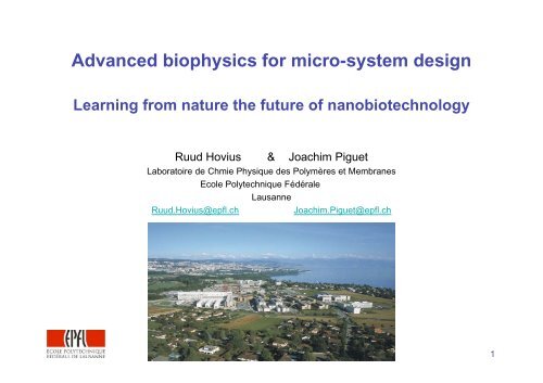
Advanced biophysics for micro-system design<br />
Learning from nature the future of nanobiotechnology<br />
Ruud Hovius & Joachim Piguet<br />
Laboratoire de Chmie Physique des Polymères et Membranes<br />
Ecole Polytechnique Fédérale<br />
Lausanne<br />
Ruud.Hovius@epfl.ch<br />
Joachim.Piguet@epfl.ch<br />
1
Structure of course<br />
Part I - Molecules and Labeling<br />
• Introduction<br />
÷ Molecular interactions<br />
÷ Molecular machines<br />
÷ Fluorescence<br />
• Labeling proteins<br />
÷ During biosynthetic<br />
Green fluorescent protein<br />
Non-natural amino acids<br />
÷ After biosynthesis<br />
Enzyme or substrate fusion<br />
Binding partners<br />
Chemical reactivity<br />
÷ Covalent vs reversible<br />
Part II - Single molecule imaging and<br />
spectroscopy<br />
• Single molecule detection<br />
÷ Theory<br />
÷ Experiment<br />
• Wide-field imaging<br />
÷ Molecular motions<br />
÷ Interactions & catalysis<br />
• Confocal detection<br />
÷ Fluctuation analysis<br />
÷ Multi-parameter fluorescence<br />
• Nano-devices<br />
2
What does one see in a living cell<br />
What changes<br />
• Activity<br />
• Structure<br />
• Location<br />
• Concentration<br />
• Diffusion<br />
• Transport<br />
• Interactions<br />
3
Molecular interactions - Biological networks<br />
• The cell is extremely complex<br />
many intertwined signaling and metabolic multi-dimensional networks<br />
a strong spatial-temporal organization<br />
4
Cells - Very complicated reaction conditions<br />
• Highly heterogeneous - compartments with very different contents<br />
• Transport and separation<br />
• Size-dependent effects on diffusion, kinetics and thermodynamics<br />
• Non-equilibrium state<br />
• Many components - 5’000 different gene products<br />
- many proteins have similar functions<br />
=> Systems biology :<br />
Description in space (x,y,z) and time (t) of a cell, an organism or even biotope<br />
Goodsell, Nat Chem Biol 3 (2007) p.681<br />
5
Cells - Highly crowded<br />
6
Molecular interactions - Reductionist’s approach<br />
• The cell is extremely complex<br />
⇒ Simplification, e.g.<br />
⇒ Isolated membranes<br />
⇒ Purified proteins<br />
⇒ Bio-inspired systems<br />
7
Biological networks - Molecular interactions<br />
• Interacting neurons<br />
• Synaptic organization<br />
Signaling events within a cell<br />
⇒ Right place<br />
⇒ Right time<br />
⇒ Proper arrangement<br />
⇒ Specificity & right partners<br />
Hammond “Cell & Mol Neurobiology”<br />
Kim, Nat Rev Neurosci 5 (2004) 772<br />
through reversible and adjustable<br />
molecular interactions.<br />
8
<strong>Protein</strong>s - Modular assembly of functional domains<br />
• <strong>Protein</strong>s must be multi-functional<br />
- specific interaction<br />
- regulation of interaction<br />
- effector<br />
• <strong>Protein</strong>s are in general not monolithic globules<br />
but rather have a beads-on-a-string multi-domain structures,<br />
where each domain has a specific function<br />
• Domains: small (70-140 amino acids) autonomously folding sequences<br />
specific function e.g. catalysis or binding<br />
> 1’000 type of domains present in human genome<br />
• Linear combination of a few domains yields an infinite variation of proteins<br />
with each unique properties<br />
“Modular <strong>Protein</strong> Domains” Cesareni e.a. (Eds) 2005 Wiley<br />
9
Reversible & adjustable protein interactions<br />
M-M Zhou, Bhattacharyya Ann Rev Bc 2006<br />
10
Reversible & adjustable protein interactions<br />
Example 1: The SH2 domain family<br />
- binds specifically to the protein sequence: ….-pY-x-x-hydrophobic-…<br />
where pY is a tyrosine phosphorylated by the action of a specific kinase:<br />
- The domains have a conserved structure and<br />
- The ligands are positioned similarly.<br />
- Specificity is detemined by the 3rd amino acid<br />
after -pY- determines specificity<br />
11
Reversible & adjustable protein interactions<br />
Growth factor receptors are also called receptor tyrosine kinases. They are present<br />
on the surface of cells as monomers.<br />
Upon ligand binding the receptors dimerize and trans-phosphorylate eachother’s<br />
tyrosines => activation of cellular signalling<br />
12
Reversible & adjustable protein interactions<br />
Example 2: The PH domain family<br />
- the human genome encodes about 250 PH domains<br />
some bind to PIP 2 ,<br />
an abundant lipid in the plasma membrane of cells<br />
13
Imaging PIP 2 in vivo using PH-GFP chimeras<br />
• PIP 2<br />
is rapidly broken down upon activation of e.g. the angiotensin receptor<br />
• A fusion protein PH_GFP, composed of a green fluorescent protein (GFP) and a<br />
PH domain binds to PIP 2<br />
in the plasma membrane<br />
• Angiotensin addition leads to PIP 2<br />
breakdown<br />
=> PH-GFP can not bind to the membrane anymore<br />
PI(4,5)P 2<br />
specific domain:<br />
- [PI(4,5)P 2<br />
] high in membrane<br />
- Angiotensin promotes breakdown<br />
Balla, TiPS 2000<br />
14
Domain combination - Variations on a theme<br />
“Combinatorial” protein design to create proteins with new properties<br />
15
Molecular interactions exploiting domains<br />
• Organizing molecules<br />
• Scaffolds allow regulation and integration of signals and<br />
organization of signaling molecules in space and time<br />
• Coincidence of events is important for both control and out-put<br />
16
Scaffolds - regulation and integration<br />
17
Modified scaffolds => modified output<br />
Comparison of pheromone and osmo-sensing in yeast<br />
organisation by different scaffolds => different cellular response<br />
INPUT<br />
Chimeric scaffold<br />
OUTPUT<br />
18
<strong>Protein</strong> interactions: Genome wide approaches<br />
• Classical methods to determine protein-protein interactions:<br />
e.g. 2-hybrid, GST-pull down, phage display<br />
⇒ protein interaction maps<br />
⇒“simplistic” view of proteins<br />
19
<strong>Protein</strong> interactions: Genome wide approaches<br />
• Domains:<br />
- approx 1’000 types of domains in genome<br />
÷ for about 750 types, 1 or more structures known<br />
- about 10’000 types of domain-domain interactions are predicted<br />
÷ 20% have been structurally characterized<br />
=> Use functional and structural information to predict<br />
- interactions between domains, and thus between proteins<br />
- identify interactions that<br />
÷ can happen simultaneously<br />
÷ are sterically overlapping, and thus excluding<br />
- estimate by modeling<br />
÷ affinities<br />
÷ rate constants<br />
20
<strong>Protein</strong> interactions: Genome wide approaches<br />
21
<strong>Protein</strong> interactions: Genome wide approaches<br />
• An example for the protein Ras:<br />
22
Interacting molecules - Nanoscopic building blocks<br />
Constructing supra-molecular functional entities using nature’s components.<br />
=> in solution or on surfaces<br />
Commonly used constructing materials:<br />
Streptavidin<br />
Biotin<br />
Immunoglobulins Antigens <strong>Protein</strong> A or G<br />
Oligonucleotides<br />
23
Interacting molecules: Streptavidin<br />
• Tetrameric protein from Streptomyces avidinii, about 65 kDa (approx 5x5x5 nm)<br />
• each monomer binds very tightly to biotin : K d<br />
~ 10 -14 M or ΔG ~ 32 kT.<br />
Biotin, also known as vitamin H or B7<br />
O<br />
H<br />
HN<br />
NH<br />
H<br />
S<br />
COOH<br />
• Avidin is a glycoprotein from bird eggs with comparable structure and properties.<br />
It prevents biotin absorption in the gastrointestinal tract.<br />
• Biotin can easily be linked chemically to many substances.<br />
24
Interacting molecules: Monovalent streptavidin<br />
Streptavidin is a tetrameric protein<br />
- Danger of cross-linking biotinylated molecules<br />
=> Design a “Monovalent streptavidin” using<br />
“dead” subunits carrying mutations N23A,S27D &S45A => K a = 10 3 M -1<br />
1”alive” + 3 “dead” => K d<br />
~ 2.10 -13 M<br />
Howarth, Nature Meth 3 (2006) 267<br />
25
Interacting molecules: Streptavidin<br />
How to bind proteins to streptavidin<br />
=> biotinylation either chemical of biosynthetically<br />
=> fusion with a Streptavidin-tag<br />
Analysis of peptide libraries identified peptide with good affinities for streptavidin<br />
• Strep-tag WSHPQFEK-COOH K d<br />
= 72 µM<br />
K d<br />
= 1 µM to engineered streptavidin<br />
Voss & Skerra, <strong>Protein</strong> Eng 10 (1997) 975<br />
• SBP-tag :<br />
K d<br />
= 2.5 nM<br />
MDEKTTGWRGGHVVEGLAGELEQLRARLEHHPQGQREP<br />
Keefe, Prot Exp Purif 23 (2001) 440<br />
26
Interacting molecules: Immunoglobins<br />
A binding partner to almost anything, the immune system uses immunoglobins,<br />
also called antibodies:<br />
• the antigen is bound to the variable regions<br />
• genetic recombination yield infinite variations<br />
Antibody of IgG class<br />
• Each domain is approx 50 kDa and 5 nm long.<br />
• Dissociation constants are nM for good antibodies.<br />
27
Interacting molecules: Immunoglobins<br />
Potential disadvantages:<br />
• large size (150 kDa) steric problems<br />
• 2 binding sites<br />
cross-linking<br />
Solution: make it smaller!!<br />
=> Enzymatic treatment => Genetic engineering<br />
F ab<br />
- fragments<br />
scFV-fragments<br />
28
Interacting molecules: Antibody-binders<br />
• Bacterial have developed antibody-binding proteins as a means of defense<br />
<strong>Protein</strong> A is a cell wall protein found in Staphylococcus aureus,<br />
4 binding sites for heavy chain on F c<br />
with a K d<br />
= 10 -8 M<br />
<strong>Protein</strong> G is a cell wall protein from G. streptococci,<br />
2 F c<br />
binding sites<br />
<strong>Protein</strong> L is from Peptostreptococcus magnus,<br />
binds to Ig’s with a particular kappa-light chain<br />
These proteins are often used to coat surfaces for further antibody Y<br />
immobilization.<br />
Y Y Y Y<br />
29
Interacting molecules: Oligonucleotides<br />
Oligonucleotides are polymers made of the 4 (ribo)nucleobases : A C G T<br />
Complementary antiparallel strands hybridize through base pairing and sequence<br />
recognition<br />
30
Interacting molecules: Oligonucleotides<br />
Complementary strands are kept together with many weak H-bonds<br />
=> temperature sensitive<br />
DNA double strands dissociate at a ginven temperature: the melting temperature T m<br />
GATC<br />
|||| melts at 12 °C<br />
CTAG<br />
31
Interacting molecules: Oligonucleotides<br />
Base pairing & sequence recognition can be used e.g. to construct<br />
complicated structures:<br />
165x165 nm<br />
Rothemund, Nature 440 (2006) 297<br />
Scale: 1 to 2.10 14<br />
32
Interacting molecules - Nanoscopic building blocks<br />
Alternative new proteins: See reviews<br />
•<br />
•<br />
Advantages are:<br />
• small<br />
• easier to optimize against any target<br />
• can be expressed in large amounts<br />
33
Interacting molecules - Nanoscopic building blocks<br />
Organic molecules specifically recognizing oxide surfaces:<br />
=> Selective Molecular Assembly Patterning (SMAP)<br />
e.g. - alkane phosphates (DDP) => Nb 2<br />
O 5<br />
or Ta 2<br />
O 5<br />
- Poly-l-lysine-PEG (PLL-PEG) => SiO 2<br />
N. Spencer, ETH-Zürich<br />
34
Interacting molecules: LEGO for the advanced<br />
35
Interacting molecules: LEGO for the advanced<br />
• The cell is extremely complex<br />
36
Molecular machines<br />
⇒ Linear motors<br />
⇒ Rotational motors<br />
⇒ Energy conversion<br />
⇒ <strong>Protein</strong> factories<br />
⇒ Transport systems<br />
⇒ Channels<br />
Motors walk on filament<br />
Kinesin on microtuble<br />
Filament slide on immobilized motors<br />
Myosin on Actin<br />
Typical speed:<br />
Step size:<br />
about 1 µm/s<br />
about 10 nm<br />
37
Molecular machines<br />
⇒ Linear motors<br />
⇒ Rotational motors<br />
⇒ Energy conversion<br />
⇒ <strong>Protein</strong> factories<br />
⇒ Transport systems<br />
⇒ Channels<br />
Bacterial flagella<br />
Energy conversion: Electro-chemical >> mechanical<br />
Number of H + per revolution: ~ 1000 (~ 6000 kT)<br />
Maximum rotation rate: 300 Hz (H + ) or 1700 Hz (Na + )<br />
Maximal speed:<br />
1 µm/s<br />
Diameter:<br />
50 nm<br />
38
Molecular machines<br />
⇒ Linear motors<br />
⇒ Rotational motors<br />
⇒ Energy conversion<br />
⇒ <strong>Protein</strong> factories<br />
⇒ Transport systems<br />
⇒ Channels<br />
ATPase uses H +- gradient to produce ATP: 3-5 H + needed per ATP<br />
Energy conversion: Electro-chemical >> mechanical >> chemical<br />
Dimensions:<br />
Diameter about 10 nm<br />
Rotations:<br />
> 3000 rpm => >10’000 ATP/min<br />
39
Molecular machines<br />
⇒ Linear motors<br />
⇒ Rotational motors<br />
⇒ Energy conversion<br />
⇒ <strong>Protein</strong> factories<br />
⇒ Transport systems<br />
⇒ Channels<br />
Energy conversion:<br />
Speed:<br />
Chemical => chemical<br />
Few residues per second<br />
40
Molecular machines<br />
⇒ Linear motors<br />
⇒ Rotational motors<br />
⇒ Energy conversion<br />
⇒ <strong>Protein</strong> factories<br />
⇒ Transport systems<br />
⇒ Channels<br />
In vitro<br />
Energy conversion:<br />
Speed:<br />
In vivo<br />
Chemical => motion<br />
µm/s<br />
41
Molecular machines<br />
⇒ Linear motors<br />
⇒ Rotational motors<br />
⇒ Energy conversion<br />
⇒ <strong>Protein</strong> factories<br />
⇒ Transport systems<br />
⇒ Channels<br />
closed ⇔ open<br />
Kuo, Structure 13 (2005) 1463<br />
Voltage-gated potassium channels<br />
Permeability:<br />
10 6 ions/s<br />
Selectivity:<br />
only potassium<br />
Conversion:<br />
potential => current<br />
42
Biomolecular nanotechnology => Nanomachines<br />
÷ Molecular interactions<br />
÷ Molecular machines<br />
=> Nanobiomachines<br />
NASA/Rutgers<br />
Arizona<br />
43
Biomolecular nanotechnology => Nanomachines<br />
Nanobiomachines: A recent example of a miniaturized flee circus<br />
Hiratsuka, PNAS 103 (2006) 13618<br />
44
Where to study the biological objects<br />
• In vitro<br />
+ Purified, well controlled system<br />
+ Easy to label<br />
- Out of cellular matrix/context<br />
• In vivo<br />
+ In the cellular context<br />
+ Can be labelled biosynthetically<br />
- Hard to access<br />
- Very complex<br />
- Difficult to control<br />
45
Method of choice: Fluorescence<br />
• Non-invasive => in vivo<br />
• Sensitive => down to a single molecule<br />
• On line => direct info from ns to days<br />
• High information content<br />
– Location, movement & distance<br />
– Microenvironment<br />
– Molecular interaction<br />
• Many parameters<br />
– Intensity<br />
– Spectral shape<br />
– Lifetime<br />
– Anisotropy<br />
– Position<br />
46
Method of choice: Fluorescence<br />
What is needed<br />
• Molecular and cellular biology<br />
Design, engineering, expression, purification of desired molecules<br />
• Instrumentation and methodology<br />
Acquisition and data treatment, interpretation<br />
• Labeling<br />
Specific introduction of non-perturbing probes<br />
47
Labeling is needed for identification<br />
=> Discriminate from matrix and between molecules of interest<br />
- Cells are full of fluorescent molecules<br />
=> Detect the parameter of interest<br />
Ideal:<br />
• Specific labeling of molecular of interest within a living cell<br />
• Stoichiometric<br />
• Non-perturbing<br />
• Bright and stable labels<br />
48
What kind of molecules to label<br />
• Sugars Glycomics<br />
– Cell recognition<br />
– Protection<br />
• Lipids Lipidomics<br />
– Membrane barrier<br />
– Signaling<br />
• Nucleic acids Genomics<br />
– Genetic information<br />
– Catalysis<br />
• <strong>Protein</strong>s Proteomics<br />
– Catalysis<br />
– Machines<br />
• Small molecules Metabolomics<br />
Pharmacolomics<br />
49
A classical fluorophore:<br />
Fluorescein<br />
Some basics on fluorescence<br />
• Wavelength of excitation < emission => Stokes’ shift<br />
• Lifetime τ of the excited state ns - ms<br />
• Extinction coefficient: probability of absorbance 10 4 - 10 6 M -1 cm -1<br />
• Quantum yield q : photons emitted/absorbed 0 - 1<br />
• Total number of photons before photo-damage 10 4 - 10 6<br />
• Rate of emission ≤ 2.5 10 6 s -1<br />
50
Photoselection and anisotropy of fluorescence<br />
Steady-state anisotropy r is defined by:<br />
with the rotational correlation time<br />
=> r depends also on mass.<br />
51
Photoselection and anisotropy of fluorescence<br />
• Applications: • Binding of small ligand A to big receptor B:<br />
A* + B A*B<br />
• Rotation & orientation of molecules<br />
e.g. ATPase or flagella<br />
52
Some basics on fluorescence<br />
Fluorescence Resonance Energy Transfer (FRET) efficiency E depends<br />
on:<br />
- Overlap of emission D and absorption A<br />
- Distance between D and A<br />
=> E = 0.5 at the Föster radius R o<br />
5 Å < R o<br />
< 80 Å<br />
53
Some basics on fluorescence<br />
• Applications: • Molecular interactions: D* + A° D*A°<br />
• Structural changes:<br />
D*-A° D* ---A°<br />
S. Weiss, Science (1999)<br />
54
Labeling, with what<br />
The probe should be:<br />
- Small is beautiful<br />
- Bright<br />
- Stable<br />
- Non-perturbing<br />
- ..<br />
The labelling should be/allow:<br />
- Specific<br />
- Stoichiometric<br />
- Inside or on surface of cell<br />
- Permanent or reversible<br />
Both should match the lifetime of the<br />
process of interest<br />
E. A. Jares, Nat BioTech 21 (2003) 1387<br />
55
Labeling, with what<br />
- unspecific<br />
- unspecific<br />
- blinking<br />
+ specific<br />
• <strong>Protein</strong>s + (bio)synthetically - large<br />
+ specific - post-labeling<br />
56
Labeling with organic dyes<br />
Very large choice of (reactive) dyes commercially available from e.g.<br />
Invitrogen (Molecular Probes )(Alexa)<br />
GE (Amersham) (Cy dye)<br />
Toronto Research Chemicals<br />
Chromeon<br />
Atto-tec (Atto)<br />
Biotium<br />
Dyomics<br />
FluoProbes<br />
57
Labels: Organic dyes<br />
Hard to choose form the many dyes available => try many!!<br />
Important points:<br />
• Wavelengths of excitation & emission<br />
• Type of matrix, e.g. living cell or silicon waver<br />
• Interference with functionality of the molecule to be labeled<br />
e.g. labelled acetylcholine<br />
depending on spacer length<br />
either agonist or antagonist<br />
Baindur, Drug Development Research 33 (1994) 373<br />
58
Labels: Organic dyes<br />
Hard to choose form the many dyes available => try many!!<br />
Important points:<br />
• Stability of dye &<br />
observation time needed<br />
www.atto-tec.com<br />
• Hydrophobicity of probe<br />
Dye molecules contain large conjugated, aromatic systems<br />
=> aggregation<br />
=> unspecific binding<br />
• “Bad ones” Bodipy’s, Dy-655, rhodamines, ATTO647N<br />
• Nonspecific interactions<br />
with membranes e.g. Cy5<br />
with mitochondria e.g. Atto647N<br />
59
Labels: Quantum dots<br />
Semiconductor crystals of nm-size show:<br />
• Size dependant absorption and emission wavelengths<br />
=> colors can be tuned by varying size<br />
• Great brightness<br />
• Narrow emission spectra<br />
• Highly photostability<br />
• Broad absorption spectra => allows multiplexing.<br />
CdSe/CdS<br />
- Large size & stoichiometry of modification<br />
Bruchez, Science 281 (1999) 2012<br />
60
How to get them<br />
• Home made<br />
Labels: Quantum dots<br />
=> quite tedious<br />
• Commercial sources<br />
available with e.g. streptavidin or -COOH surface modification<br />
=> batch to batch variations,<br />
especially in blinking properties<br />
Where to get them from<br />
Invitrogen (Quantum dot)<br />
Evident<br />
Biopixels<br />
61
Labels: Quantum dots in imaging<br />
• Single AMPA receptor imaging on neurons<br />
- post-biosynthetic labeling upon bionylation.<br />
PSD-95-YFP & AMPA receptors with QD605<br />
1- PSD-95-YFP, then overlaid with DIC image<br />
2- plus QD605 movie (speeded up 3-fold)<br />
3- QD605 alone.<br />
Howarth, PNAS (2005) 1388<br />
62
Quantum dots for supra-molecular entities<br />
• Scaffold<br />
• Multi-functional building block<br />
Medintz, Nat Materials 2 (2003) 630<br />
Geissbuehler, Angew. Chem. 44 (2005) 1388<br />
63
How to label<br />
Methods:<br />
Modality:<br />
• Biosynthetically • Covalent<br />
- GFP<br />
- Non-natural amino acids • Reversible<br />
• Post-biosynthetically<br />
- Enzymes<br />
- Small tags<br />
- Ligands<br />
• Chemically<br />
- Reactive functional groups<br />
64
Reviews<br />
• General<br />
Marks & Nolan “Chemical labeling strategies for cell biology”<br />
Nature Methods 3 (2006) 591<br />
Prescher & Bertozzi “Chemistry in living systems”<br />
Nature Chem Biol 1 (2005) 13<br />
Hovius e.a. “Fluorescent Labelling of Membrane <strong>Protein</strong>s in Living Cells”<br />
in "Structural Genomics on Membrane <strong>Protein</strong>s” CRC Press (2006) 199<br />
• Fluorescent proteins<br />
Tsien, R.Y. The green fluorescent protein.<br />
Annu. Rev. Biochem., 67, 509–544,1998.<br />
Zimmer, M. Green fluorescent protein (GFP): applications, structure, and related<br />
photophysical behavior.<br />
Chem. Rev., 102, 759–781,2002.<br />
• Nonnatural amino acidsand suppressor tRNA<br />
Wang, Xie & Schultz “Expanding the genetic code”<br />
Annu Rev Biophys Biomol Struct 35 (2006) 225<br />
65
Biosynthetic labeling<br />
• Labeling during biosynthesis of the protein<br />
Advantages:<br />
- in the cell<br />
- can be targeted to compartment of wish<br />
- on the protein of wish<br />
- site-specific<br />
Disadvantages:<br />
- limited choice of labels<br />
- limited quality<br />
- compatibility with protein<br />
- incomplete targeting<br />
66
Biosynthetic labeling: Fluorescent proteins<br />
Green Fluorescent <strong>Protein</strong> from the<br />
jellyfish Aequorea victoria<br />
• 26 kDa protein with a β-barrel structure<br />
• Chromophore within barrel => very stable!!<br />
R. Y. Tsien<br />
M. Zimmer, Chem Rev (2002)<br />
67
Biosynthetic labeling: Fluorescent proteins<br />
• Many color variants either by mutagenesis or from other species<br />
=> Very differing spectroscopic properties !!<br />
Ex (nm) Em (nm) Abs coeff (M - Q τ (ns) r o φ (ns) brillance<br />
1<br />
cm -1 )<br />
eCFP 434 477 26'000 0.4 1-1.5 (50 %)<br />
0.2<br />
3.3-4.0<br />
eBFP 380 440 31'000 0.18 3 0.11 Bleaches fast<br />
eYFP 514 527 84'000 0.61 3.7 1.0 Bleaches fast !<br />
Long dark states<br />
eGFP 489 508 55'000 0.6 2.5 0.33 24 0.64 pH sensitive<br />
DsRed 558 583 22'5 - 27'000 0.25-0.4 4 ±0.15 Aggregates<br />
far reds ±590 ±640<br />
A. Miyawaki, Nat. Neurosci Sept 2003, S1<br />
68
Biosynthetic labeling: Fluorescent proteins<br />
• Fusion to protein of interest<br />
N- or C-terminal, internal<br />
• Can be targetted to all organelles<br />
Clontech<br />
• Not (too) toxic<br />
R. Y. Tsien<br />
69
Biosynthetic labeling: Fluorescent proteins<br />
Combination of several spectral variants facilitates multi-color-imaging<br />
up to 6 colors in parallel<br />
• E.g. using an LSM510 equipped with a 32 channel META detector<br />
and excitation at 458 nm.<br />
T. Kogure, Nat Biotech 24 (2006) 577<br />
70
Biosynthetic labeling: Fluorescent proteins<br />
Timer, a mutant red fluorescent protein, which slowly changes color.<br />
In vitro<br />
In vivo: Following protein expression<br />
and transport in time<br />
A. Terskikh, Science 290 (2000) 1585<br />
71
Fluorescent proteins : Photoactivation<br />
• GFP Mutant T203H: Irradiation at 413nm => inversed absorbance spectrum<br />
=> has a 100-fold contrast upon excitation at 480 nm!!<br />
• In vitro: Immobilized GFP<br />
Excitation at 488 nm<br />
Activation at 413<br />
=> Photo-lithography<br />
G. H. Patterson, Science 297 (2002) 1873<br />
72
Fluorescent proteins : Switching<br />
• mCherry mutant can be switched on and off repetitively!!<br />
Experiment: live E. coli’s expressing the proteins & using a Hg-lamp.<br />
Stiel (2008) Biophys J 2989<br />
73
Biosynthetic labeling: Fluorescent proteins<br />
Molecular interactions or conformational changes can be detected by FRET.<br />
Combining fluorescent proteins one can fine-tune the distance dependence<br />
of FRET to the system under investigation.<br />
G. H. Patterson, Anal Bc 284 (2002) 438<br />
74
Biosynthetic labeling: Fluorescent proteins<br />
Molecular interactions or conformational changes can be detected by FRET.<br />
e.g. a sensor of tyrosine phosphorylation:<br />
Sato, Nat Biotech (2002) 278<br />
75
Fluorescent proteins: Transducers in sensors<br />
Variations on a theme to detect molecular interactions, changes of<br />
conformation or of analyte concentrations.<br />
J. Zhang, Nat Rev MolCelBiol 3 (2002) 906<br />
76
Biosynthetic labeling: Fluorescent proteins<br />
Fluorescent protein based sensors have been used to measure:<br />
[Ca 2+ ] A. Miyawaki. Nature 388 (1997) 882<br />
[H + ] J. Llopis, PNAS 95 (1998) 6803<br />
[Cl - ] S. Jayaraman, JBC 275 (2000) 6047<br />
cAMP M. Zaccolo, Nature Cell Biology 2 (2000) 25<br />
[Zn 2+ ] K. K. Jensen, Bc 40 (2001) 938<br />
PK-C activity J. D. Violin. J Cell Biol 161 (2003) 899<br />
Tyr-kinase Sato, Nat Biotech (2002) 278<br />
Redox potential C. T. Dooley, JBC 279 (2004) 22284<br />
Androgens Awais, Angew Chem 45 (2006) 2707<br />
and many, many other analytes!!<br />
77
Biosynthetic labeling: Fluorescent proteins<br />
GFP analogues have revolutionized life sciences.<br />
But they are not perfect:<br />
Non-ideal fluorophores<br />
Wrongly targeted proteins<br />
Stoichiometry of co-expressed interacting fusion proteins<br />
Moreover, many fluorescent proteins and their use have been patented<br />
=> commercial applications need permission &<br />
license fees have to be paid.<br />
Fortunately monthly new fluorescent proteins are being found in<br />
different species !!<br />
78
Biosynthetic labeling: Nonnatural amino acids<br />
Normal protein synthesis<br />
DNA<br />
mRNA<br />
Stop codon<br />
Stop codon<br />
Each amino acid is encoded in the DNA by 3<br />
bases forming a codon.<br />
The end of a protein is indicated by the stopcodons.<br />
P. G. Schultz, D. Dougherty<br />
79
Biosynthetic labeling: Nonnatural amino acids<br />
Site-specific introduction of artificial amino acids<br />
with a suppressor tRNA complementary to the stop codon “AUC”<br />
UGA<br />
Stop codon<br />
P. G. Schultz, D. Dougherty<br />
80
Biosynthetic labeling: Nonnatural amino acids<br />
Sofar, only small, relatively<br />
weak fluorophores could be<br />
introduced directly:<br />
• Dansyl<br />
• NBD<br />
• Coumarine<br />
• Bodipy FL<br />
Wang, Annu Rev Biophys Biomol Struct 35 (2006) 225<br />
81
Biosynthetic labeling: Nonnatural amino acids<br />
Site-specific incorporation of NBD-amino acids in a G protein-coupled receptor.<br />
• In vivo NBD was introduced at different sites<br />
• A rhodamine labeled ligand was bound to the receptor<br />
• Distances between labels was measured by FRET<br />
=> A model of the ligand-receptor complex.<br />
G. Turcatti, JBC 271 (1996) 19991<br />
82
Biosynthetic labeling: Nonnatural amino acids<br />
Problems<br />
• nnaa-tRNA<br />
- difficult to synthesis<br />
- hydrolysis in cell<br />
- low yield<br />
- high background<br />
• Only in vitro or in oocyte<br />
- tedious<br />
- mammalian cell system wished<br />
• Only small, no so ideal fluorophores<br />
- limited scope of application<br />
however:<br />
• Biotin-containing nnaa’s => labeled streptavidin<br />
• Chemo-selective nnaa’s<br />
83
Biosynthetic labeling: Nonnatural amino acids<br />
Introduction of a chemo-selective aldehyde containing nnaa:<br />
- reacts specifically with hydrazine derivatives of fluorophores.<br />
In vivo demonstration: m-acetyl-L-phenylalanine<br />
was introduced in LamB using suppressor tRNA<br />
methods in E. coli, and post-biosynthetically<br />
labeled with 3 hydrazine derivatives.<br />
Z. Zhang, Bc 42 (2003) 6735<br />
84
Biosynthetic labeling: Nonnatural amino acids<br />
Introduction of a chemo-selective alkynyl amino acids, which react specifically<br />
with azido-derivatives. (or the inverse!):<br />
+<br />
Met Phe Azido-coumarine<br />
In vivo demonstration:<br />
labeling of barnstar in expressed E. coli,<br />
and post-biosynthetically<br />
• - o/n labeling<br />
• - random<br />
Beatty, J Am Chem Soc 127 (2005) 14150<br />
85
Post-biosynthesis labeling of a genetic tag<br />
The protein of interest can be genetically modified to contain a<br />
sequence that can be labelled once the protein has been made.<br />
Such a sequence can be:<br />
<strong>Protein</strong><br />
Enzyme<br />
Substrate<br />
Binding site<br />
Peptide<br />
Binding partner<br />
Substrate<br />
86
Post-biosynthesis labeling: Enzyme tag - hAGT<br />
• O 6 -Alkylguanine-DNA Alkyltransferase (hAGT) is a DNA-repair “enzyme”.<br />
• The nucleotide-modifying LABEL is covalently bound to AGT.<br />
-/+ Not all substrates are membrane permeable.<br />
- Cells need to be devoid of endogenous interfering AGTs => Mutant versions of AGT<br />
Keppler, Nat BioTech 21 (2003) 86 Commercially available as SNAP-tag from<br />
87
Post-biosynthesis labeling: Enzyme tag - hAGT<br />
• Development of AGT-mutants:<br />
- no more interference of endogenous AGT<br />
- “CLIP” recognizing cytosine-based substrates<br />
Keppler (2003)Nat BioTech; (2004)PNAS; Juillerat (2005) ChemBioChem; Gautier (2008)Chem & Biol<br />
88
Post-biosynthesis: Substrate protein - Carrier proteins<br />
Intermediates in cellular biosynthesis are in several cases attached to so-called<br />
“carrier proteins” (CP) to enhanced their solubility or preserve reactive form.<br />
÷ Carrier proteins are small 70-100 residues, compact and fold autonomously.<br />
÷ Enzymes couple the CoA-activated intermediates () to the carrier proteins<br />
+<br />
CP<br />
PPTase<br />
CP<br />
4’-phosphopantetheine (PP) group of coenzyme A (CoA) is coupled covalently to<br />
an OH-group of a serine on CP by PP-transferase (PPTase).<br />
Modification of the SH-group of CoA has no or little effect on the reaction.<br />
± Only cell surface molecules<br />
Examples are:<br />
Peptide Carrier <strong>Protein</strong>s (PCP) J. Yin, Chem & Biol 12 (2005) 999<br />
Acyl Carrier <strong>Protein</strong>s (ACP) George, JACS 126 (2004) 8896<br />
89
Post-biosynthesis: Substrate protein - Carrier proteins<br />
Important for labeling:<br />
• Genetically fuse CP to protein of interest<br />
• Express in cells<br />
• Incubate with fluorescently labeled CoA and PPTase<br />
• Wash out free substrate before imaging<br />
-+ Surface protein labeling only<br />
as labeled CoA and PPTase are membrane impermeable.<br />
+- CoA-fluorophores nicely water-soluble.<br />
Commercially available from<br />
90
Post-biosynthesis: Substrate protein - ACP<br />
Two-color labeling for FRET: Comparison with fluorescent proteins<br />
NK1_eCFP NK1_EYFP Merge<br />
FPs:<br />
Co-transfect<br />
mixed DNA<br />
ACP:<br />
Label with<br />
mixed CoA’s<br />
ACP-NK1_Cy3 ACP-NK1_Cy5 Merge<br />
=> ACP is by far better for investigations on cell surface proteins.<br />
Meyer, PNAS 103 (2006) 2138 & FEBS (2006)<br />
91
Post-biosynthesis: Substrate protein - Carrier proteins<br />
Possibility for specific labeling of several proteins using both ACP and PCP fusions,<br />
employing specific PPT enzymes:<br />
AcpS from E. coli will only modify ACP<br />
Sfp from B. subtilis will modify both ACP & PCP<br />
Example: cell wall proteins in yeast labeled with Cy3 on ACP and Cy5 on PCP.<br />
Vivero-Pol, JACS 127 (2005) 12770<br />
92
Post-Biosynthetic labeling: Peptide substrate - Biotin-ligase<br />
• Fusion of biotin acceptor sequence GLNDIFEAQKIEWHE to protein of<br />
interest<br />
• Enzymatic coupling of a biotin analogue which reacts specifically with<br />
O<br />
hydrazine derivatives of fluorophores.<br />
O<br />
N<br />
N<br />
H<br />
S<br />
S<br />
Biotin ligase<br />
Ketone 1<br />
ATP<br />
H 2 N<br />
pH 6-7<br />
N<br />
H<br />
O<br />
+ Highly specific for Biotin tag<br />
+ Only surface exposed proteins<br />
- Hydrazine coupling slow<br />
- Bond can hydrolyse<br />
Chen, Nature Meth (2005)<br />
93
Post-Biosynthetic labeling: Peptide substrate - Biotin-ligase<br />
Alternatively, coupling with normal biotin and labelling with QD-streptavidin<br />
- Huge size of label<br />
Howarth, PNAS (2005)<br />
94
Post-Biosynthetic labeling: Peptide tags - Epitopes<br />
Epitopes are sequences of 5 to about 20 amino acids against<br />
which antibodies can be raised or engineered. These<br />
sequences can be introduce into protein of wish.<br />
Examples are:<br />
HA Tyr - Pro - Tyr - Asp - Val - Pro - Asp -Tyr - Ala<br />
VSV Tyr - Thr - Asp - Ile - Glu - Met - Asn - Arg - Leu - Gly - Lys<br />
FLAG Asp -Tyr -Lys -Asp - Asp - Asp - Asp - Lys<br />
His6 His - His - His - His - His - His<br />
Fluorescently labelled specific & high affinity antibodies are<br />
commercially available.<br />
+ Small tags<br />
+ Specific & high affinity<br />
+ Any color<br />
+- Multiple labels per antibody<br />
- Antibodies are huge<br />
95
Post-Biosynthetic labeling: Peptide tags - FlAsH binding<br />
• The biarsenical FlAsH is virtually non-fluorescent<br />
• Binding to a CCxxCC motif results in a 100-fold fluorescence increase<br />
• Reversed by addition of ethylenedithiol (EDT)<br />
+ Works also in vivo<br />
- Binding is slow and has a low affinity<br />
- Need for high concentration of EDT and non-fluorescent dyes.<br />
B. A. Griffin, Science 281 (1998) 269<br />
96
Post-Biosynthetic labeling: Peptide tags - FlAsH binding<br />
FlAsH labelling, an example:<br />
Dual color pulse labeling of CCxxCC-connexin using<br />
- 1 st pulse of FlAsH<br />
- 2 nd pulse of ReAsH later in time<br />
J. Zhang, Nat Rev MolCelBiol 3 (2002) 906 &<br />
G. Gaietta, Science 296 (2002) 503<br />
97
Post-Biosynthetic labeling: Peptide tags - FlAsH binding<br />
Improvement of peptide tag<br />
1998: Initial ..CC-xx-CC.. peptide α-helix<br />
=> background = specific needed a lot of thiol reagents<br />
2002: Then ..CC-PG-CC.. loop<br />
=> less non specific<br />
2005: Now: ..FLN CC-PG-CC-MEP..<br />
=> 1.7-fold increase quantum yield<br />
=> 20-fold increased resistance dithiol reagents<br />
- can be washed with 0.75 mM dimercaptopropanol to<br />
suppress non-specific binding<br />
- Always tested on highly expressed proteins<br />
e.g. actin or concentrated proteins like connexins<br />
- Still very slow 1-2 hours<br />
Martin, Nat Biotech 23 (2005)1308<br />
98
Post-Biosynthetic labeling: Peptide tags - FlAsH binding<br />
Improvement of the system:<br />
2007: Development of AsCy3<br />
+ more stable and brighter than previous probes<br />
+ binds specifically to ..CC-AEAA-CC..<br />
and not to ..CC-xx-CC..<br />
+ binds faster than FlAsH and ReAsH<br />
+ orthogonal double labeling possible by using both AsCy3 and FlAsH<br />
and their respective peptide tags CC-xx-CC and CC-AEAA-CC<br />
Cao, JACS 129 (2007) 8672<br />
99
Post-Biosynthetic labeling: Peptide tags - NTA-Probes<br />
Principle: Selective binding of NTA-Ni 2+ to oligohistidine sequences<br />
+ Wide choice of chromophores: Quenching, co-localisation, FRET,<br />
membrane permeability..<br />
+ Small label<br />
-+ Moderate affinity<br />
Guignet (2004) Nature Biotech<br />
100
Post-Biosynthetic labeling: Peptide tags - NTA-Probes<br />
NTA-I binding to a His 6 -tagged GFP is demonstrated in vitro by FRET.<br />
+ Fast binding and disociation constant K d = 3 µM<br />
+- Fully reversible by EDTA<br />
Guignet (2004) Nature Biotech<br />
101
Post-Biosynthetic labeling: Peptide tags - NTA-Probes<br />
Reversible labelling of proteins on oligo-histidine tags by NTA-conjugates<br />
Lata<br />
2005 JACS<br />
K d : t 1/2 :<br />
mono-NTA 5’000 nM 5 sec fits to endurance of Cy5<br />
tris-NTA 0.1 nM 30 min need a quantum dot<br />
or a good organic dye<br />
102
Imaging single 5HT3R receptors using NTA-probes<br />
• NTA-Atto 647<br />
Different modes of diffusion were observed<br />
Guignet, ChemPhysChem 2007<br />
103
Labeling of receptors with fluorescent ligands<br />
Example: Conjugation of acetylcholine with dansyl to probe the<br />
muscle-type acetylcholine receptor<br />
104
Reversible Sequential single cell ligand binding assays<br />
A single cell expressing the acetylcholine receptor is pulse labeled &<br />
washed repeatedly many times<br />
Schreiter, ChemBioChem 2005<br />
105
Reversible Sequential labeling with receptor ligands<br />
A single cell is pulse labeled repeatedly under different conditions,<br />
e.g. changing concentrations of competing non-fluorescent ligand<br />
=> Pharmacological profiling on a single cell<br />
Schreiter, ChemBioChem 2005<br />
106
Post-Biosynthetic: Chemical labelling<br />
Reactive groups in proteins:<br />
• -SH of Cys<br />
• ε-NH 2<br />
of Lys or N-terminus<br />
• -OH of Ser<br />
• -COOH of C-terminus,<br />
Asp or Glu<br />
Abundance of amino acids:<br />
• Cys low on outside<br />
• Lys high<br />
• Ser + Thr high<br />
• Asp + Glu high<br />
Special cases:<br />
• N-terminal Ser<br />
• N-terminal Cys<br />
Accessibility<br />
• Surface exposed<br />
• Burried<br />
+ large choice of dyes<br />
- limited site-specificity, especially in vivo<br />
⇒ Often performed in vitro using purified proteins!!<br />
⇒ the only option is Cys labeling !!<br />
107
Chemical labeling of cysteines<br />
Maleimide and methylthiosulfonates (MTS) are usefull to react<br />
with SH-groups in aqueous conditions and on live cells.<br />
+ large commercial offer<br />
- bulky thioeter linkage<br />
+- in general slow to react<br />
- require large excess of reagent<br />
- unwanted side reactions<br />
Methylthiosulfonate<br />
OH<br />
S<br />
O<br />
- limited commercial offer<br />
- hydrolyses in water<br />
+ react extremely rapid<br />
+ highly selective<br />
+ quantitative without excess<br />
+ reversible<br />
108
Post-Biosynthetic: Site-specific single Cys-labeling<br />
.Strategy:<br />
i) Remove native Cys<br />
ii) Introduce single Cys<br />
iii) Fluorescence labeling of Cys<br />
iv) Fluorescence measurements<br />
NB: - step i) is not always needed<br />
- steps i) and ii) might kill protein<br />
- step iii) too!!<br />
109
Post-Biosynthetic: Cys-labeling of cell surface receptor<br />
Rhodamine-MTS labeling of a single Cys-mutant in channel of the receptor.<br />
• red:<br />
• green:<br />
rhodamine labeling<br />
fluorescent receptor ligand<br />
=> Specific labeling.<br />
Guignet<br />
110
Channel gating monitored by fluorescence<br />
Addition of agonist induces a reversible change in fluorescence intensity.<br />
1.4x10 6<br />
Agonist<br />
Wash<br />
Fluorescence intensity<br />
1.3<br />
1.2<br />
1.1<br />
1.0<br />
0<br />
100<br />
200<br />
300<br />
Time (s)<br />
400<br />
500<br />
600<br />
=> Structural rearrangements during channel gating.<br />
Guignet<br />
111
Post-Biosynthetic labeling: Comparison<br />
Method Tag Partner In cell Rev Fast<br />
HaloTag 33 kDa < 1 kDa Y N m - h<br />
hAGT 207 aa < 1 kDa Y N m<br />
DHFR 18 kDa < 1 kDa Y Y m - d<br />
FKBP 12 kDa < 1 kDa Y N/Y m - h<br />
ACP (PCP) 77 aa < 1 kDa + E N N m - h<br />
Epitope 5-30 aa 150 kDa N N/Y m - h<br />
Biotin-ligase 15 aa < 1 kDa + E N N m - h<br />
NTA 6-10 aa < 1 kDa Y N/Y s<br />
FlAsh 6-10 aa < 1 kDa Y N/Y m - d<br />
React (nn)AA 1 aa < 1 kDa N N m - h<br />
Ligand - 1 - 1000 kDa Y Y s - m<br />
112
Reversible vs covalent labeling<br />
Covalent<br />
+ once it’s there it is there to stay<br />
- upon photo bleaching your labeled molecule is invisible<br />
Reversible<br />
Depending on affinity and off rate complexes can have a<br />
• short lifetime of seconds to minutes<br />
- Cannot wash or label in advance<br />
+ Upon photobleaching can be replaced<br />
+ Repetitive labeling under same or different conditions<br />
• long lifetime of hours to days => almost as covalent labeling<br />
113
Labeling<br />
The ideal label and labeling method do not exist.<br />
=> What do you would like to learn and where<br />
÷ Localisation and movement ÷ In live cells<br />
÷ Molecular interactions ÷ Reconstituted systems<br />
÷ Structural changes<br />
=> Each protein & each process needs an individual approach.<br />
=> Combine orthogonal methods for multiple specific labeling.<br />
=> Beware of nonspecific labeling and artefacts.<br />
=> Combine measuring techniques.<br />
=> Do lots of careful experiments.<br />
114


