Breast Discharge: Ultrasound and Doppler Evaluation - NCI
Breast Discharge: Ultrasound and Doppler Evaluation - NCI
Breast Discharge: Ultrasound and Doppler Evaluation - NCI
You also want an ePaper? Increase the reach of your titles
YUMPU automatically turns print PDFs into web optimized ePapers that Google loves.
264<br />
RESULTS<br />
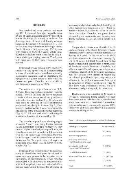
One hundred <strong>and</strong> seven patients, their mean<br />
age 42±13 years <strong>and</strong> their ages ranged between<br />
23 <strong>and</strong> 65 years, presenting either by uniorifical<br />
breast discharge (54 cases) or multi orifice<br />
discharge (one of them with bloody or serosanguous<br />
discharge) (53 cases) (Table 1). Duct<br />
ectasia was the predominant pathology, identified<br />
in 88 cases, their ages range 23-53 years,<br />
with mean age 33.8±11.6 years. While intraductal<br />
mass lesions were identified in only 17<br />
cases, their ages ranging between 27-65 years,<br />
with mean age: 45±12 years. Two cases were<br />
normal.<br />
<strong>Ultrasound</strong> proved to have 100% <strong>and</strong> 82.4%<br />
sensitivity <strong>and</strong> specificity in differentiating<br />
intraductal mass from non mass lesions, namely<br />
inspissated secretions <strong>and</strong> in identifying the<br />
benign or malignant nature of these masses.<br />
Colour <strong>and</strong> power <strong>Doppler</strong> raises specificity<br />
<strong>and</strong> diagnostic accuracy to 100%.<br />
The mean size of papillomas was (8.7±<br />
4.3mm). They lied within 3.2±2.1cm from the<br />
nipple. They all fulfilled the above described<br />
criteria with the exception of two papillomas<br />
with rather irregular outline (Fig. 2). A vascular<br />
stalk could be identified in 4 cases <strong>and</strong> minimal<br />
peripheral vascularity in 3 cases (Fig. 3), Ductoscopy,<br />
performed for 1 case, confirmed the<br />
site <strong>and</strong> size of the ultrasound detected papilloma<br />
(Fig. 4). 3D US was performed confirming the<br />
intraductal location of a lesion (Fig. 5).<br />
The intraductal papillomas showing atypia<br />
measured 5 <strong>and</strong> 11mm, being located between<br />
12 <strong>and</strong> 27mm from the nipple respectively. They<br />
showed higher vascularity than papillomas, the<br />
vessels are arranged in haphazard distribution<br />
(Fig. 6). The case proved to be ductal hyperplasia<br />
with atypia, was discovered during annual<br />
follow-up for fibrocystic breast changes, an<br />
intraductal mass 5mm is seen 27mm from the<br />
nipple (Fig. 7).<br />
<strong>Ultrasound</strong> was complementary to mammography<br />
in identifying malignant intraductal lesions,<br />
one case proved to be invasive ductal<br />
carcinoma, on mammography it was reported<br />
as BIRADS 3, on ultrasound an intraductal mass<br />
with wall irregularity was seen, microcalcification<br />
are seen within the mass but obscured on<br />
Soha T. Hamed, et al.<br />
mammogram by lobulated dilated duct (Fig. 8).<br />
Other masses were ranging in size 4-14mm, no<br />
definite ductal dilatation was seen in two of<br />
them. On colour <strong>Doppler</strong>, malignant lesion<br />
showed higher vascularity, non tapering r<strong>and</strong>omly<br />
dispersed vessels except in small 4mm<br />
mass.<br />
Simple duct ectasia was identified in 88<br />
cases according to the above described criteria.<br />
Mammography showed tubular retroareolar<br />
density in 8 cases, in the rest of cases, there<br />
was just an increase in retroareolar density; By<br />
US In 51 cases, bilateral dilated thin walled<br />
ducts are ranging in caliber from 3-8mm, some<br />
of the ducts showed Intra-ductal mobile, non<br />
adherent movable echogenic secretions were<br />
sometimes identified. In two cases, echogenic<br />
ball like lesions were identified resembling<br />
intraductal pappilomas, yet, they were non<br />
adherent to the wall <strong>and</strong> no colour flow could<br />
be detected on <strong>Doppler</strong> application (Fig. 9).<br />
The ducts were completely normal on both<br />
ultrasound <strong>and</strong> galactography in two cases.<br />
Ductography was requested in 20 cases, in<br />
five cases, intraductal filling defects were seen<br />
three cases proved to be intraductal mass lesions,<br />
other two cases were inssipisated secretions<br />
with no malignancy. Ductography showed 100%<br />
sensitivity <strong>and</strong> 60% specificity in diagnosing<br />
intraductal filling defect.<br />
Table (1): Pathological diagnosis of uni-orificial discha.<br />
Pathological diagnosis<br />
Intraductal carcinoma<br />
Intraductal papilloma<br />
(including intraductal<br />
papillomatosis, one case)<br />
Intra ductal papilloma/<br />
hyperplasia with atypia<br />
Localized duct ectasia<br />
Bilateral diffuse duct ectasia<br />
Normal<br />
Total<br />
No. of cases %<br />
6 (5.6%)<br />
8 (7.5%)<br />
3 (2.8%)<br />
37 (35%)<br />
51 (47.7%)<br />
2 (1.8%)<br />
107 (100%)


