Breast Discharge: Ultrasound and Doppler Evaluation - NCI
Breast Discharge: Ultrasound and Doppler Evaluation - NCI
Breast Discharge: Ultrasound and Doppler Evaluation - NCI

Create successful ePaper yourself
Turn your PDF publications into a flip-book with our unique Google optimized e-Paper software.
266<br />
Soha T. Hamed, et al.<br />
(A)<br />
(B)<br />
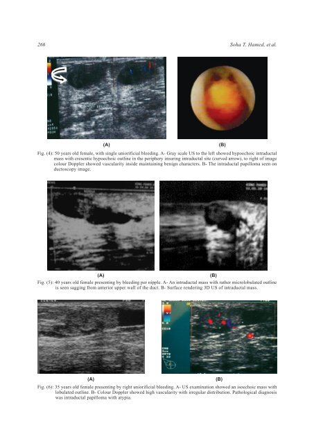
Fig. (4): 50 years old female, with single uniorificial bleeding. A- Gray scale US to the left showed hypoechoic intraductal<br />
mass with cresentic hypoechoic outline in the periphery insuring intraductal site (curved arrow), to right of image<br />
colour <strong>Doppler</strong> showed vascularity inside maintaining benign characters. B- The intraductal papilloma seen on<br />
ductoscopy image.<br />
(A)<br />
Fig. (5): 40 years old female presenting by bleeding per nipple. A- An intraductal mass with rather microlobulated outline<br />
is seen sagging from anterior upper wall of the duct. B- Surface rendering 3D US of intraductal mass.<br />
(B)<br />
(A)<br />
Fig. (6): 35 years old female presenting by right uniorificial bleeding. A- US examination showed an isoechoic mass with<br />
lobulated outline. B- Colour <strong>Doppler</strong> showed high vascularity with irregular distribution. Pathological diagnosis<br />
was intraductal papilloma with atypia.<br />
(B)