Breast Discharge: Ultrasound and Doppler Evaluation - NCI
Breast Discharge: Ultrasound and Doppler Evaluation - NCI
Breast Discharge: Ultrasound and Doppler Evaluation - NCI

You also want an ePaper? Increase the reach of your titles
YUMPU automatically turns print PDFs into web optimized ePapers that Google loves.
<strong>Breast</strong> <strong>Discharge</strong>: <strong>Ultrasound</strong> & <strong>Doppler</strong> <strong>Evaluation</strong><br />
267<br />
(A)<br />
(B)<br />
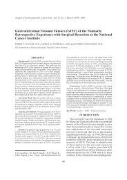
Fig. (7): 35 years old female with long history of breast discharge &developed uniorificial bleeding. A- A small mass with<br />
ill defined outline is seen with proximal ductal dilatation. B- On colour <strong>Doppler</strong> a small vascular stalk is seen.<br />
Pathology was florid ductal hyperplasia with focal atypia.<br />
(A)<br />
(B)<br />
(C)<br />
(D)<br />
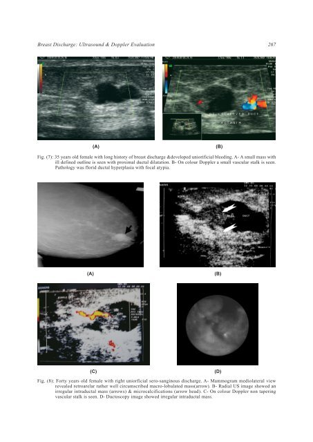
Fig. (8): Forty years old female with right uniorficial sero-sanginous discharge. A- Mammogram mediolateral view<br />
revealed retroarelar rather well circumscribed macro-lobulated mass(arrow). B- Radial US image showed an<br />
irregular intraductal mass (arrows) & microcalcifications (arrow head). C- On colour <strong>Doppler</strong> non tapering<br />
vascular stalk is seen. D- Ductoscopy image showed irregular intraductal mass.