Breast Discharge: Ultrasound and Doppler Evaluation - NCI
Breast Discharge: Ultrasound and Doppler Evaluation - NCI
Breast Discharge: Ultrasound and Doppler Evaluation - NCI

You also want an ePaper? Increase the reach of your titles
YUMPU automatically turns print PDFs into web optimized ePapers that Google loves.
<strong>Breast</strong> <strong>Discharge</strong>: <strong>Ultrasound</strong> & <strong>Doppler</strong> <strong>Evaluation</strong><br />
265<br />
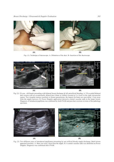
(A)<br />
Fig. (1): Technique of ductoscope. A- Dilatation of the duct. B- Insertion of the ductoscope.<br />
(B)<br />
(A)<br />
Fig. (2): 54 year - old female presenting with bilateral breast discharge & left uniorificial bleeding. A. US revealed bilateral<br />
duct ectasia with an exceptionally dilated duct (9mm in caliber) located at 9 o’clock in the right retroareolar<br />
region. Two irregular masses isoechoic to breast parenchyma, the largest (6x3mm) are seen about 1.2cm away<br />
from the nipple (arrows). B. Power <strong>Doppler</strong> application showed a central vascular stalk in the larger lesion.<br />
Diagnosis of intraductal papilloma was confirmed by both FNAB <strong>and</strong> post duct excision revision of the pathology<br />
specimen.<br />
(B)<br />
(A)<br />
Fig. (3): Two different cases of intraductal papillomas presenting by uni orificial bloody nipple discharge. Both lesions<br />
appeared isoechoic. A. Mass was seen 2.8cm from the nipple. B. A central vascular stalk was identified on Power<br />
<strong>Doppler</strong>. Diagnosis was confirmed after FNAB.<br />
(B)