Reproductive Ecology and Body Burden of Resident ... - The Love Lab
Reproductive Ecology and Body Burden of Resident ... - The Love Lab
Reproductive Ecology and Body Burden of Resident ... - The Love Lab

You also want an ePaper? Increase the reach of your titles
YUMPU automatically turns print PDFs into web optimized ePapers that Google loves.
Otolith preparation <strong>and</strong> microchemical analysis using laser-ablation ICP-MS<br />
Each otolith was soaked in deionized water for 1-3 hours, cleaned with a s<strong>of</strong>t nylon brush in a deionized<br />
water bath, rinsed, <strong>and</strong> air dried in clean, covered plastic multiwell trays. A transverse section was cut from<br />
each otolith using a low-speed rotary diamond blade saw <strong>and</strong> mounted on a plastic slide with metal-free<br />
epoxy resin (Epo-Thin, Buehler). Plastic slides were cleaned in metal-free soap <strong>and</strong> rinsed in distilled water<br />
(resistivity>2 M cm). Each section was polished to the center <strong>of</strong> the otolith core using a lapping wheel <strong>and</strong><br />
9, 3, <strong>and</strong> 1 μm 3M diamond polishing films. <strong>The</strong> sections were broad, 0.5-1mm between cuts, to keep the<br />
polished surface, which was to be assayed, free <strong>of</strong> the epoxy used to bond the base <strong>of</strong> the section to the slide.<br />
After polishing, decontamination steps described by Warner et al. (2005) were performed in a clean<br />
laboratory. Each otolith section mounted on a slide was individually soaked in equal parts <strong>of</strong> 30% H 2<br />
0 2<br />
<strong>and</strong><br />
0.1 mol l -1 NaOH for 30 minutes, rinsed with ultrapure water (N-pure, resistivity >18.1M cm), individually<br />
soaked in 5 separate 5-minute baths <strong>of</strong> ultrapure water, rinsed again in ultrapure water, <strong>and</strong> air-dried<br />
in a HEPA-filtered Class 100 laminar flow hood. <strong>The</strong> otoliths were placed separately in multi-welled tissue<br />
culturing trays <strong>and</strong> metal-free airtight bags until the microchemical analysis. All materials that came into<br />
contact with the otoliths during the decontamination process (e.g., glassware, plastic trays, plastic instruments)<br />
were cleaned in metal-free soap, rinsed five times in distilled water (resistivity>2 M cm), soaked in 1<br />
N mol l -1 trace metal grade HCl overnight, <strong>and</strong> rinsed 10 times with ultrapure water.<br />
We used laser-ablation inductively coupled plasma mass spectrometry (LA ICP-MS) to sample a discrete<br />
portion <strong>of</strong> the sectioned otolith nearest the edge. Otolith material was vaporized by a 213 nm solid<br />
state Nd:YAg laser ablation system (New Wave UP213). Helium carrier gas swept the sample <strong>of</strong> ablated particles<br />
into a spray chamber where Argon gas was mixed with the aerosol before entering the ICP-MS torch.<br />
<strong>The</strong> sample was analyzed on a Finnigan MAT Element 2-sector field ICP-MS. An imaging system was used<br />
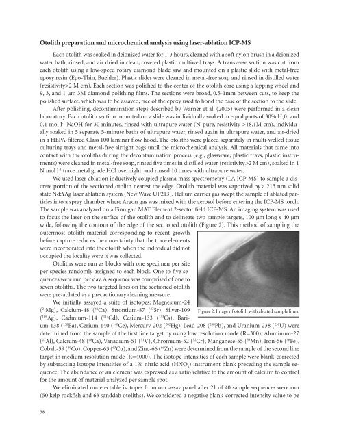
to focus the laser on the surface <strong>of</strong> the otolith <strong>and</strong> to delineate two sample targets, 100 µm long x 40 µm<br />
wide, following the contour <strong>of</strong> the edge <strong>of</strong> the sectioned otolith (Figure 2). This method <strong>of</strong> sampling the<br />
outermost otolith material corresponding to recent growth<br />
before capture reduces the uncertainty that the trace elements<br />
were incorporated into the otolith when the individual did not<br />
occupied the locality were it was collected.<br />
Otoliths were run as blocks with one specimen per site<br />
per species r<strong>and</strong>omly assigned to each block. One to five sequences<br />
were run per day. A sequence was comprised <strong>of</strong> one to<br />
seven otoliths. <strong>The</strong> two targeted lines on the sectioned otolith<br />
were pre-ablated as a precautionary cleaning measure.<br />
We initially assayed a suite <strong>of</strong> isotopes: Magnesium-24<br />
( 24 Mg), Calcium-48 ( 48 Ca), Strontium-87 ( 87 Sr), Silver-109 Figure 2. Image <strong>of</strong> otolith with ablated sample lines.<br />
( 109 Ag), Cadmium-114 ( 114 Cd), Cesium-133 ( 133 Cs), Barium-138<br />
( 138 Ba), Cerium-140 ( 140 Ce), Mercury-202 ( 202 Hg), Lead-208 ( 208 Pb), <strong>and</strong> Uranium-238 ( 238 U) were<br />
determined from the sample <strong>of</strong> the first line target by using low resolution mode (R=300); Aluminum-27<br />
( 27 Al), Calcium-48 ( 48 Ca), Vanadium-51 ( 51 V), Chromium-52 ( 52 Cr), Manganese-55 ( 55 Mn), Iron-56 ( 56 Fe),<br />
Cobalt-59 ( 59 Co), Copper-63 ( 63 Cu), <strong>and</strong> Zinc-66 ( 66 Zn) were determined from the sample <strong>of</strong> the second line<br />
target in medium resolution mode (R=4000). <strong>The</strong> isotope intensities <strong>of</strong> each sample were blank-corrected<br />
by subtracting isotope intensities <strong>of</strong> a 1% nitric acid (HNO 3<br />
) instrument blank preceding the sample sequence.<br />
<strong>The</strong> abundance <strong>of</strong> an element was expressed as a ratio relative to the amount <strong>of</strong> calcium to control<br />
for the amount <strong>of</strong> material analyzed per sample spot.<br />
We eliminated undetectable isotopes from our assay panel after 21 <strong>of</strong> 40 sample sequences were run<br />
(50 kelp rockfish <strong>and</strong> 63 s<strong>and</strong>dab otoliths). We considered a negative blank-corrected intensity value to be<br />
38