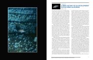
Figure 2. A) Ovary <strong>of</strong> Pacific s<strong>and</strong>dab showing hydrated oocyte (left) <strong>and</strong> yolk deposition in follicle (lower right); B) ovary <strong>of</strong> Pacific s<strong>and</strong>dab showing older postovulatory follicle (center); C) ovary <strong>of</strong> Pacific s<strong>and</strong>dab showing atretic follicle (center). Note enlarged height <strong>of</strong> granulosa cell layer <strong>and</strong> ingested yolk granules. All images at 160 X. 75
Discussion In general, we observed relatively little evidence <strong>of</strong> conspicuous reproductive impairment in Pacific s<strong>and</strong>dab from both platforms <strong>and</strong> natural sites. Overall, most fish contained hydrated oocytes <strong>and</strong> were about to spawn <strong>and</strong> most contained oocytes with smaller yolks <strong>and</strong> thus were likely to spawn again. Many fish exhibited minor atresia. Atresia is the spontaneous degeneration <strong>of</strong> an oocyte at any stage in its development <strong>and</strong> it occurs at low frequency throughout the ovarian cycle. For instance, background (i.e., non-test) levels <strong>of</strong> minor atresia in laboratory-raised zebrafish, Danio rerio, were 58% (Rossteuscher et al. 2008). <strong>The</strong> frequency <strong>of</strong> minor atresia typically increases toward the end <strong>of</strong> the spawning cycle when follicles that initiated, but did not complete, yolk deposition degenerate (Goldberg, 1981). We observed relatively few instances <strong>of</strong> pronounced atresia, a condition that is only occasionally observed in unstressed populations. As an example, pronounced atresia was only found in 2% <strong>of</strong> female Atka mackerel, Pleurogrammus monopterygius, taken from the relatively pristine Aleutian Isl<strong>and</strong>s (McDermott et al. 2007). A wide range <strong>of</strong> environmental stressors, including heavy metals (Pierron et al. 2008), endocrine disrupters (Pollino et al. 2007), <strong>and</strong> starvation <strong>and</strong> lipid-poor diets (Hunter <strong>and</strong> Macewicz 1985; Sherwood et al. 2007) can cause pronounced atresia. <strong>The</strong> level <strong>of</strong> pronounced atresia we documented was much less than that found in a study <strong>of</strong> longspine combfish, Zaniolepis latipinnis, <strong>and</strong> yellowchin sculpin, Icelinus quadriseriatus, from the vicinity <strong>of</strong> sewage outfalls in Santa Monica Bay <strong>and</strong> <strong>of</strong>f Palos Verdes, southern California, or from a control site in Santa Monica Bay (Cross et al. 1984). That study documented high levels <strong>of</strong> pronounced atresia in 28-49% <strong>of</strong> females from around sewage outfalls <strong>and</strong> 42-44% from a control site (that was apparently also heavily polluted). It might be argued that there was some reduction in the reproductive capacity <strong>of</strong> fish from Rincon as fewer fish from that site harbored 1) hydrated oocytes, 2) those smaller oocytes destined for later spawning, <strong>and</strong> 3) post-ovulatory oocytes. Furthermore, 16% <strong>of</strong> female Pacific s<strong>and</strong>dab from Rincon contained ovaries with pronounced atresia. We note that our sample size from Rincon was relatively small, encompassed only one year, <strong>and</strong> the factors responsible for the reduction in ovarian output in these females remain to be determined. Acknowledgments M. McCrea <strong>and</strong> M. Nishimoto collected the specimens <strong>and</strong> prepared the fish for shipment. We thank A. Bull <strong>and</strong> D. Schroeder for their support <strong>of</strong> this research. This research was funded by MMS Cooperative Agreement No.1435-01-05-CA-39315. 76