TraumaCad Admin's Guide - Voyant Health
TraumaCad Admin's Guide - Voyant Health
TraumaCad Admin's Guide - Voyant Health

You also want an ePaper? Increase the reach of your titles
YUMPU automatically turns print PDFs into web optimized ePapers that Google loves.
<strong>TraumaCad</strong> <strong>Admin's</strong> <strong>Guide</strong><br />
Scaling X-Rays<br />
5<br />
Calibration Spheres – <strong>Guide</strong>lines on Correct<br />
Placement<br />
Introduction<br />
It is a known fact that there is an inherent magnification factor associated with<br />
radiographic images. When in a softcopy viewing environment, this must be taken into<br />
account during the pre-planning of orthopedic templating procedures.<br />
Depending on the radiographic technique and the anatomical area of interest, the<br />
magnification factor associated with a particular radiograph can vary by between 105% -<br />
120% when compared with its true size.<br />
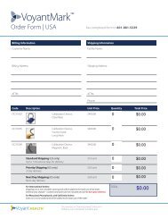
<strong>Voyant</strong>Mark – Calibration Sphere<br />
<strong>Voyant</strong> <strong>Health</strong>’s spherical metal X-ray marker scaling device is intended to provide a<br />
scale for plain X-rays. The scaling sphere should be placed at the bone height. The bone<br />
and sphere should then be the same distance from the X-ray plate and the X-ray source.<br />
When an X-ray image is scaled using a ruler or simple object, it is impossible to verify<br />
whether the scale was placed in the correct plane at the time of acquisition. The<br />
advantage of the sphere is that it is three-dimensional and its diameter is constant from<br />
any angle that the X-ray is taken.<br />
Note: Measurements performed on uncalibrated images are in pixel units, while in<br />
calibrated images they are in millimeters.<br />
50