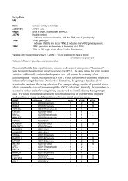
WAVLD Symposium Handbook_V4.indd - csiro
WAVLD Symposium Handbook_V4.indd - csiro
WAVLD Symposium Handbook_V4.indd - csiro

You also want an ePaper? Increase the reach of your titles
YUMPU automatically turns print PDFs into web optimized ePapers that Google loves.
World Association of Veterinary Laboratory Diagnosticians – 13 th International <strong>Symposium</strong>, Melbourne, Australia, 11-14 November 2007<br />
CANINE RABIES IN SOUTH AFRICA: IDENTIFICATION OF A NEW LINEAGE IN LIMPOPO AND A<br />
RECENT SPREAD INTO THE FREE STATE PROVINCE<br />
Introduction<br />
Chuene Ernest Ngoepe a, * , Gugulethu Zulu a , Claude Sabeta a , Louis Nel b<br />
a Rabies Unit, Onderstepoort Veterinary Institute, Private Bag X05, Onderstepoort, 0110<br />
b University of Pretoria, Microbiology and Plant Pathology, 0002 Pretoria, South Africa<br />
In 2005, there was a drastic and unexpected increase of dog rabies cases in the Limpopo province.<br />
Laboratory confirmed cases increased from 5 in 2004 to 35 in 2005 and to 100 in 2006. The outbreak of<br />
rabies in domestic dogs was followed by a human rabies outbreak in which at least 30 human deaths were<br />
confirmed between 2005 and 2006. In contrast, the Free State province has been historically associated with<br />
endemic rabies in the yellow mongoose Cynictis penicillata (Snyman, 1940; Swanepoel et al. 1993). Due to<br />
spillover events, rabies viruses of the mongoose biotype were recovered from domestic dogs. More recently,<br />
there has been increased number of rabies cases of the canid biotype reported in domestic dogs in this<br />
province. The objectives of this study were; 1. to establish the exact source of infection in the human rabies<br />
cases and the origin of the rabies virus lineage responsible for the recent rabies outbreak in Limpopo, and 2.<br />
to trace the origin of canid rabies and assess the public health threat of mongoose rabies (in dogs) in the<br />
Free State province.<br />
Material & methods<br />
A molecular epidemiological study was therefore performed on a cohort of 98 rabies viruses recovered from<br />
domestic dogs between 1995 and 2007 from the Free State province and 52 rabies viruses recovered from<br />
domestic dogs, jackals and humans from Limpopo and southern Zimbabwe. The cytoplasmic domain of the<br />
glycoprotein and the G-L intergenic region of the rabies viruses in this study sample were amplified and<br />
sequenced. Phylogenetic trees were reconstructed from an alignment of a 592-bp region of the genome<br />
under investigation.<br />
Results<br />
Case 1: Phylogenetic analysis revealed that human rabies viruses were closely related to those obtained<br />
from domestic dogs in the same locality and the rabies viruses from Limpopo were closely related to viruses<br />
obtained from southern Zimbabwe.<br />
Case 2: The phylogenetic analyses segregated the rabies viruses in this study sample into two main<br />
clusters; the genetically compact canid rabies biotype and a second group of heterogeneous viruses of the<br />
mongoose rabies biotype. From the data, it could be demonstrated that viruses of the canid rabies group<br />
were recently introduced into this part of South Africa.<br />
Discussions & conclusions<br />
The reasons for the emergence and rapid dissemination of the new dog rabies strain in Limpopo are not very<br />
clear. It appears that common rabies infection cycles persist between domestic dogs and jackals in southern<br />
Zimbabwe and northern South Africa. It is evident that the new canid group belongs to the same<br />
epidemiological cycle circulating in dogs in both the Free State province and across the international border<br />
with Lesotho. Our results confirm that spillover of the mongoose biotype into domestic dogs lead to dead-end<br />
infections. In comparison to mongoose rabies that is endemic here, canid rabies has emerged to become of<br />
much greater importance to the public and veterinary health sectors of this region.<br />
References<br />
1. Swanepoel, R., Barnard, B. J. H., Meredith, C. D., Bishop, G. C., Bruckner, G. K., Fogging, C. M.,<br />
Hubschle, O. J. B., 1993. Rabies in Southern Africa. Onderstepoort Journal of Veterinary Research, 60: pp.<br />
325-346.<br />
2. Snyman, P.S., 1940. The study and control of vectors of rabies in South Africa. Onderstepoort Journal of<br />
Veterinary Science Animal Husbandry, 66: pp. 296-307<br />
Mon 12 November<br />
Mon 12 November<br />
World Association of Veterinary Laboratory Diagnosticians – 13 th International <strong>Symposium</strong>, Melbourne, Australia, 11-14 November 2007<br />
CAPRIPOXVIRUS TROPISM AND SHEDDING: A QUANTITATIVE TIME-COURSE STUDY IN<br />
EXPERIMENTALLY INFECTED SHEEP AND GOATS<br />
T R Bowden 1,* , S L Babiuk 2,3 , M P Anderson 1 , G R Parkyn 2 , R P Kitching 2 , J S Copps 2 and D B Boyle 1<br />
1 CSIRO Livestock Industries, Australian Animal Health Laboratory, Private Bag 24, Geelong, Victoria 3220, Australia<br />
2 Canadian Food Inspection Agency, National Centre for Foreign Animal Disease, 1015 Arlington Street, Winnipeg,<br />
Manitoba R3E 3M4, Canada<br />
3 University of Manitoba, Department of Immunology, Basic Medical Sciences Building 730 William Avenue, Winnipeg,<br />
Manitoba R3E 0W3, Canada.<br />
Introduction<br />
Sheeppox virus (SPPV) and goatpox virus (GTPV), which are members of the genus Capripoxvirus in the<br />
family Poxviridae, cause systemic disease in sheep and goats that is characterized by fever, generalized<br />
skin nodules, lesions in the respiratory and gastrointestinal tracts and lymph node enlargement. SPPV and<br />
GTPV are endemic in north and central Africa, the Middle East, central Asia and the Indian subcontinent,<br />
where they are a cause of significant morbidity and mortality in susceptible sheep and goats. Despite the<br />
considerable threat that these viruses pose to small ruminant production and to global trade in sheep, goats<br />
and their products, knowledge of the underlying pathogenesis of sheeppox and goatpox has long been<br />
deficient. To increase understanding of the pathogenesis of these diseases, we undertook quantitative timecourse<br />
studies in sheep and goats, utilizing real-time PCR and traditional virological assays, following<br />
intradermal inoculation of Nigerian SPPV or Indian GTPV in their respective homologous hosts.<br />
Materials & methods<br />
Nine sheep and nine goats were inoculated intradermally with either 10 4.9 TCID50 of Nigerian SPPV (sheep)<br />
or 10 4.4 TCID50 of Indian GTPV (goats). To enable absolute quantitation of viral DNA concentrations in blood,<br />
swabs and tissue samples, we developed a quantitative real-time PCR TaqMan assay to amplify and detect<br />
a short product from within ORF074 of capripoxvirus genomes. Multiplexed detection of either an<br />
endogenous or exogenous control, depending on the sample type, provided a means of verifying the<br />
absence of PCR inhibitors in samples tested. Isolation of infectious virus from clinical materials was routinely<br />
attempted by inoculation of samples, in duplicate, onto 80-90% confluent ovine testis cell monolayers.<br />
Capripoxvirus-positive specimens were subsequently titrated on replicate cell monolayers to determine<br />
sample infectivity.<br />
Results<br />
Viral DNA concentrations in blood, swabs, urine, faeces and solid tissues varied by as many as six orders of<br />
magnitude. Viraemia, determined by real-time PCR and virus isolation, cleared within two to three weeks<br />
post inoculation. Peak shedding of viral DNA and infectious virus in nasal, conjunctival and oral secretions<br />
occurred between days 10 and 14 post inoculation, and persisted at low levels for up to an additional three to<br />
six weeks. The highest infectious titres in nasal, conjunctival and oral swabs were observed in goats, and<br />
correlated with the greater severity of disease observed in these animals. Although gross lesions were<br />
evident in multiple organs at necropsy, highest viral titres were detected in skin as well as in lung and<br />
discrete sites within oronasal tissues and gastrointestinal tract. Of all tissues tested, skin was consistently<br />
positive throughout the sampling period.<br />
Discussion & conclusions<br />
Our studies have provided novel insights regarding replication, dissemination and shedding of<br />
capripoxviruses in their natural hosts, the pathogenesis of which is similar to smallpox and monkeypox<br />
where greatest viral replication occurs in the skin. The consistently high concentrations of viral DNA in skin,<br />
along with its high infectivity, suggest that mechanical transmission by biting insects might occur more<br />
frequently than previously thought. Furthermore, long term shedding of virus from mucosal surfaces<br />
illustrates the potential for some animals to remain infectious for up to two months after the onset of fever.<br />
These findings should facilitate development of improved strategies for detection and control of capripoxvirus<br />
infections in sheep and goats.