WAVLD Symposium Handbook_V4.indd - csiro
WAVLD Symposium Handbook_V4.indd - csiro
WAVLD Symposium Handbook_V4.indd - csiro

You also want an ePaper? Increase the reach of your titles
YUMPU automatically turns print PDFs into web optimized ePapers that Google loves.
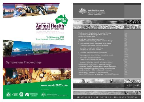
<strong>Symposium</strong> Proceedings<br />
HOSTS GOLD SPONSOR<br />
13TH INTERNATIONAL WORLD ASSOCIATION OF<br />
VETERINARY LABORATORY DIAGNOSTICIANS SYMPOSIUM<br />
11–14 November 2007<br />
Crown Promenade Melbourne AUSTRALIA<br />
www.wavld2007.com
WELCOME<br />
It is our pleasure to welcome you to the 2007 World Association of Veterinary Laboratory Diagnosticians biennial symposium<br />
held in conjunction with the World Animal Health Organisation (OIE). The timing of this meeting could not be more<br />
appropriate given the challenges brought by Avian Infl uenza, Bluetongue and Foot and Mouth Disease in Europe, Africa and<br />
Asia and, here in Australia, Equine Infl uenza. Equally, the attention given by regulatory authorities to biosafety, biosecurity,<br />
bio-terrorism and select agent management issues continues to occupy us.<br />
The organising committee have brought together a wide array of internationally renowned speakers. We would like to thank<br />
our generous sponsors for enabling us to achieve this.<br />
The fi rst day focuses on trans-boundary animal diseases with specialised sessions on avian infl uenza, foot and mouth<br />
disease and bluetongue. Prompted by the current epidemic in Australia, there is also a special session on equine infl uenza.<br />
Day Two belongs to the OIE and, chaired by Professor Steve Edwards, provides an insight into a wide array of new approaches<br />
to disease diagnosis. On Day Three we focus on issues that are critical to current laboratory management,<br />
including the use of new and developing technologies.<br />
Of course no meeting of this size can proceed without a social program and we hope that you will be well entertained.<br />
We have great pleasure in inviting you to our conference dinner, highlighted by a presentation by the Nobel Laureate<br />
Professor Peter Doherty. But let’s not forget that Melbourne is a beautiful city, well known for its many attractions and vast<br />
range of restaurants for you to enjoy.<br />
Whether a visitor from overseas or Australia, we welcome you to Melbourne for what we hope will be a most memorable<br />
Scientifi c and Social Program.<br />
Martyn Jeggo Peter Kirkland<br />
Joint Chair <strong>WAVLD</strong> 2007 Joint Chair <strong>WAVLD</strong> 2007<br />
Director President<br />
CSIRO Livestock Industries AAVLD<br />
Australian Animal Health Laboratory<br />
CONTENTS<br />
WELCOME & CONTENTS ..............................................................................................................................................3<br />
SPONSOR ACKNOWLEDGEMENT & COMMITTEE..........................................................................5<br />
INVITED SPEAKERS ..................................................................................................................................................8 – 9<br />
PROGRAM .......................................................................................................................................................................10 – 13<br />
POSTER LISTING ....................................................................................................................................................16 – 17<br />
EXHIBITION FLOORPLAN ........................................................................................................................................18<br />
EXHIBITOR LISTING .......................................................................................................................................................19<br />
SOCIAL PROGRAM & DINING IN MELBOURNE .................................................................20 – 21<br />
<strong>WAVLD</strong> ORAL PRESENTATIONS ......................................................................................................22 – 122<br />
<strong>WAVLD</strong> POSTER PRESENTATIONS ...........................................................................................123 – 225<br />
<strong>WAVLD</strong> 2007, 11 – 14 November 2007 | 3 4
SPONSORSHIP<br />
SPONSORS<br />
<strong>WAVLD</strong> 2007 extends its appreciation to the following sponsors for their support:<br />
GOLD SPONSOR<br />
SILVER SPONSORS<br />
Includes Sponsorship of:<br />
SYMPOSIUM DINNER<br />
MORNING OR AFTERNOON TEAS<br />
DELEGATE SPONSORSHIP<br />
Welcome Reception<br />
ORGANISING COMMITTEE<br />
Dr Martyn Jeggo<br />
Co-Convenor<br />
Dr Peter Kirkland<br />
Co-Convenor<br />
Dr Peter Daniels<br />
Secretary<br />
Dr Russell Graydon<br />
Local Arrangements Committee Chair<br />
Delegate Name<br />
Badge & Lanyard<br />
Poster Area &<br />
Internet Cafe<br />
<strong>Symposium</strong> Cafe<br />
<strong>WAVLD</strong> 2007 SYMPOSIUM MANAGERS<br />
The Meeting Planners<br />
91–97 Islington Street<br />
Collingwood Victoria 3066<br />
tel: +61 3 9417 0888<br />
fax: +61 3 9417 0899<br />
email: wavld2007@meetingplanners.com.au<br />
W W W . M E E T I N G P L A<br />
N E R S . C O M . A U<br />
<strong>WAVLD</strong> 2007, 11 – 14 November 2007 | 5<br />
The pace of leadership.<br />
Institut Pourquier and IDEXX Laboratories are proud to join forces to sponsor<br />
the World Association of Veterinary Laboratory Diagnosticians.<br />
Renowned medical research and<br />
development since 1878.<br />
A wholly owned subsidiary<br />
of IDEXX Laboratories, Inc.<br />
© 2007 IDEXX Laboratories, Inc. All rights reserved. • 7406-00
The science of innovation.<br />
Institut Pourquier joined IDEXX Laboratories in March 2007 and together will<br />
continue unparalleled research, development and support of the most advanced<br />
animal health diagnostics, services and solutions in the world.<br />
Innovative production animal<br />
services since 1984. A worldwide<br />
leader in veterinary, food<br />
and water biotechnology.<br />
INVITED SPEAKERS<br />
Dr Tammy Beckham<br />
Dr Beckham received her Doctorate of Veterinary<br />
Medicine from Auburn University in 1998. While still<br />
at Auburn, she furthered her education through a<br />
Doctoral program, which allowed her to conduct<br />
research in support of her dissertation at the U.S.<br />
Army Medical Research Institute for Infectious<br />
Diseases (USAMRIID), in Frederick, MD. Dr Beckham received her<br />
Doctorate in 2001. Following completion of her Doctorate,<br />
Dr Beckham fostered her interest in diagnostic assay<br />
development and validation through positions with the<br />
Department of Homeland Security and the USDA Animal and<br />
Plant Health Inspection Service (APHIS), Foreign Animal Disease<br />
Diagnostic Laboratory (FADDL). Dr Beckham recently became<br />
the Head of the Profi ciency and Validation Services Section at<br />
FADDL. In this position, Dr Beckham coordinates all aspects of<br />
FADDL’s role as a reference laboratory for the National Animal<br />
Health Laboratory Network (NAHLN). This includes overseeing<br />
the validation of assays; identifying, evaluating, and developing<br />
new and emerging technologies; and developing and organizing<br />
training and profi ciency testing programs.<br />
Professor Corrie Brown<br />
Corrie Brown received her B.Sc. in Animal<br />
Behavior from McGill University and her DVM<br />
from Ontario Veterinary College at the University<br />
of Guelph (1981). She completed a combined<br />
residency/PhD in Comparative Pathology at<br />
the University of California at Davis. Board<br />
certifi cation (ACVP) and PhD were both attained in 1986. She was<br />
an assistant professor of pathology at Louisiana State University<br />
briefl y before joining the U.S. Department of Agriculture at Plum<br />
Island, where, as Head of the Pathology Section, she specialized<br />
in the diagnosis and pathogenesis of foreign animal diseases.<br />
In 1996, she joined the University of Georgia College of Veterinary<br />
Medicine as Professor and Head of the Department of Veterinary<br />
Pathology. She currently serves as Coordinator of International<br />
Veterinary Medicine for the College of Veterinary Medicine. In<br />
2003, she was honored with the university’s highest teaching<br />
award, being named a Josiah Meigs Distinguished Teaching<br />
Professor. Her professional interests are in infectious diseases<br />
of food-producing animals, emerging diseases, and international<br />
veterinary medicine. She has published or presented over 250<br />
scientifi c papers and has testifi ed to Congress on issues involving<br />
agroterrorism. She has served on many industrial and federal<br />
panels, and been a technical consultant to numerous foreign<br />
governments on issues involving infectious diseases and animal<br />
health infrastructure.<br />
8 | <strong>WAVLD</strong> 2007, 11 – 14 November 2007<br />
Dr Ilaria Capua<br />
Dr Ilaria Capua graduated in Veterinary Medicine<br />
with honours (110/110 e lode ), University of<br />
Perugia , in February 1989. Obtained post<br />
graduate qualifi cations as a specialist in Animal<br />
Health and hygiene from Pisa University in 1991<br />
and PhD from University of Padua. She is currently<br />
Head of the Virology Department at Istituto Zooprofi lattico<br />
Sperimentale delle Venezie, Padova, Italy and Head of the<br />
National, FAO and OIE Reference Laboratories for Avian Infl uenza<br />
and Newcastle disease. Between 1999 and 2006 she has been<br />
directly involved in managing several AI and ND epidemics and<br />
in 2000 developed the “DIVA”-Differentiating Vaccinated from<br />
Infected Animals strategy, based on heterologous vaccination, to<br />
combat AI. This strategy, the fi rst ever developed to combat AI by<br />
vaccination still enabling trade of products resulted in eradication<br />
of AI at that time in Italy and was approved by the EC and by OIE.<br />
During her career as a veterinary virologist her work has been<br />
recognized with her nomination as OIE and FAO expert for AI and<br />
ND. Since 1995 she has been involved with the EU Commission,<br />
participating in meetings and working groups on viral diseases of<br />
poultry and mammals, including AI, ND, FMD, CSF and Crimean-<br />
Congo Haemorrhagic Fever. In 2005 she has been nominated<br />
the Chairman of OFFLU- the newly established OIE/FAO network<br />
on Avian Infl uenza. This network has the role of supporting<br />
developing countries in managing the AI crisis and offering<br />
veterinary expertise to complement the international efforts of<br />
the medical community in managing the pandemic threat posed<br />
by AI.<br />
Laureate Professor Peter Doherty<br />
Laureate Professor Peter Doherty shared the<br />
Nobel Prize in Physiology or Medicine in 1996<br />
with Swiss colleague Rolf Zinkernagel, for their<br />
discovery of how the immune system recognises<br />
virus-infected cells. He was Australian of the Year<br />
in 1997, and has since been commuting between<br />
St Jude Children’s Research Hospital in Memphis and the<br />
Department of Microbiology and Immunology at the University<br />
of Melbourne. His research is mainly in the area of defence<br />
against viruses. He regularly devotes time to delivering public<br />
lectures, writing articles for newspapers and magazines and<br />
participating in radio discussions. Peter Doherty graduated from<br />
the University of Queensland in Veterinary Science and became a<br />
veterinary offi cer. Moving to Scotland, he received his PhD from<br />
the University of Edinburgh Medical School. He is the fi rst person<br />
with a veterinary qualifi cation to win a Nobel Prize.
INVITED SPEAKERS CONTINUED<br />
Professor Steve Edwards<br />
Steve qualifi ed as a veterinarian from Cambridge<br />
University, England, in 1972. He spent four years<br />
in clinical farm animal practice in the Welsh<br />
border country before returning to university<br />
at Edinburgh to study for an MSc in Tropical<br />
Veterinary Science. This stimulated his continuing<br />
passion for laboratory-based diagnostic investigation work. He<br />
then spent two years working in the UK Government’s overseas<br />
aid programme in El Salvador, Central America, working<br />
alongside local counterparts to identify the disease constraints<br />
to animal production. Steve joined the Central Veterinary<br />
Laboratory (now VLA) at Weybridge in 1980 working on virology<br />
research. From 1991 Steve brought his interest and expertise<br />
in laboratory diagnostics to the OIE Standards Commission,<br />
eventually becoming President of that group (now renamed<br />
Biological Standards Commission) in 2003, a position he still<br />
holds. He took over as Director and Chief Executive of the VLA in<br />
2000 on the retirement of his predecessor. He has led the Agency<br />
through a period of consolidation, with an expansion of its external<br />
collaborative links, as well as coping with an apparently neverending<br />
succession of animal disease issues, including classical<br />
swine fever, foot and mouth disease, tuberculosis, BSE, rabies,<br />
Newcastle disease, and avian infl uenza.<br />
Dr Luis Rodriguez<br />
Dr Rodriguez obtained his DVM in 1979 at the<br />
National University of Costa Rica. From 1980 to<br />
1985 he attended graduate school at the University<br />
of Wisconsin-Madison. In 1982 obtained a M.Sc.<br />
and in 1985 a Ph.D. in veterinary science with<br />
emphasis in Animal Virology working in molecular<br />
virology of bovine herpesvirus 1. From 1985 to 1995 worked a the<br />
School of Veterinary Medicine, National University of Costa Rica<br />
where he taught veterinary virology and carried out research<br />
focusing on vesicular stomatitis. He established the Tropical<br />
Disease Research Program and obtained several grants from<br />
international organizations to carry out research in pathogens of<br />
human and veterinary importance. From 1995 –1997 worked at<br />
the Special Pathogens Branch, Division of Viral and Rickettsial<br />
Diseases of the Centers for Disease Control and Prevention, in<br />
Atlanta Georgia. In 1997 joined the USDA – ARS Foot-and Mouth<br />
Disease Research Unit Plum Island as lead scientist in charge<br />
of vesicular stomatitis research working on genomics and<br />
pathogenesis of VSV and in 2002 Dr Rodriguez became Research<br />
Leader of the Foot and Mouth Disease Research Unit. In 2004 he<br />
also established the Foreign Animal Disease Research Unit. Dr<br />
Rodriguez personal current research focuses on studying the early<br />
phases of FMDV infection and pathogenesis and insecttransmission<br />
of VSV in cattle.<br />
Professor Mark Rweyemamu<br />
A Tanzanian veterinarian. Former Head of the<br />
Infectious Diseases Programme of the Food and<br />
Agriculture Organization (FAO) of the United<br />
Nations at its Headquarters in Rome. From 1994<br />
to his retirement from FAO in 2002, he was the<br />
inaugural Head of the FAO special programme on<br />
infectious diseases, known as the Emergency Prevention System<br />
for Animal and Plant Pests and Diseases (EMPRES) which<br />
included the coordination of the Global Rinderpest Eradication<br />
Programme (GREP). Before moving to FAO Headquarters, he<br />
had set up the Pan African Veterinary Vaccine Centre in Addis<br />
Ababa, on behalf of FAO and the Organisation of African Unity<br />
(now African Union). He has worked in both governmental<br />
and industrial settings. In government: as Virologist and Chief<br />
Veterinary Research Offi cer for the Tanzanian Government<br />
and as Head of the Virus Diseases at the then East African<br />
Veterinary Research Organisation, Muguga, Kenya. In industry<br />
he has been the Director of Veterinary Vaccine Research for<br />
Pfi zer International and Head of FMD Vaccine Research for<br />
the Wellcome Foundation. His area of interest is on the major<br />
infectious diseases of animals, known as transboundary animal<br />
diseases. He is now a private consultant and a Visiting Professor<br />
at the Royal Veterinary College, University of London.<br />
Dr Linfa Wang<br />
Dr Linfa (Lin-Fa) Wang is a Senior Principal<br />
Research Scientist at the CSIRO Australian Animal<br />
Health Laboratory (AAHL), and a project leader in<br />
the Australian Biosecurity Cooperative Research<br />
Centre (AB-CRC) for Emerging Infectious Diseases.<br />
Dr Wang is a member of the WHO SARS Scientifi c<br />
Research Advisory Committee and participated<br />
in the WHO-FAO-Chinese Government joint mission to China on<br />
animal reservoir of the SARS-CoV and potential transmission<br />
to humans in August 2003. His research group played key<br />
roles in the discovery and molecular analysis of Hendra and<br />
Nipah viruses, which led to the establishment of a new genus<br />
Henipavirus to accommodate the newly discovered highly lethal<br />
zoonotic paramyxoviruses of bat origin. Dr Wang completed his<br />
science degree in 1982 at the East China Normal University,<br />
Shanghai, followed by a PhD in Biochemistry (Molecular Biology)<br />
from the University of California, Davis, USA in 1986, where he<br />
went on to become a Postdoctoral Research Fellow with the<br />
Department of Biochemistry. After moving to Australia in 1989,<br />
Dr Wang spent 18 months working at Monash University and<br />
then joined CSIRO in 1990. Dr Wang has more than 140 scientifi c<br />
publications to his name along with four patents and numerous<br />
conference abstracts. He is currently serving on fi ve international<br />
editorial boards for publications in the areas of virology,<br />
biotechnology and immunotechnology.<br />
Crown Promenade, Melbourne, Australia | 9<br />
PROGRAM<br />
Monday 12 November<br />
0700 Registration Open<br />
0815 – 1030 Opening Session<br />
Room: Promenade 1 & 2<br />
Welcome Address<br />
Martyn Jeggo & Peter Kirkland<br />
Opening<br />
Minister Joe Helper<br />
Plenary Sessions – Global Risks and Challenges<br />
Chair: Peter Daniels<br />
Prof Mark Rweyemamu<br />
Future Risks from Infectious Disease of Animals<br />
Prof Corrie Brown<br />
New & Emerging Diseases<br />
Dr Ilaria Capua<br />
Avian Infl uenza – Challenges and Opportunities for the Veterinary Profession<br />
Dr Linfa Wang<br />
Zoonotic Viruses of Bat Origin – Discovery and Diagnosis<br />
1030 – 1100 Morning Tea & Exhibition/Poster Viewing<br />
Location: Exhibition Area<br />
1100 – 1230 Room: Promenade 1 & 2 Room: Promenade 3<br />
Concurrent Session 1.1<br />
New & Emerging Diseases/Wildlife<br />
Chair: Dr Linfa Wang Chair: Dr Simone Warner<br />
Dr Kim Halpin<br />
Experimental models for Henipavirus infection: bats, cats and<br />
pseudo-rats<br />
Prof Paul Gibbs<br />
Emergence of canine infl uenza in the USA: developments since<br />
discovery in 2004 and the diagnostic challenge<br />
Ms Deborah Finlaison<br />
Investigations of viral myocarditis in pigs associated with a novel<br />
pestivirus.<br />
Mrs Melinda Frost<br />
Characterisation of a novel pestivirus associated with an outbreak<br />
of stillbirths and pre-weaning in pigs due to myocarditis<br />
Prof Stefan Vilcek<br />
How many pestiviruses circulate in nature? Pestiviruses in<br />
free-living animals: present status and problems<br />
Dr Julia Ridpath<br />
Characterization and detection of BVDV-related reproductive<br />
disease in white tail deer<br />
1230 – 1330 Lunch<br />
Location: Exhibition Area<br />
1330 – 1455 Room: Promenade 1 & 2 Room: Promenade 3<br />
Concurrent Session 1.2<br />
FMD<br />
Concurrent Session 2.1<br />
Molecular Diagnostic Assays – I (Bacterial diseases)<br />
Mrs Melissa Higgins<br />
Development of an improved identifi cation matrix for<br />
Aeromonas salmonicida<br />
Dr Ian Marsh<br />
An improved PCR-based test for QX disease confi rmation in<br />
Sydney Rock Oysters<br />
Dr Adrian Whatmore<br />
The development of SNP discrimination assays on a real-time<br />
PCR platform as a tool for the rapid identifi cation and speciation<br />
of Brucella.<br />
Dr Graeme Eamens<br />
Detection of Mycoplasma hyopneumoniae in pig samples using<br />
polymerase chain reaction tests<br />
Ms Kgomotso Sibeko<br />
Development and evaluation of a real-time PCR test for the<br />
detection of Theileria parva infections in cape buffalo<br />
(Syncerus caffer) and cattle<br />
Dr Wendy McDonald<br />
Detection and strain typing of Coxiella burnetii using Molecular<br />
Beacons in a real-time PCR<br />
Concurrent Session 2.2<br />
Avian Infl uenza<br />
Chair: Prof Mark Rweyemamu Chair: Dr Ilaria Capua<br />
Dr Divakar Hemadri<br />
Changing faces of foot-and-mouth disease type O Pan Asia strain<br />
India<br />
Dr Paul Kitching<br />
The 2001 UK foot-and-mouth disease outbreak:<br />
an update<br />
Speaker TBA<br />
FMD in the UK, 2007<br />
Ms Janine Muller<br />
Development and evaluation of a recombinant antibody-based<br />
competitive ELISA for FMDV<br />
antibody detection<br />
Mr Chris Morrissy<br />
International validation of a new 3abc C-ELISA for FMD serology<br />
10 | <strong>WAVLD</strong> 2007, 11 – 14 November 2007<br />
Mrs Janice Pedersen<br />
Results of 2006 wild bird surveillance for the detection of highly<br />
pathogenic avian infl uenza in the United States<br />
Dr Ian K. Barker<br />
Canada’s inter-agency wild bird infl uenza surveillance<br />
programme 2005 – 2007<br />
Dr Tze-Hoong Chua<br />
Performance evaluation of fi ve detection tests<br />
Dr Simone Warner<br />
Optimised sampling and processing for enhanced detection of<br />
avian infl uenza virus from fi eld samples<br />
Dr Michael Johnson<br />
Analysis of the sensitivity of H5N1 Isolates from the South East<br />
Asian region to Oseltamivir and Zanamivir
Monday 12 November (continued)<br />
1455 – 1525 Afternoon Tea<br />
Location: Exhibition Area<br />
1525 – 1720 Room: Promenade 1 & 2 Room: Promenade 3<br />
Concurrent Session 1.3<br />
Bluetongue & orbiviruses<br />
Concurrent Session 2.3<br />
Zoonoses, Pox viruses & other TADS<br />
Chair: Dr Ian Gardner Chair: Prof Corrie Brown<br />
Dr Carrie Batten<br />
An update on bluetongue virus in Europe<br />
Dr Heather Elliott<br />
From testing to trade: the response to the outbreak of BTV 8 in<br />
NW Europe<br />
Gudrun Delbridge<br />
Evaluation of ELISAs to detect antibodies to bluetongue virus in<br />
individual and tank milk samples<br />
Dr Sushila Maan<br />
Molecular epidemiology of bluetongue virus type 8 from the<br />
Netherlands 2006<br />
Prof Peter Mertens<br />
Sequencing and RT-PCR assays for genome segment 2 of the 24<br />
bluetongue virus serotypes: identifi cation of exotic serotypes in<br />
the Southeastern USA (1999-2006)<br />
Ms Karin Darpel<br />
Replication of bluetongue virus in the skin of<br />
infected sheep<br />
Dr Hume Field<br />
Emerging zoonoses and wildlife – a multi-disciplinary challenge<br />
Prof. Tin Tin Myaing<br />
Wild life as a source of zoonotic diseases<br />
Mr Keith Jahans<br />
Surveillance of Mycobacterium bovis infection in cattle in Great<br />
Britain during 2006<br />
Mr Claude Sabeta<br />
Spread of canid rabies into the Free State province of South Africa<br />
Dr Tim Bowden<br />
Capripoxvirus tropism and shedding: a quantitative time-course<br />
study in experimentally infected sheep and goats<br />
Dr David Boyle<br />
An indirect ELISA, based upon recombinant capripoxvirus<br />
antigens, for serological detection of sheeppox, goatpox and<br />
lumpy skin disease<br />
Mrs Eva Veronesi<br />
Validation of a diagnostic technique for the detection and<br />
quantifi cation of bluetongue virus in adult biting midges<br />
(Culicoides spp: Diptera: Ceratopogonidae)<br />
1745 – 1915 Special Plenary Sessions – Equine Infl uenza<br />
Room: Promenade 1 & 2<br />
Chair: Dr Bob Biddle<br />
Dr Peter Timoney<br />
Changing dynamics in the global distribution of equine diseases<br />
Richard Newton<br />
Global Overview<br />
Graeme Garner<br />
Australian Situation<br />
Dr Peter Daniels<br />
Diagnosis and Characterisation of Equine Infl uenza (EI) in Australia: A/equine/Sydney/2888-8/2007 H3N8<br />
Dr Peter Kirkland<br />
Equine infl uenza in NSW – challenges and highlights from a laboratory perspective<br />
1915 – 2115 Welcome Reception<br />
Location: Exhibition Area<br />
Crown Promenade, Melbourne, Australia | 11<br />
Tuesday 13 November<br />
0830-0945 OIE Biotechnology <strong>Symposium</strong><br />
Room: Promenade 1 & 2<br />
Co-Chairs: Prof Steve Edwards & Dr Gideon Brückner<br />
Welcome Address<br />
Prof Steve Edwards<br />
Dr Yi-Seok Joo<br />
Simple rapid, on-site (pen-side) detection of animal pathogens<br />
Dr Deborah Middleton<br />
Application of Biotechnology to Infections within Wildlife Hosts<br />
0945 – 1015 Morning Tea<br />
Location: Exhibition Area<br />
1015 – 1215 Dr Gideon Bruckner<br />
The OIE Concept of Twinning between Laboratories<br />
Dr Michael Macintosh<br />
Diagnostics of Emerging Infectious Diseases by Microarray<br />
Prof Noboru Inoue<br />
Development of Loop-Mediated Isothermal Amplifi cation (LAMP) Technique for Diagnosis of African Trypanosomosis<br />
Dr Alex Hyatt<br />
Diagnostic Electron Microscopy: Historical review and future<br />
1215 – 1315 Lunch<br />
Location: Exhibition Area<br />
1315-1445 Dr Norbert Bannert<br />
Overview of electron microscopy and its role in infectious disease diagnosis<br />
Dr Yves Robinson<br />
Telepathology: its role in disease diagnosis in meat hygiene<br />
Dr Paul Monaghan<br />
Understanding Host-Pathogen Interactions: Confocal and Live Cell Imaging in Veterinary Science<br />
1445 – 1515 Afternoon Tea<br />
Location: Exhibition Area<br />
1515-1645 Dr Brad J Marsh<br />
Multi-Resolution Spatio-Temporal Analysis of Mammalian Cells Reconstructed in 3D by Electron Microscope Tomography<br />
Dr Simon Carpenter<br />
Detecting and interpreting arboviral infections in insects<br />
Dr Hez Hird<br />
Molecular methods for speciation<br />
1900 – 2400 <strong>Symposium</strong> Dinner<br />
Location: RACV Club<br />
Wednesday 14 November<br />
0815 – 1000 Plenary Sessions – Preparing for the Future<br />
Room: Promenade 1 & 2<br />
Chair: Dr Russell Graydon<br />
Dr Martyn Jeggo<br />
Labs of the Future<br />
Dr Luis Rodriguez<br />
Biosafety and Biosecurity Concerns Of High Security Animal Pathogen Laboratories<br />
Dr Tammy Beckman<br />
Diagnostic Technologies for Veterinary Laboratories of the Future<br />
Prof Steve Edwards<br />
Can we believe Laboratory Test Reports?<br />
1000 – 1030 Morning Tea<br />
Location: Exhibition Area<br />
1030 – 1230 Room: Promenade 1 & 2 Room: Promenade 3<br />
Concurrent Session 1.4<br />
Concurrent Session 2.4<br />
New Technologies & Platforms<br />
Quality Assurance<br />
Chair: Dr Tammy Beckham Chair: Dr Luis Rodriguez<br />
Mrs Barbara Martin<br />
The United States National Animal Health Laboratory Network<br />
(NAHLN)<br />
Dr Bill Colston<br />
Developing an approach for rapid identifi cation of emerging<br />
biological threats<br />
Dr Juliet Dukes<br />
Viral identifi cation using microarrays<br />
Dr. Julia Ridpath<br />
Assay platforms for the rapid detection of viral pathogens by the<br />
ultrahigh sensitivity monitoring of antigen-antibody binding<br />
Prof Lawrence Wangh<br />
LATE-PCR with PrimeSafeTM – maximum diagnostic information<br />
from a closed-tube<br />
Dr Xingwang Fang<br />
A complete workfl ow for nucleic acid based high throughput<br />
pathogen detection<br />
Dr Carmelo Volpe<br />
An advanced fi eld deployable sample preparation and PCR system<br />
Dr Rod Reece<br />
The Australian Animal Pathology Standards program:<br />
preservation and digitisation of the collection, and access via<br />
‘new’ technologies<br />
12 | <strong>WAVLD</strong> 2007, 11 – 14 November 2007<br />
Dr. Paul Kitching<br />
Labs of the future – a cultural shift in Canada for foreign animal<br />
disease (FAD) detection<br />
Dr Craig Carter<br />
Laboratory-based early animal disease detection utilizing a<br />
prospective scan statistic<br />
Dr Philip Wakeley<br />
Development, validation and implementation of molecular<br />
diagnostics tests for important pathogens of farmed animals and<br />
wildlife – a pragmatic approach<br />
Dr Jane Oakey<br />
Quality assurance and control issues with PCR as a diagnostic tool<br />
Mr Lindsay Pritchard<br />
A review of training in molecular diagnostic techniques for avian<br />
infl uenza in South East Asia<br />
Dr Kim Halpin<br />
Development and consolidation of an EQA programme for the<br />
molecular diagnosis of avian infl uenza<br />
Dr. Hermann Unger<br />
How useful are test kits when applied in different target<br />
population: producer and end-user responsibilities from a<br />
developing country perspective<br />
Dr Mark M Williamson<br />
Laboratory management techniques for responding to client<br />
demands: diagnosis of swine pneumonia.
Wednesday 14 November (continued)<br />
1230 – 1330 Lunch<br />
Location: Exhibition Area<br />
1330-1500 Room: Promenade 1 & 2 Room: Promenade 3<br />
Concurrent Session 1.5<br />
Diagnostic assays – ELISA & molecular<br />
Concurrent Session 2.5<br />
Biosecurity, IT, & LIMS<br />
Chair: Dr Lorna Melville Chair: Dr Michael Johnson<br />
Dr Simon Anthony<br />
Complete sequence analyses of the genome of epizootic<br />
haemorrhagic disease virus (EHDV): the design of<br />
diagnostic assays.<br />
Dr Jovita Fernández<br />
New advances in the molecular diagnosis of African horse<br />
sickness (AHS)<br />
Dr Carmina Gallardo<br />
New approaches in molecular and serological diagnosis of<br />
African swine fever (ASF) on new emerging variants<br />
Dr Karen Harmon<br />
Rapid high-throughput real-time RT-PCR testing for PRRSV:<br />
problems and solutions<br />
Dr Roger Ayling<br />
Development and initial evaluation of a lateral fl ow device for the<br />
rapid detection of contagious bovine pleuropneumonia.<br />
1500 – 1530 Afternoon Tea<br />
Location: Exhibition Area<br />
1530 – 1700 Room: Promenade 1 & 2 Room: Promenade 3<br />
Concurrent Session 1.6<br />
Molecular Diagnostic assys- II (Viral)<br />
Dr Scott Fitzgerald<br />
Benefi ts realized from the fi rst biosafety level 3 large animal<br />
necropsy facility in a state diagnostic laboratory in North America<br />
Dr Steve Weber<br />
Linking multiple veterinary laboratory information<br />
management systems to support the National Animal Health<br />
Laboratory Network<br />
Dr Paul Collery<br />
Implementation, function and benefi ts of a laboratory information<br />
management system in a national veterinary laboratory service<br />
Dr Peter Durr<br />
Beyond the laboratory gate – enhancing a laboratory information<br />
management system (LIMS) for exotic animal disease<br />
preparedness<br />
Dr Paul-Michael Agapow<br />
The ReLaIS project: an epi-informatics constellation<br />
Concurrent Session 2.6<br />
Epidemiology & Surveillance<br />
Chair: Dr Gary Horner Chair: Dr Peter Daniels<br />
Mrs Katarzyna/Kasia Bachanek-Bankowska<br />
High throughput diagnostic PCR for bluetongue virus<br />
Dr William C. Wilson<br />
Nucleic acid diagnostic tools for early detection of<br />
arthropod-borne animal viral diseases.<br />
Dr Dennis Gomez<br />
Molecular detection of betanodaviruses from subclinically<br />
infected aquarium fi shes and invertebrates<br />
Dr Frank Wong<br />
Purifi cation and characterisation of a herpes-like virus infecting<br />
Australian populations of abalone, Haliotis spp.<br />
Dr Giovanni Cattoli<br />
Development of a real-time duplex TaqMan-PCR assay for the<br />
detection of equine rhinitis A and B viruses in clinical specimens<br />
1715 – 1730 <strong>Symposium</strong> Close<br />
Ms Kerri Tyrrell<br />
Effects of dynamic ‘communal’ interactions on disease processes<br />
and individual agent diagnosis where multiple infectious agents<br />
are present in sheep<br />
Dr Fraser Hill<br />
Use of molecular and epidemiological information for the costeffective<br />
diagnosis of BVD infection in New Zealand dairy cattle<br />
Eng Sandra Benevides<br />
Assessing the within herd prevalence of antibody positive cows to<br />
bovine herpevirus 1 using an Indirect ELISA on bulk tank milk<br />
Mr Ronald Coilparampil<br />
Evaluation of a commercially available ELISA for detection<br />
antibodies to liver fl uke (Fasciola hepatica) in pooled bovine serum.<br />
Dr Ashraf Ahmed<br />
Zoo animals as a potential reservoir of gram-negative bacteria<br />
harboring integrons and antimicrobial resistance genes<br />
Dr Nguyen Van Long<br />
Porcine reproductive & respiratory syndrome (PRRS) –<br />
a new challenge<br />
Crown Promenade, Melbourne, Australia | 13 14<br />
A new direction for animal health.<br />
Introducing animal health solutions from a proven technology leader.<br />
With traditional procedures, simplicity, speed and precision are often compromised. That’s why more labs<br />
are switching to superior PCR-based reagents for everyday veterinary and agricultural applications.<br />
PCR helps simplify procedures and accelerate time to results with unparalleled sensitivity and specifi city.<br />
Only Applied Biosystems, with over 25 years of experience, provides one source for a complete molecular<br />
solution. With automated sample prep, superior molecular reagents and master mixes, and easy-to-use<br />
instruments for PCR analysis, we’ve got you covered — from sample to signal.<br />
Go molecular.<br />
For a free AgPath-ID One-Step<br />
RT-PCR Reagent, go to<br />
appliedbiosystems.com/animalhealth.<br />
For research use only. Not for use in diagnostic procedures. Not licensed by the USDA.<br />
Applied Biosystems, and AB (Design) are registered trademarks and AgPath-ID is a trademark<br />
of Applera Corporation or its subsidiaries in the US and/or certain other countries. The<br />
AgPath-ID OneStep RT-PCR Kit is compatible with the 5’ nuclease detection and ds<br />
DNA-binding dye processes covered by patents owned or licensable by Applied Biosystems.<br />
No license under these patents is conveyed expressly, by implication, or by estoppel to the<br />
purchaser by the purchase of this product. Further information on purchasing licenses may<br />
be obtained by contacting the Director of Licensing, Applied Biosystems, 850 Lincoln Centre<br />
Drive, Foster City, California 94404, USA. © 2007 Applied Biosystems. All rights reserved.
<strong>WAVLD</strong> POSTER PRESENTATIONS<br />
Poster No. Author Poster Title<br />
1 Ann Nordengrahn Proximity ligation assay – detection of individual microbial pathogens<br />
2 Wayne Jorgensen Improved detection of bovine reproductive disease pathogens<br />
3 Marinda Oosthuizen Identifi cation of a novel Babesia species from sable, roan and giraffe by means of the Reverse Line Blot (RLB)<br />
hybridization assay<br />
4 Maria Jose Dus Santos Detection of BLA in frozen semen samples by PCR assay<br />
5 Ian Marsh A simple internal PCR control for Mycobacterium avium subsp. paratuberculosis constructed by PCR techniques<br />
6 Graeme Eamens Investigating shedding rates and the proportion of animals shedding Mycobacterium avium subsp paratuberculosis in<br />
cattle and goat herds<br />
7 Darcy Myers A streamlined workfl ow for rapid and sensitive detection of Mycobacterium avium subsp. paratuberculosis (MAP) in<br />
bovine faecal samples by real-time PCR<br />
8 Jose Blanco Automated DNA isolation: an advantageous tool in molecular diagnostic of dermatophytosis<br />
9 Sarah North Development of real time PCRs to detect important bacterial pathogens of the horse.<br />
10 Masoud Ghorbanpoor<br />
Najafabadi<br />
Enhanced cultural sensitivity of Staphylococcus aureus from bovine mastitis milk by sample freezing<br />
11 Lourdes Corpus Standardisation of PCR for identifi cation of Listeria monocytogenes in fresh cheese<br />
12 Lone Hoj Molecular ID of vibrio isolates<br />
13 Samantha Gibbs Indonesian veterinary laboratory capacity building project for highly pathogenic avian infl uenza<br />
14 Manuela Dalla Pozza Management of a DIVA vaccination programme for the control of low pathogenicity avian infl uenza in Italy<br />
15 Adam Foord Adaptation of real time PCR assays for detection of new and evolving avian infl uenza virus strains<br />
16 Hans Heine Identifi cation of a novel hemagglutinin cleavage site in a highly pathogenic avian infl uenza H7 from North Korea<br />
17 Anna Lecoq Preliminary validation of a commercial avian infl uenza N1 antibody competitive ELISA kit that can be used as part of a<br />
DIVA strategy<br />
18 Giovanni Cattoli Inactivation of avian infl uenza viruses<br />
19 Huaguang Lu A rapid laboratory diagnosis of H5N1 avian infl uenza in Saudi Arabia<br />
20 Masoud Hosseini Seroprevalence of H9N2 antibody in poultry farm and slaughter-house workers of Iran using HI test<br />
21 Ross Lunt Advancing the role of a regional reference laboratory for foot–and-mouth disease in South East Asia<br />
22 Melanie Chitray Heterogeneity in the outer capsid-coding region of foot-and-mouth disease virus A and O serotypes in Africa<br />
23 Claro Mingala Quantifi cation of water buffalo (Bubalus bubalis) cytokine expression in response to inactivated foot-and-mouth<br />
disease vaccine using real-time PCR assay<br />
24 Chris Morrissy Development of an improved capability in support of national biosecurity for the surveillance and control of foot &<br />
mouth disease in cattle and pigs in Vietnam.<br />
25 Simon Anthony Nucleotide sequence analyses of epizootic haemorrhagic disease virus genome segments encoding the outer capsid<br />
proteins: a serological and genetic re-evaluation of serotypes.<br />
26 Rahana Dwarka Phylogenetic analysis of African swine fever viruses from South Africa, Mozambique and Tanzania for the period<br />
2001 – 2007<br />
27 Scott Fitzgerald Efforts to control and eradicate bovine tuberculosis in a wildlife reservoir in Michigan, USA: a preliminary success story<br />
28 Ji-hyung Kim Detection of major bacterial and viral pathogens from trash fi sh for cultured fl ounder<br />
29 Amir Hamir Experimental transmission of transmissible spongiform encephalopathies<br />
30 Elizabeth Houston TSE surveillance in Australia: a regional laboratory perspective<br />
31 Francisco Blanco Viera BSE surveillance in Argentina<br />
Crown Promenade, Melbourne, Australia | 15<br />
<strong>WAVLD</strong> POSTER PRESENTATIONS (continued)<br />
Poster No. Author Poster Title<br />
32 Michael Zalunardo Complete automation of a BSE post mortem diagnostic test<br />
33 Sanjay Kapil Emergence of canine parvovirus 2c in North America: 2006<br />
34 Tin Tin Myaing Prevalence study of parasitic infestation in Myanmar timber elephants<br />
35 Hermann Unger The concept of early detection and rapid response to TAD’s by APH, IAEA<br />
36 Emma Stubberfi eld Brucellosis in marine mammals: Veterinary Laboratories Agency involvement<br />
37 Magdalena Dunowska Prevalence of methicillin resistant Staphylococcus aureus (MRSA) in a veterinary teaching hospital<br />
38 Ross Lunt A virus neutralization test for the detection of antibody to Australian bat lyssavirus<br />
39 Chris Morrissy Development of a porcine circovirus diagnostic capability at AAHL allowing the detection and nucleotide sequence<br />
analysis of PCV from disease outbreaks.<br />
40 Babak Asghari Study of brucellosis among abattoir workers in Isfahan,Iran<br />
41 Mihai Turcitu Genetic variability analysis of rabies virus serotype 1 on Romanian territory<br />
42 Jianning Wang Inter-laboratory evaluation of a real-time PCR assay for detection of bovine herpesvirus 1 in bovine semen<br />
43 Petrina Young The Australian National Quality Assurance Program<br />
44 James Watson LESTER: The use of a simulation tool to enable detailed evaluation of laboratory capacity to respond to an outbreak.<br />
45 Jacek Gwozdz Reproducibility of results in batches of three ELISA kits for the diagnosis of Johnes’s disease in cattle:<br />
recommendations for kit evaluation criteria<br />
46 Ann Nordengrahn Lateral fl ow technology – pen-side test for the detection of foot-and-mouth disease<br />
47 Kerri Tyrrell Development of a high throughput, robotics-based, ELISA for the detection of antibodies specifi c for parasitic worm<br />
species in sheep<br />
48 Rohit Mistry LATE-PCR detection of foot and mouth disease virus using the BioSeeq II<br />
49 Lawrence Wangh A highly multiplexed RT-LATE-PCR assay for detection of H5N1 and other subtypes of infl uenza virus<br />
50 Mikhayil Hakhverdyan One-step primer-probe energy transfer (PriProET) real-time RT-PCR detection of SVDV using Rotor-Gene 6000<br />
51 Giovanni Cattoli Development and validation of a real time PCR assay for the simultaneous detection of avian infl uenza viruses<br />
belonging to the H5, H7 and H9 subtypes<br />
52 John White An evaluation of commercially available nucleic acid extraction methods for the processing and subsequent qPCR<br />
analysis of samples infected with avian infl uenza virus.<br />
53 Angela Burrell High throughput isolation and detection of North American and European porcine reproductive and respiratory virus by<br />
qRT-PCR<br />
54 Songhua Shan Multiplex PCR for NDV & AI<br />
55 Songhua Shan Detection of CSFV in meat by qPCR<br />
56 Richard Hodgson Sample preparation methods for high quality nucleic acid isolation from a variety of veterinary samples<br />
57 Richard Hodgson Evaluation of real-time PCR based assays for the detection and quantitation of different diseases in animals<br />
58 Luis Samartino Advances in fl uorescence polarization for diagnosis of bovine brucellosis in milk<br />
59 Jing Zhang Effects of different populations on an assessment of the sensitivity and specifi city of an antigen ELISA for BVDV<br />
60 Michael Zalunardo Development of a new avian infl uenza antibody blocking ELISA<br />
61 Mirabos Aminjonov Diagnostics of parasitic diseases of agricultural animals in Uzbekistan<br />
62 Ann Nordengrahn Targeted selective treatment of Ostertagia ostertagi – the use of an ELISA as a diagnostic tool for the control of the<br />
parasite in cattle.<br />
63 Irene Alvarez Validation of an agar gel immunodiffusion and western blot assays for the diagnosis of equine infectious anaemia using<br />
a recombinant p26 protein<br />
64 Wayne Jorgensen New diagnostic assays for the detection and monitoring of coccidiosis in poultry.<br />
65 Gerónimo Gutiérrez Accurate detection of bovine leukaemia virus infection using recombinant P24-ELISA and PCR<br />
66 Annie Rodolakis Is the Coxiella burnetii Nine Mile strain a suitable antigen for detection of Q fever antibodies of ruminants by ELISA?<br />
67 Katsuhiko Fukai Differentiation of foot-and-mouth disease virus (FMDV) infected pigs from vaccinated pigs using a western blotting<br />
assay based on baculovirus expressed FMDV 2C and 3D antigens<br />
68 Ross Lunt Development of a commercial ELISA for the detection of serum antibody to Akabane virus<br />
69 Anna Lecoq Preliminary validation of a new multi-species competitive ELISA kit for the detection of antibodies directed against<br />
West Nile virus: ID Screen ® West Nile Competition<br />
70 Anna Lecoq Validation of a new commercial kit for the detection of antibodies directed against Leishmania infantum in canine sera:<br />
ID Screen ® Leishmaniasis indirect<br />
16 | <strong>WAVLD</strong> 2007, 11 – 14 November 2007
<strong>WAVLD</strong> POSTER PRESENTATIONS (continued)<br />
Poster No. Author Poster Title<br />
71 Maria Jose Dus Santos Development of an ELISA kit to detect and evaluate bovine viral diarrhoea virus antigen in the critical stages of<br />
vaccine production<br />
72 Hinrich Voges Fine-tuning a bulk milk EBL ELISA as primary screening tool for all EBL-free herds in New Zealand<br />
73 Hinrich Voges Adapting a DNA tissue sampler as an aid for effi cient BVDV antigen screening<br />
74 Hinrich Voges Validation of an indirect bulk milk BVDv antibody ELISA for sero-prevalence investigations amongst New Zealand<br />
dairy herds<br />
75 Hinrich Voges Preliminary evaluation of Neospora caninum ELISA kits for use with New Zealand milk samples<br />
76 Seong-in Lim ELISA based on recombinant Erns protein mixtures for serelogical diagnosis of classical swine fever<br />
77 Misako Konishi Development and evaluation of ELISA for detecting antibodies against CAEV<br />
78 Jody Hobson-Peters A diagnostically useful peptide to detect West Nile virus infection in horses<br />
79 Maria Virginia Meikle Novel antigens for the diagnosis of bovine tuberculosis<br />
80 Anna Lecoq Preliminary validation of a new commercial ELISA kit for the detection of antibodies directed against Chlamydia abortus<br />
81 Anna Lecoq Preliminary validation of a new commercial diagnostic test for the detection of anti-equine infectious anaemia virus<br />
antibodies in horse sera<br />
82 James Conland Application of immunomagnetic bead technology for improved diagnosis of classical swine fever virus in a low<br />
technology setting<br />
83 Oday Sami Beyon Laboratory evaluation of the appropriate vaccination programs against Newcastle disease in broiler chickens under<br />
fi eld conditions in Mosul province (Iraq).<br />
84 Xingnian Gu Australian strains of bovine herpesvirus 1 are genetically homogenous<br />
85 Richard Clough Interference in disease differentiation in pigs by pestivirus cross-reactions<br />
86 Rodney Davis Venereal disease and infertility in cattle due to bovine herpesvirus type 5.<br />
87 Bruce Corney An unusual case of infectious laryngotracheitis virus detection in a backyard poultry fl ock<br />
88 Georgina Ibata Detection of bovine lymphotropic herpesvirus in cases of non-responsive post partum metritis in dairy herds in the UK<br />
89 Andrew Cheung Reproduction of PMWS of high mortality with a porcine circovirus type 2-group 1 isolate<br />
90 Jasbir Singh Seroprevalence of porcine reproductive and respiratory syndrome in Malaysia<br />
91 Edla Arzey Investigations of nodavirus infections of Australian bass<br />
92 Masao Kamiya Diagnosis and countermeasure of alveolar echinococcosis in red foxes utilizing local resources in Japan<br />
93 Jose Trinipil Lagapa Smart fi eld sampling for diagnosis of Echinococcosis in wildlife defi nitive host using GIS-based maps<br />
94 Laura Hernández<br />
Andrade<br />
Mycoplasma mycoides subsp.capri associated with goat respiratory disease and high fl ock mortality<br />
95 Sandra Romero Reservoir of West Nile virus and another encephalitis virus in Mexican bats (Desmodus rotundus)<br />
96 Jianming Tan Purifi cation of a herpes-like virus in abalone<br />
97 Gian Mario De Mia Genetic heterogeneity of bovine viral diarrhoea virus isolates from Italy: identifi cation of new BVDV-1 genotypes<br />
98 Stefan Vilcek Genetic characterisation of border disease virus isolated from chamois<br />
99 Mihai Turcitu Molecular characterisation of Newcastle disease viruses isolated from recent outbreaks in Romania<br />
100 Claude Sabeta Molecular epidemiology of rabies in bat-eared foxes (Otocyon megalotis) in South Africa<br />
101 Claude Sabeta Identifi cation of a new dog rabies lineage in northern South Africa<br />
Crown Promenade, Melbourne, Australia | 17<br />
<strong>WAVLD</strong> 12 – 14 NOVEMBER 2007<br />
LEVEL 1, CROWN PROMENADE MELBOURNE<br />
18 | <strong>WAVLD</strong> 2007, 11 – 14 November 2007
<strong>WAVLD</strong> 2007 EXHIBITOR LIST<br />
Booth Company<br />
1 Enfer Diagnostics<br />
2 Corbett Life Science<br />
3 Laboratorios Hipra<br />
4 Lab Diagnostics<br />
5 Pathtech Pty Ltd<br />
6 Bio-X Diagnostics<br />
7 Department Primary Industries Research Victoria<br />
(DPI) Silver Sponsor<br />
8 National Association of<br />
Testing Authorities (NATA)<br />
9–10 Gribbles Vet Pathology<br />
11 InterAct<br />
12 Veterinary Laboratories Agency<br />
Silver Sponsor<br />
Booth Company<br />
13 QIAGEN Pty Ltd<br />
14 Smiths Detection Silver Sponsor<br />
15 Australian Biosecurity CRC<br />
16 Applied Biosytems<br />
Silver Sponsor<br />
17 Animal Genetics Inc<br />
18 Svanova<br />
19 Inverness Medical<br />
20 ID Vet<br />
21 CSIRO Livestock Industries<br />
22–23 The Department of Agriculture, Fisheries<br />
and Forestry (DAFF) Gold Sponsor<br />
24 IDEXX Laboratories<br />
25 Ai Scientifi c<br />
Crown Promenade, Melbourne, Australia | 19<br />
SOCIAL PROGRAM<br />
SYMPOSIUM WELCOME RECEPTION<br />
Proudly Sponsored by<br />
Date: Monday, 12th November 2007<br />
Time: 7.15pm – 9.15pm<br />
Venue: Crown Promenade, Pre Function and Exhibition Area<br />
Additional Tickets: A$80.00 per guest<br />
Inclusive of <strong>Symposium</strong> Full and Day Registration<br />
Delegates are invited to attend the <strong>WAVLD</strong> 2007<br />
<strong>Symposium</strong> Welcome Reception to be held at Crown<br />
Promenade. It will be a great chance to come together with<br />
old friends and colleagues and make new acquaintances,<br />
whilst enjoying canapés and drinks. Additional tickets are<br />
available for partners and work colleagues.<br />
DEPARTMENT OF<br />
PRIMARY INDUSTRIES<br />
Science<br />
SYMPOSIUM DINNER<br />
Proudly Sponsored by<br />
Date: Tuesday, 13th November 2007<br />
Time: 7.00pm – 12.00am<br />
Venue: RACV Club – Level 17, 501 Bourke Street, Melbourne<br />
Tickets: A$100.00 per guest<br />
The <strong>Symposium</strong> Dinner will be held at RAVC Club, level<br />
17 – offering spectacular views of Melbourne skylines and<br />
Port Phillip Bay . Come and enjoy an evening of delicious<br />
food, good wine and great company. You will be able to<br />
socialise and network with old and new friends alike.<br />
Please note: The <strong>Symposium</strong> Dinner is not included in<br />
the registration fee for delegates. If you wish to purchase<br />
tickets, please visit the registration desk.<br />
How to get to the venue: Directions have been provided in<br />
the delegate satchel and are posted on the message board.<br />
Directions are also available at the registration desk.<br />
DPI’s science assists<br />
Victoria’s primary<br />
industries to improve their<br />
productivity and respond<br />
to the challenges of climate<br />
change and drought.<br />
DPI employs more<br />
scientists than any other<br />
organisation in Victoria.
DINING IN MELBOURNE<br />
Melbourne’s melting pot of cultures is refl ected in its<br />
variety of restaurants, cafes, bistros and bars. Fashionable,<br />
eclectic and eccentric – Melbourne’s dining spots offer<br />
a dizzying spread of the world’s great cuisines, serving<br />
meals from the substantial and classic to the truly exotic.<br />
In the city, you can enjoy afternoon tea in the genteel<br />
surroundings of a nineteenth-century hotel, watch and be<br />
watched in buzzing laneway cafés and bars, or handpick<br />
a bottle of Yarra Valley chardonnay at the latest überchic<br />
hangout. Head out a little further and explore one of<br />
Melbourne’s specialist eating destinations – Carlton for<br />
Italian classics or Fitzroy for tantalising Spanish tapas.<br />
LYGON STREET, CARLTON<br />
Enticing aromas of Italy lead you along the footpaths of<br />
Carlton as you enjoy some of Australia’s’ best hospitality<br />
in one of the friendliest cities in the world. Lygon Street is<br />
a must-see part of Carlton, one of the favourite suburbs of<br />
inner Melbourne. Casual or otherwise, the choice is yours<br />
and the restaurateurs of Lygon Street are great at providing<br />
terrifi c food, quickly almost any time of the day or night.<br />
What’s attracting<br />
all the attention on<br />
stand 14?<br />
Maybe it’s the opportunity to see the worlds fi rst<br />
truly portable, veterinary diagnostic laboratory?<br />
Perhaps it’s the prospect of meeting the most<br />
innovative and advanced manufacturer<br />
of detection equipment?<br />
Or is it the chance to win the latest GPS<br />
Navigation system?<br />
Make sure you visit us on stand 14 and see for<br />
yourself how we may have the answer to the world’s<br />
fi ght against virulent diseases such as Avian Flu and<br />
Foot and Mouth.<br />
www.smithsdetection.com/vet<br />
BRUNSWICK STREET, FITZROY<br />
If you were after one area of Melbourne that best refl ects<br />
the city’s soul, it could be Brunswick Street, Fitzroy. This<br />
cafe and food precinct bordering on the CBD is very much<br />
like Melbourne in that it is tolerant and accommodating of<br />
all types of people. Here you can be Bohemian, poor, rich,<br />
alternative, trendy, young or old. Brunswick Street is art<br />
with a capital A; the people who live here ooze art. It’s the<br />
best selection of small, unpretentious cafes serving tasty<br />
food of any food precinct in Melbourne<br />
SOUTHGATE, MELBOURNE CITY<br />
Southgate is home to 17 of Melbourne’s favourite and<br />
most awarded restaurants, bars and cafes. Where better<br />
to sit and relax with world class food, wine and the most<br />
spectacular views of the Melbourne skyline and the Yarra<br />
River. We have more awards than any other precinct in<br />
Melbourne; making Southgate arguably Melbourne’s<br />
premier dining destination.<br />
21<br />
13th International World Association of Veterinary<br />
Laboratory Diagnosticians Syposium<br />
11 - 14 November 2007<br />
Monday 12 November
World Association of Veterinary Laboratory Diagnosticians – 13 th International <strong>Symposium</strong>, Melbourne, Australia, 11-14 November 2007<br />
0815 - 1030 Plenary Session - Global Risks and Challenges Mon<br />
FUTURE RISKS FROM INFECTIOUS DISEASES OF ANIMALS<br />
12<br />
Mark M. Rweyemamu - Tanzania and Former FAO<br />
Visiting Professor of Virology, Department of Pathology and Infectious Diseases,<br />
The Royal Veterinary College, Hawkshead Lane, North Mymms, Hatfield AL9 7TA, England, UK. mark.rweyemamu@btinternet.com<br />
November<br />
Abstract:<br />
During the last 2 decades there have been incidents of high profile epidemics of human and animal diseases.<br />
These reflect trends in the global distribution of the major infectious diseases. The UK driven Foresight<br />
global study 1 on infectious diseases of humans, animals and plants identified common underlying risk drivers<br />
as (i) Culture and governance, including legislation and systems of government; (ii) Technology and<br />
innovation; (iii) Conflict and law; (iv) Human activity and social pressures; (v) Economic factors – including<br />
globalisation; and (vi) Climate change. Probably the most acute human activity and social pressure with<br />
respect to animal agriculture is that which the study by FAO, IFPRI and ILRI had termed the livestock<br />
revolution. This is characterised by an unprecedented demand for livestock products, a rapid growth in the<br />
livestock sector and an increased risk for animal disease epidemics. Animal agriculture now represents<br />
the fastest growing component of agriculture. An aspect of social pressures is the increasing human<br />
settlement encroachment on previously separated wildlife habitats resulting in an increasing humanlivestock-wildlife<br />
interface.<br />
Several risk assessment studies lead to a conclusion that major infectious diseases are normally absent from<br />
highly industrialised countries while they remain endemic in developing countries. The pattern of disease<br />
spread is no longer limited to episodic spread in localities or to neighbouring countries but has also led to<br />
wider global dissemination. Thus the recent patterns of international spread of foot-and-mouth disease,<br />
classical swine fever, avian influenza and bluetongue have illustrated the importance of transboundary<br />
animal diseases to the global livestock industry and to trade in livestock commodities beyond their endemic<br />
settings. Another trend has been the association of the majority of emerging human diseases with the<br />
animal source. Most such diseases have also originated from developing countries.<br />
Thus the overall conclusion of the Foresight study was that:<br />
• Many existing diseases will remain important, but new diseases will emerge in the<br />
future – noting that in the last 25 to 30 years some 80% of new/emerging infectious<br />
diseases of humans had originated from animals;<br />
• Major infectious diseases are endemic in Africa and Asia;<br />
• Substantial advances in infectious disease prevention and management will be made<br />
through integration of research across sectors (human, animal, plant) and disciplines<br />
(natural and social science);<br />
• New technological systems for early detection, identification and monitoring of<br />
infectious diseases have the potential to transform our capabilities in managing future<br />
disease risks, especially if challenges of international development are met;<br />
• Societal contexts will be crucial in realising the benefits of the new technological<br />
systems.<br />
Therefore, future research on the Detection, Identification and Monitoring (DIM) of infectious diseases needs<br />
to have a special focus on developing countries, preferably through inter-institutional partnerships between<br />
those in highly industrialised countries, where expertise resides but major infectious diseases are not<br />
endemic and those in developing countries, where such diseases are endemic and thereby the risk to global<br />
animal agriculture and human health. This research should be viewed as a contribution to the international<br />
pubic good and essential to the concept of ONE WORLD, ONE HEALTH, ONE MEDICINE.<br />
1 Foresight DIID study www.foresight.gov.uk. Professor Rweyemamu was the Lead Coordinator for the African strand of the study<br />
Mon 12 November<br />
World Association of Veterinary Laboratory Diagnosticians – 13 th International <strong>Symposium</strong>, Melbourne, Australia, 11-14 November 2007<br />
NEW AND EMERGING DISEASES<br />
Corrie Brown, DVM, PhD, University of Georgia, Athens, Georgia, USA<br />
Emerging disease has become almost a household term. Fully three-quarters of all the emerging diseases<br />
in the last decade came from animals. The world is averaging at least one new extensive emerging disease<br />
every year. Today, the unrelenting forces of globalization, fueled effectively by the expanding human<br />
population and the institution of the World Trade Organization, have ensured that pathogens can travel<br />
easily from one species to another and one continent to another. No health issue can remain local for long.<br />
The steady parade of new pathogens, beginning with HIV, moving from a primate reservoir into a few and<br />
then millions of humans globally, mobilized the field of infectious diseases in a massive way. The almost<br />
simultaneous emergence of Salmonella DT104 and E. coli O157:H7 underscored large gaps in<br />
understanding between human medicine and animal husbandry. Subsequently, the march of bovine<br />
spongiform encephalopathy through Europe in the Trojan cow of exported meat and bone meal further<br />
emphasized the need for connectedness between animal and human health. More recently, the emergence<br />
of Nipah virus and avian influenza, to the intense consternation of both agricultural and public health<br />
communities, has mobilized synergistic efforts in earnest. Ebola and SARS are two other headline agents<br />
that have sparked panic in humans after moving out of their animal reservoirs.<br />
Bioterror threats have received star billing recently with extensive funding provided to the biomedical<br />
community for directed study. Of the bioterror agents on the US Department of Health and Human Services<br />
List, almost all are pathogens of, or have their origins in, animals. One medicine reigns here as well. The list<br />
of emerging zoonotic disease and targeted bioterror pathogens has so many interconnections that<br />
classifying the two individually is a challenge. They are as connected as the figures in a Jackson<br />
Pollock drawing.<br />
Elucidating the pathogenesis of some of these emerging and bioterror pathogens will help greatly in devising<br />
effective control and intervention measures. Veterinarians have studied many of these diseases, e.g.,<br />
anthrax, smallpox, Nipah, SARS, and avian influenza, using suitable animal models, with resulting expansion<br />
of available information. Findings from a few of these studies will be presented.
World Association of Veterinary Laboratory Diagnosticians – 13 th International <strong>Symposium</strong>, Melbourne, Australia, 11-14 November 2007<br />
AVIAN INFLUENZA – CHALLENGES AND OPPORTUNITIES FOR THE VETERINARY PROFESSION<br />
I. Capua OIE, FAO and National Reference Laboratory for Newcastle Disease and Avian Influenza<br />
Istituto Zooprofilattico Sperimentale delle Venezie, Viale dell’Università 10 35020 – Legnaro, Padova, Italy<br />
D. Alexander Unaffiliated Consultant Virologist<br />
Notifiable Avian influenza is an OIE listed disease that has become a disease of great importance both for<br />
animal and human health. The increased relevance of AI in the fields of animal and human health, has<br />
highlighted the lack of scientific information on several aspects of the disease, which has hampered the<br />
adequate management of some of the recent crises. Millions of animals have died, and there is growing<br />
concern over the loss of human lives and over the management of the pandemic potential.<br />
Until 1999, Highly Pathogenic Avian Influenza (HPAI) was considered a rare disease, with only 18 primary<br />
outbreaks reported since 1959. At the turn of the millennium, HPAI initiated a series of outbreaks with<br />
different characteristics to what had been previously seen. The Italian H7N1 1999-2000, and Dutch H7N7<br />
2003 caused outbreaks of unprecedented magnitude were only a prelude to the ongoing H5N1 crisis. The<br />
latter appears to represent the possibly the greatest threat to animal health, with very serious implications for<br />
public health that the veterinary community has ever been called to face.<br />
Avian influenza has become widespread in vast areas including Asia, the Middle East, Europe and Africa.<br />
This opportunity given to the virus has greatly increased its potentials, affecting the health of wild and<br />
domestic animals and of humans. Currently, human health is affected both in terms of the reduction of food<br />
security and of the infection of humans as a prelude to the emergence of a new pandemic virus.<br />
Highly Pathogenic avian influenza is believed to originate from an H5 or H7 precursor of low pathogenicity<br />
harboured in wild birds, primarily Anseriformes. The introduction of this progenitor into domestic poultry has<br />
caused, on several occasions the mutation of the low pathogenicity virus to a highly pathogenic mutant<br />
containing multiple basic aminoacids at the cleavage site of the haemagglutinin molecule. This mutation<br />
enables the virus to become systemic, replicate in vital organs and bring about the death of the bird.<br />
Historically, highly pathogenic avian influenza caused a self-limiting disease as most birds affected died as a<br />
result of systemic infection. The virus infected poultry, did not infect wild birds and was apathogenic for<br />
waterfowl. The latter was only very rarely infected.<br />
In the Asian context, the persistence of a highly pathogenic H5N1 virus in mixed rural poultry, in an area with<br />
a high population density has generated a unique set of opportunities for the virus. It has closely<br />
encountered avian and mammalian species, to which it has been able to adapt and is in a continuous effort<br />
to be more efficient. The rural farming environment, typically developed in the open has determined the spillover<br />
of the virus to wild birds. So far, only few species have been shown to be infected but the<br />
epidemiological consequences of this unique situation are impossible to predict.<br />
The co-existence of susceptible species is inducing the development of mutations which often result in a<br />
greater capability of causing clinical disease resulting in death of the host.<br />
The virus is actively circulating in birds reared for agricultural purposes in Asia, the Middle East and Africa<br />
and in the Eurasian wild bird population. It is now able to cause a 50% fatality rate in humans and has<br />
acquired the capability of killing domestic and wild waterfowl. It has also affected other atypical hosts such<br />
as felines.<br />
The crucial issue in resolving this situation is to limit the circulation of the virus in the animal reservoir, as this<br />
represents a never-ending source of virus. Although specific tools are available, the infrastructure and<br />
economic conditions of most of the affected areas are insufficient to react to the emergency. At the rural<br />
level, basic hygienic measures are rarely respected in animal husbandry and no concept of disease control<br />
measures is known. The social behaviour of the rural human population includes habits which facilitate the<br />
spread of infection, within the same village and, through trade, to other villages. International interventions<br />
are therefore to be focused primarily on education programmes and on veterinary support to farmers.<br />
The scientific veterinary community has the duty and responsibility to address the complex issue of AI<br />
ecology and epidemiology in the countries it affects – which in turn is essential to the development of<br />
adequate control strategies.<br />
The medical, veterinary and agricultural scientific communities are challenged with a virus that is moving in a<br />
tri-dimensional fashion, modifying itself as it adapts to different species and reassorting with other influenza<br />
viruses of avian and potentially mammalian origin, as it infects new species. A significant collaborative and<br />
financial effort in a transparent scientific environment are required to generate data and ideas contributing to<br />
the eradication effort.<br />
Until the extensive circulation of the virus is limited in the avian reservoir, avian influenza will continue to<br />
remain an issue for food security and a global threat for animal and human health.<br />
The veterinary community should take ownership of this responsibility as it has the knowledge and<br />
understanding to offer sustainable solution for the management of this infection.<br />
Mon 12 November<br />
Mon 12 November<br />
World Association of Veterinary Laboratory Diagnosticians – 13 th International <strong>Symposium</strong>, Melbourne, Australia, 11-14 November 2007<br />
ZOONOTIC VIRUSES OF BAT ORIGIN – DISCOVERY AND DIAGNOSIS<br />
Linfa Wang<br />
CSIRO Livestock Industries, Australian Animal Health Laboratory, Geelong, Victoria 3220<br />
Bats, probably the most abundant, diverse and geographically dispersed vertebrates on earth, have recently<br />
been shown to be the reservoir hosts of a variety of zoonotic viruses responsible for severe human and<br />
animal disease outbreaks, some with very high mortality. These include some of the most deadly viruses to<br />
emerge in recent history, such as Hendra and Nipah viruses, SARS coronaviruses and Ebola virus. For the<br />
last decade, our group has been working on bat zoonotic viruses belonging to three different virus families:<br />
Paramyxoviridae, Coronaviridae and Reoviridae. Examples will be given for each of the three groups of<br />
viruses, emphasizing on different technology platforms used in virus discovery and development of novel<br />
diagnostic strategies.
World Association of Veterinary Laboratory Diagnosticians – 13 th International <strong>Symposium</strong>, Melbourne, Australia, 11-14 November 2007<br />
1100 - 1230 Concurrent Session 1.1 - New & Emerging Diseases/Wildlife Mon<br />
EXPERIMENTAL MODELS FOR HENIPAVIRUS INFECTION: BATS, CATS AND PSEUDO-RATS.<br />
12<br />
Bruce A. Mungall, Deborah J. Middleton, Kim Halpin, Peter Daniels, and John Bingham.<br />
Australian Animal Health Laboratory, CSIRO Livestock Industries, 5 Portarlington Rd, Geelong, Australia<br />
Introduction:<br />
November<br />
Hendra virus (HeV) and Nipah virus (NiV), comprising the genus Henipavirus, family Paramyxoviridae, have<br />
been responsible for numerous outbreaks of zoonotic disease in livestock and humans. Bats of the genus<br />
Pteropus have been identified as the reservoir hosts for these viruses. However, how virus spills over from<br />
the reservoir host has remained a mystery. Nipah virus has continued to re-emerge in Asia, specifically<br />
Bangladesh, since 2001, with case fatality rates as high as 92%. As there are no currently available vaccines<br />
or antivirals indicated for Henipavirus infection, possible therapeutic and prophylactic options in the treatment<br />
and prevention of Nipah virus disease has been seen as a priority in this field of research. In addition to<br />
novel emerging pathogens, Henipaviruses are also BSL4 agents that possess several biological features<br />
that make them highly adaptable for use as bioterror agents. Clearly, there is a high level of public health<br />
importance attributed to Henipaviruses, and by inference, a considerable risk associated with new<br />
emergence events by other unknown or as yet, uncharacterised paramyxoviruses.<br />
Material & methods:<br />
Understanding the amplifying host, the routes of transmission, the type of susceptible human hosts, and the<br />
epicentres for zoonotic and human transmissions is crucial in the control of zoonotic infections. There have<br />
been multiple recent paramyxovirus emergence events, many of these involving bats. Some of these newly<br />
emerged viruses are highly pathogenic (henipaviruses), some are moderately pathogenic (Menangle, PoRV,<br />
PPMV) while many are of unknown pathogenicity (Tioman, Mapuera, SalV, TPMV). The development or<br />
characterization of animal models to study these newly identified viral zoonoses is important for<br />
understanding their pathogenic features and in the development of therapeutics or vaccines. In order to<br />
address the different angles of Henipavirus research, we have developed a range of experimental animal<br />
models including bats, pigs, cats, guinea pigs and ferrets, all amenable to BSL4 conditions. This<br />
presentation describes these models.<br />
Results:<br />
While the human symptoms of NiV infection range from fever and headache to severe acute febrile<br />
encephalitis [1], the porcine disease is primarily a febrile respiratory illness with or without neurological signs<br />
[3]. In both humans and pigs, NIV pathogenesis appears to be related to a systemic vasculitis [3, 5] and<br />
experimental infection of cats, hamsters and ferrets has revealed a similar underlying pathology [3, 4, 6]<br />
(Middleton, unpublished data). While guinea pigs have also been experimentally infected with HeV [2]<br />
(Halpin et al., Manuscript in preparation) and NiV (Halpin et al., Manuscript in preparation), the pathology<br />
differed significantly in several respects from the human cases and from naturally and experimentally<br />
infected horses. NiV and HeV do not cause disease in mice even after subcutaneous administration, and<br />
there is no serological evidence for NiV in rodents in Malaysia. However, and not surprisingly, HeV and NiV<br />
will kill mice if administered intracranially but the lack of natural infection precludes the establishment of<br />
useful rodent models.<br />
Discussions & conclusions:<br />
While the cat represents the animal model in which the pathology most closely resembles the lethal disease<br />
course in humans, and will provide an excellent model for some studies, the ferret provides an excellent<br />
more economical model for preliminary therapeutic testing. The high level of similarity between the two<br />
viruses enables therapeutic studies to be carried out for both pathogens in parallel in ferrets followed by cats.<br />
References:<br />
1. Goh et al., Clinical features of Nipah virus encephalitis among pig farmers in Malaysia. N Engl J<br />
Med, 2000. 342(17): p. 1229-35.<br />
2. Hooper et al., Lesions of experimental equine morbillivirus pneumonia in horses. Veterinary<br />
Pathology, 1997. 34: p. p312-322.<br />
3. Middleton etal., Experimental Nipah virus infection in pigs and cats. J Comp Pathol, 2002. 126(2-3):<br />
p. 124-36.<br />
4. Mungall et al., Feline model of acute nipah virus infection and protection with a soluble glycoproteinbased<br />
subunit vaccine. J Virol, 2006. 80(24): p. 12293-302.<br />
5. Wong et al., Nipah virus infection: pathology and pathogenesis of an emerging paramyxoviral<br />
zoonosis. Am J Pathol, 2002. 161(6): p. 2153-67.<br />
6. Wong et al., A golden hamster model for human acute Nipah virus infection. Am J Pathol, 2003.<br />
163(5): p. 2127-37.<br />
Mon 12 November<br />
World Association of Veterinary Laboratory Diagnosticians – 13 th International <strong>Symposium</strong>, Melbourne, Australia, 11-14 November 2007<br />
THE EMERGENCE OF CANINE INFLUENZA IN THE USA: THE RELATIONSHIP TO<br />
EQUINE INFLUENZA AND DEVELOPMENTS SINCE DISCOVERY IN 2004.<br />
P.C. Crawford 1 , T.L. Anderson 1 , W.L. Castleman 1 , M.T. Long 1 , R.O. Donis 2 , T.E. Chambers 3 ,<br />
E.P.J. Gibbs 1* .<br />
1 College of Veterinary Medicine, University of Florida, Gainesville, FL, USA.<br />
2 Division of Viral and Rickettsial Diseases, Centers for Disease Control and Prevention, Atlanta, GA, USA.<br />
3 OIE International Reference Laboratory for Equine Influenza, Gluck Equine Research Center, University of Kentucky, Lexington, KY,<br />
USA.<br />
In 2004, an influenza virus was isolated from an outbreak of severe respiratory diseases in racing<br />
greyhounds at a track in Florida. Several dogs died of pulmonary haemorrhage. An influenza virus was<br />
isolated and the entire genome was sequenced. It was concluded that all eight genes had originated from<br />
equine influenza virus (H3N8), consistent with cross-species transfer from horse to dog. The details of the<br />
discovery of canine influenza and the characterization of the virus have been published (Crawford et al.<br />
2005, Science 310, 482-485). This paper reviews the features of subsequent outbreaks of canine influenza<br />
in Florida and other states within the USA, and reports on the susceptibility of horses to the canine isolate.<br />
Since 2004, canine influenza has been confirmed in multiple outbreaks of respiratory disease at greyhound<br />
tracks throughout the USA and the virus has spread into the pet dog population, particularly those dogs<br />
housed in shelters. The disease has been confirmed by serology and virus isolation in pet dogs in 25 states<br />
in the contiguous USA. Molecular characterizations of recent isolates indicate that the virus is continuing to<br />
evolve. Horses experimentally inoculated with the canine virus were infected as evidenced by<br />
seroconversion and virus replication in pulmonary tissues, but clinical disease was mild.<br />
The paper will discuss the relevance of this discovery to the recent epidemic of equine influenza in Australia.
World Association of Veterinary Laboratory Diagnosticians – 13 th International <strong>Symposium</strong>, Melbourne, Australia, 11-14 November 2007<br />
INVESTIGATIONS OF VIRAL MYOCARDITIS IN PIGS ASSOCIATED WITH A NOVEL PESTIVIRUS<br />
*D.S. Finlaison, P.D. Kirkland, R.W. Cook, M. Srivastava, K.R. King, M.J. Frost<br />
Virology Laboratory, Elizabeth Macarthur Agricultural Institute, NSW Department of Primary Industries<br />
Introduction<br />
In June 2003 a syndrome of sudden death in sucker pigs, elevated stillbirth percentages, increased<br />
preweaning losses and to a lesser extent increased mummification rates were recognised on a property in<br />
NSW, Australia 1 . Pathological changes included acute to subacute multifocal, nonsuppurative myocarditis<br />
and myonecrosis. In addition, elevated immunoglobulin G (IgG) was identified in almost 50% of stillborn<br />
piglets indicating an in utero infection of viral aetiology was the likely cause. The term porcine myocarditis<br />
syndrome (PMC) was used to describe this disease. A wide range of recognised viral and bacterial<br />
pathogens were excluded during diagnostic investigations and numerous attempts to isolate a virus in cell<br />
culture were unsuccessful. The aim of this study was to produce infected tissues suitable for identification<br />
and characterisation studies, to reproduce the lesions of the PMC, and to investigate the pathogenesis of the<br />
disease.<br />
Materials & methods<br />
Direct foetal inoculation following exposure of the uterus by laparotomy was used to inoculate a proportion of<br />
foetuses in each of 22 litters with tissue extracts collected from foetuses and stillborn piglets on the affected<br />
farm. Three gestational ages were chosen for inoculation and the effects of 6 different inocula were<br />
examined. Gilts were euthanased between 7 and 28 days post-inoculation. Foetuses were screened for<br />
evidence of exposure to an infectious agent by measurement of serum IgG and a section of heart was<br />
examined for histopathological lesions consistent with PMC. A peroxidase linked assay and real-time RT-<br />
PCR were used to examine the antibody response and quantify viral replication in the dam and foetus.<br />
Results<br />
Three inocula were identified as infectious on the basis of elevated IgG in inoculated foetuses. An increase<br />
in IgG was observed in those foetuses greater than 70 days of gestation at inoculation and euthanased at<br />
least 17 days post-inoculation. There was evidence of in utero spread of the agent based on IgG results.<br />
Virus-like crystalline arrays were observed in the tissues from which infectious inocula were derived and in<br />
the hearts of infected foetuses. Unequivocal lesions of PMC were not reproduced. Serum collected from<br />
foetuses in one infected litter was utilised for sequence independent single primer amplification and a novel<br />
pestivirus identified 2 . PCR results provide additional evidence of in utero viral transmission with most<br />
foetuses in litters inoculated with infectious inocula positive for Bungowannah virus RNA by 17 days postinoculation.<br />
Virus was still detected in foetuses at 28 days post-inoculation despite the presence of antibody.<br />
Viraemia in dams receiving infectious inocula resolved with the development of antibody.<br />
Discussion & conclusions<br />
Direct foetal inoculation proved to be a valuable method for amplification and identification of a novel<br />
pestivirus that had previously not been detected by cell culture systems. This study also provides preliminary<br />
information on the replication of this novel pestivirus in the porcine foetus. Further studies will be required to<br />
reproduce the disease.<br />
References<br />
1. McOrist, S., Thornton, E., Peake, A., Walker, R., Robson, S., Finlaison, D., Kirkland, K., Reece, R., Ross,<br />
A., Walker, K., Hyatt, A. and Morrissy, C., 2004. An Infectious myocarditis syndrome affecting late-term and<br />
neonatal piglets. Aust. Vet. J. 82, 509-511.<br />
2. Kirkland, P.D., Frost, M.J., Finlaison, D.S., King, K.R., Ridpath, J.F., Gu, X., 2007. Identification of a novel<br />
virus in pigs-Bungowannah virus: A possible new species of pestivirus. Virus Res. 129, 26-34.<br />
Mon 12 November<br />
Mon 12 November<br />
World Association of Veterinary Laboratory Diagnosticians – 13 th International <strong>Symposium</strong>, Melbourne, Australia, 11-14 November 2007<br />
CHARACTERISATION OF A NOVEL PESTIVIRUS ASSOCIATED WITH AN OUTBREAK OF<br />
STILLBIRTHS AND PRE-WEANING DEATHS IN PIGS DUE TO MYOCARDITIS.<br />
*M.J. Frost 1 , D.S. Finlaison 1 , P.D. Kirkland 1 , R. Cook 1 , M. Srivastava 1 , K.R. King 1 , J.F. Ridpath 2 , X.Gu 1 .<br />
1 Virology Laboratory, Elizabeth Macarthur Agricultural Institute, Camden, NSW, Australia<br />
2 USDA, National Animal Disease Center, Ames, Iowa, USA.<br />
Introduction<br />
A syndrome of stillbirths and preweaning losses with myocarditis occurred on 2 Australian pig farms in<br />
2003 1 . While extensive investigations excluded a wide range of known agents, a foetal inoculation study 2<br />
confirmed an infectious agent was present and likely to be viral. This paper describes the<br />
identification/characterisation of this agent.<br />
Material & methods<br />
Sequence independent single primer amplification (SISPA) 3 was undertaken on nucleic acid extracted from<br />
a pool of foetal serum. PCR primers matching terminal sequences of RNA fragments detected by SISPA<br />
were used to amplify intervening RNA segments. PCR products were cloned, sequenced and compared with<br />
data in Genbank. Viral replication in PK-15A cells was detected by PCR and immunoperoxidase (IPX)<br />
staining using convalescent pig serum. Antigenic characterisation was undertaken by IPX using a large<br />
collection of monoclonal antibodies and polyclonal antisera.<br />
Results<br />
Viral RNA sequences identified by SISPA and PCR included the 5’UTR, N pro and E2 coding regions of a<br />
pestivirus 4 . There was also limited sequence from the p7, E rns , NS5A and NS5B regions of the genome. The<br />
entire genome was subsequently sequenced. Genetic analyses and the construction of dendrograms show<br />
that this novel virus is genetically distinct from all of the recognised pestiviruses. Sequence homology with<br />
the four currently defined pestivirus species ranges from 45% to 64.91%. In addition it does not cluster with<br />
other divergent viruses such as the giraffe, HoBi or pronghorn strains of pestivirus. Antigenic<br />
characterisation studies showed that this virus is also antigenically remote from pestiviruses in each of the<br />
recognised species. No IPX staining was observed with any of the pan-reactive monoclonal antibodies.<br />
There was, however, limited cross reactivity between Bungowannah virus antigens and polyclonal antisera<br />
against some strains of BVDV2 and CSFV in IPX and virus neutralisation assays.<br />
Discussion & conclusions<br />
The viral RNA sequences identified are consistent with a novel pestivirus that has a low degree of genetic<br />
identity with recognised and proposed pestivirus species. This virus is also antigenically very different from<br />
most pestiviruses, showing no reactivity with any of the widely used pan-reactive monoclonal antibodies.<br />
Collectively these results suggest that this virus is the most divergent of the pestiviruses that has been<br />
described and warrants classification as a new species in the genus. The failure of this virus to react with<br />
commonly used monoclonal antibodies and its divergent nucleotide sequence have implications for the<br />
design and use of diagnostic assays for pestiviruses. At present it is probable that the virus would escape<br />
detection by existing diagnostic procedures.<br />
References<br />
1. McOrist S, Thornton E, Peake A, Walker R, Robson S, Finlaison D, Kirkland P, Reece R, Ross A, Walker<br />
K, Hyatt A, Morrissy C. An infectious myocarditis syndrome affecting late-term and neonatal piglets. Aust Vet<br />
J. 2004 Aug;82(8):448.<br />
2. Finlaison D.S, Kirkland P.D, Cook R.W, Sirastava M, King K.R, Frost M.J. Investigations of viral<br />
mycoarditis in pigs associated with a novel pestivirus. <strong>WAVLD</strong> 13 th International <strong>Symposium</strong> 2007<br />
3. Allander T, Emerson S.U, Engle R.E, Purcell R.H and Bukh J. A virus discovery method incorporating<br />
DNase treatment and its application to the identification of two bovine parvovirus species, Proc. Natl. Acad.<br />
Sci. 98 (2001), pp. 11609–11614.<br />
4. Kirkland P.D, Frost M.J, Finlaison D.S, King K.R, Ridpath J.F, Gu X., 2007 Identification of a novel virus in<br />
pigs—Bungowannah virus: A possible new species of pestivirus. Virus Research 129, 26–34
World Association of Veterinary Laboratory Diagnosticians – 13 th International <strong>Symposium</strong>, Melbourne, Australia, 11-14 November 2007<br />
HOW MANY PESTIVIRUSES CIRCULATE IN NATURE? PESTIRUSES IN FREE-LIVING ANIMALS:<br />
PRESENT STATUS AND PROBLEMS<br />
S.Vilcek 1* , P.F. Nettleton 2<br />
1 University of Veterinary Medicine, Kosice, Slovakia; 2 Moredun Research Institute, Pentlands Science Park, Penicuik, Midlothian EH26<br />
0PZ, UK<br />
Four pestivirus species infect domestic animals. BVDV-1 and BVDV-2 mostly infect cattle, CSFV – swine,<br />
BDV – sheep. Pestiviruses are not strictly host specific because they can infect many other animals. Genetic<br />
analysis of pestiviruses by rapid sequencing of PCR products coupled with phylogenetic analysis revealed<br />
new viral genotypes, some of them in free-living animals.<br />
Mainly BVDV specific antibodies have been reported in captive and free-living animals. Pestivirus specific<br />
antibodies were detected in over 40 animal species but the isolation of viruses from wild animals is relatively<br />
rare. It is generally known that CSFV infects not only domestic pigs but also wild boars which are significant<br />
reservoirs of this virus. The BVDV-1 isolates were already detected for example in deer, roe deer, mouse<br />
deer, lama, yak, eland, buffalo, bison. Recently, a pestivirus isolated from reindeer has been typed as BDV-2<br />
genotype (Becher et al., 2003). Pestivirus identified in chamois has been typed as BDV-4 genotype causing<br />
significant problems in chamois population in Pyrenees (Arnal et al., 2004). The first giraffe pestivirus strain<br />
was identified in 60-ties, the second giraffe strain just several years ago. Both strains belong to the<br />
unclassified pestivirus species and in the phylogenetic analysis form a giraffe pestivirus genotype (Avalos-<br />
Raminez et al., 2001). Pestivirus isolated from a pronghorn antelope found dead in Wyoming (Vilcek et al.,<br />
2005) as well as recently identified Bungowannah pestivirus in pig (Kirkland et al., 2007) are representants of<br />
two most distinct pestivirus genotypes so far.<br />
All together six pestivirus genotypes (CSFV, BVDV-1, BDV-2, BDV-4, giraffe and pronghorn antelope) have<br />
been identified in wild animal population. Viruses of BVDV-2, BDV-1 or BDV-3 genotype infecting mostly<br />
cattle and sheep have not been found in wild animals yet.<br />
Most data presented in scientific literature suggest that pestivruses are more likely transmitted from domestic<br />
animals to wildlife population. The transmission in opposite way is not exactly proved but theoretically it can<br />
not be excluded that transmission of highly virulent pestivirus strain from domestic to wild animals or vice<br />
versa could have a devastating effect on the eradication programmes as well as on wildlife population.<br />
There are potential problems with the identification of new pestiviruses:<br />
i/ selection of the correct virus for use in serological studies<br />
ii/ use of the correct cells for the isolation of new pestiviruses<br />
iii/ proper selection of RT-PCR primers for the detection of new pestivirus genomes<br />
All data suggest that pestiviruses are highly efficient viruses infecting domestic and many free-living animal<br />
species and they evolved well-adapted strategy to survive in nature. Taking into account present status with<br />
the identification of new pestiviruses we can conclude that wild animals may be infected with more<br />
pestiviruses than we have identified at present. No doubt that there is a need for more systematic research<br />
focused on the occurrence of pestiviruses in free-living animals.<br />
References<br />
Arnal, M., et al.: J. Gen. Virol., 85, 3653, 2004.<br />
Avalos-Raminez, R., et al.: Virology 286, 456, 2001.<br />
Becher, P., et al.: Virology 311, 96, 2003.<br />
Kirkland, P.D. et al. : Virus Res., 129, 26, 2007.<br />
Vilcek S., et al.: Virus Res., 108, 187, 2005.<br />
Mon 12 November<br />
Mon 12 November<br />
World Association of Veterinary Laboratory Diagnosticians – 13 th International <strong>Symposium</strong>, Melbourne, Australia, 11-14 November 2007<br />
CHARACTERIZATION AND DETECTION OF BVDV RELATED<br />
REPRODUCTIVE DISEASE IN WHITE-TAILED DEER<br />
J.F. Ridpath 1* , E.A. Driskell 2 , C.C. Chase 3 , J.D. Neill 1<br />
1 NADC/ARS/USDA, Ames, IA, 2 University of Georgia, Athens, GA, 3 South Dakota State University, Brookings, SD<br />
Introduction<br />
Bovine viral diarrhea viruses (BVDV) are the causative agent of reproductive and respiratory disease in<br />
cattle resulting in significant economic loss to the beef and dairy industries. The primary consequences of<br />
reproductive infection are due to the direct infection of the fetus and the outcome depends on the stage of<br />
gestation in which the fetal infection occurs 1 . Although abortions and weak calves have been attributed to<br />
BVDV infection in late gestation 6 , infections occurring earlier in gestation generally have greater impact.<br />
Fetal infections in cattle, occurring during the first trimester, result in fetal reabsorption, mummification,<br />
abortion or the establishment of persistently infected (PI) animals. PI cattle are considered the main vector<br />
for introduction of the virus to naïve herds.<br />
BVDV also replicates in white-tailed deer (Odocoileus virginianus) 2-5 . Free ranging white-tailed deer<br />
populations are frequently in contact with domestic cattle in the U.S., therefore, possible transfer of BVDV<br />
between cattle and deer has significant implications for proposed BVDV control programs. The goal of this<br />
study was to examine the effects of BVDV infection in pregnant white-tailed deer<br />
Material & methods<br />
Eleven white-tailed does were purchased from a commercial breeder and housed in BSL2 containment.<br />
Pregnancy status was confirmed and the calculated stage of pregnancy was based on date of contact with<br />
buck. Does were inoculated with one of two BVDV previously recovered from wild white-tailed deer. Levels<br />
of BVDV neutralizing serum antibodies were determined prior to inoculation and 21 or 35 days post<br />
inoculation. Virus isolation was performed on tissues from aborted fetuses, tissues from does that died and<br />
from blood samples from live fawns. Additionally, ear notches of live fawns were tested for BVDV antigen by<br />
antigen capture ELISA (ACE).<br />
Results<br />
Two of the does had serum antibody titers against BVDV (>512) prior to inoculation; the remaining 9 does<br />
were seronegative. Both seropositive animals gave birth to normal fawns. Of the remaining 9 seronegative<br />
animals, one was not pregnant at the start of the study, four died (death between 8 to 79 days post<br />
inoculation), one apparently readsorbed its fetus, two aborted and one gave birth to two probable PI fawns.<br />
BVDV was isolated from fetuses, maternal tissues and PI fawns. Ear notches of PI fawns were positive by<br />
ACE.<br />
Discussions & conclusions<br />
Following BVDV infection of deer we confirmed infection of fetal tissues within the first 7 – 8 days of<br />
infection. We observed abortion and mummification following infection and the birth of apparently<br />
persistently infected fawns. The fawns of does that had serum neutralizing antibodies against BVDV at the<br />
time of inoculation were protected. These observations are consistent with the clinical presentation of BVDV<br />
associated reproductive disease in cattle following exposure before 125 days gestation. Further research<br />
needs to be done to determine if the similarities between BVDV associated reproductive disease in whitetailed<br />
deer and cattle hold at later stages of gestation and to determine, more exactly, the window of fetal<br />
vulnerability for development of persistent BVDV infection in white-tailed deer.<br />
References<br />
1 Brock KV, Grooms DL, Givens MD: 2005, Reproductive disease and persistent infections. In: Bovine viral<br />
diarrhea virus: diagnossis, management and control, eds. Goyal SMRidpath JF, pp. 145-156.<br />
Blackwell Publishing, Ames, IA.<br />
2 Chase CCL, Braun LJ, Leslie-Steen P, et al.: 2007, Evidence of bovine viral diarrhea virus persistent<br />
infection in two white-tailed deer in southeastern South Dakota. Journal of Wildlife Diseases In<br />
press.<br />
3 Passler T, Walz PH, Ditchkoff SS, et al.: 2007, Experimental persistent infection with bovine viral diarrhea<br />
virus in white-tailed deer. Vet Microbiol 122:350-356.<br />
4 Ridpath JF, Mark CS, Chase CCL, et al.: 2007, Febrile response and decrease in circulating lymphocytes<br />
following acute infection of white tail deer fawns with either a BVDV1 or a BVDV2 strain Journal of<br />
Wildlife Diseases 43:In press.<br />
5 Van Campen H, Williams ES, Edwards J, et al.: 1997, Experimental infection of deer with bovine viral<br />
diarrhea virus. J Wildl Dis 33:567-573.<br />
6 Ward GM, Roberts SJ, McEntee K, Gillespie JH: 1969, A study of experimentally induced bovine viral<br />
diarrhea-mucosal disease in pregnant cows and their progeny. Cornell Vet 59:525-538.
World Association of Veterinary Laboratory Diagnosticians – 13 th International <strong>Symposium</strong>, Melbourne, Australia, 11-14 November 2007<br />
1100 - 1230 Concurrent Session 2.2 - Molecular Diagnostic Assays - I (Bacterial disease) Mon<br />
DEVELOPMENT OF AN IMPROVED IDENTIFICATION MATRIX FOR AEROMONAS SALMONICIDA<br />
12<br />
*Melissa Higgins<br />
November<br />
1 , Jeremy Carson 1 , and Nick Gudkovs 2<br />
1<br />
Department of Primary Industries and Water,<br />
2<br />
Australian Animal Health Laboratory, CSIRO, Geelong<br />
Introduction<br />
Members of the genus Aeromonas are widely distributed in aquatic environments and have been described<br />
in association with disease in both aquatic animals and humans. The genus comprises 16 species and a<br />
number of subspecies and biovars. As the number of validly described Aeromonas species is increasing,<br />
more is being revealed about the importance of these bacteria as pathogens of aquatic animals. A number<br />
of Aeromonas species are capable of causing disease in fish. One of the more notable species is<br />
Aeromonas salmonicida, the aetiological agent of furunculosis in salmonids causing significant economic<br />
losses in aquaculture worldwide. It is essential that diagnostic laboratories have the capacity to identify the<br />
subspecies and biovars of A. salmonicida. The aim of this work was to phenotypically characterise A.<br />
salmonicida including subspecies and biovars and incorporate the information into the MicroSys A24<br />
miniaturised biochemical identification system developed by The Department of Primary Industries & Water,<br />
Tasmania.<br />
Material & methods<br />
A library of 148 A. salmonicida isolates was complied and characterised for numerical taxonomy using 27<br />
tests in miniaturised format targeted for use on the Aeromonads. Phenotyping was undertaken at 25 °C for<br />
48 hours. Relationships based on similarities were assessed by cluster analysis using ClustanGraphics<br />
software. The phenotypic characteristics of each of the clusters was then used to construct a data matrix to<br />
be used in probabilistic identification using PIBWin software package<br />
Results<br />
The characterisation and subsequent analysis of the A. salmonicida library resulted in the generation of 12<br />
defined clusters. Aeromonas salmonicida ssp. salmonicida formed a discrete cluster that contained strains<br />
that were all PCR positive when tested with the Miyata primer set. The Tasmanian biovar Acheron also<br />
formed a very homogeneous cluster as did those of greenback flounder and goldfish ulcer disease strains, A.<br />
salmonicida spp achromogenes and A. salmonicida spp masoucida. The remaining five clusters<br />
represented distinct biovars of exotic atypical strains.<br />
Two identification matrices were developed from the data generated. A complete Aeromonas matrix ,<br />
AeroMat-1 capable of identifying isolates to the species level and a second matrix, AsalMat-1 for the<br />
identification of A. salmonicida subspecies and biovars<br />
Discussions & conclusions<br />
The MicroSys A24 identification panel and revised probability provide the means to identify potentially<br />
enzootic and exotic isolates of A. salmonicida and allocate them to defined biovars and subspecies. These<br />
matrices do not aim to replace the role of PCR in the primary identification of A. salmonicida rather provides<br />
a means of identifying biovars of relevance to Australia.<br />
Mon 12 November<br />
World Association of Veterinary Laboratory Diagnosticians – 13 th International <strong>Symposium</strong>, Melbourne, Australia, 11-14 November 2007<br />
AN IMPROVED PCR-BASED TEST FOR QX DISEASE CONFIRMATION IN SYDNEY ROCK OYSTERS<br />
I. Marsh 1 *, J. Go 2 , S. Austin 1 , S. Fell 1 , V. Saunders 1 and L. Reddacliff 1<br />
New South Wales Department of Primary Industries, Elizabeth Macarthur Agricultural Institute, Australia 1<br />
Faculty of Veterinary Science, University of Sydney, Camden, NSW, Australia 2<br />
Introduction<br />
Marteilia sydneyi (M. sydneyi) is the causative agent of QX disease, a parasitic disease of Sydney rock<br />
oysters (Saccostrea glomerata - SRO) that results in mass mortality events along the east coast of<br />
Australia 1 . QX disease can be diagnosed using a range of laboratory tests including: histology 2, 3 , indirect<br />
fluorescent antibody testing 4 and DNA hybridization with fluorochrome-labeled probes 5 . However, routine<br />
diagnosis is achieved using cytology 2, 3 and polymerase chain reaction (PCR) 5-7 . During recent surveillance<br />
studies of QX disease on the New South Wales coast we identified a number of issues with using a single M.<br />
sydneyi specific PCR. Firstly, without in vitro culture or experimental transmission techniques for M. sydneyi<br />
we are lacking a consistent source of positive control tissue to include in routine diagnostic testing for this<br />
disease. Secondly it is not possible to distinguish between true negative results and a reaction that has<br />
failed to amplify due to the presence of PCR inhibitory substances originating from the digestive gland or<br />
from neighboring tissues that have been included in the sample for DNA extraction. Finally, we wanted to<br />
introduce a third PCR assay to differentiate between SRO and Pacific oysters (Crassostrea gigas - PO) that<br />
may accidentally be included in a submission of SRO. Our aim was to develop a modified PCR-based QX<br />
disease testing regime to overcome these problems.<br />
Methods<br />
We developed two PCR assays that target the 16sRNA genes of the SRO and PO and examined these, plus<br />
the M. sydneyi PCR, on digestive glands of infected SRO and healthy PO. We also examined the effects of<br />
neighboring tissues, stomach and gonad, on these assays.<br />
Results<br />
The 16sRNA assays were successfully used, individually and multiplexed, to confirm the DNA extraction and<br />
differentiate between SRO and PO. We also found that the inclusion of neighboring tissues has no adverse<br />
effect on all three assays.<br />
Conclusions<br />
Here we recommend a modified PCR-based QX disease testing regime for when PCR confirmation is<br />
required to determine the presence of M. sydneyi in SRO. This testing regime overcomes much of the<br />
ambiguity of a negative PCR result when the M. sydneyi specific PCR assay is used on its own.<br />
Furthermore, it can also differentiate between SRO and PO thus increasing the overall quality control of this<br />
diagnostic test. We have attempted to multiplex the M. sydneyi and 16sRNA assays but the results indicated<br />
that the reaction was biased towards the amplification of the 16sRNA products even when M. sydneyi was<br />
present. Therefore, these assays are currently run separately until further research can be undertaken to<br />
overcome the stoichiometry conditions of the combined assay. Future work includes modifying this PCR test<br />
to a real-time PCR format.<br />
References<br />
1. Wolf, P.H. 1979. Life Cycle and Ecology of Marteilia sydneyi in the Australian Oyster, Crassostrea<br />
commercialis. Marine Fisheries Review. 41: 70 – 72.<br />
2. Bower, S.M. and Kleeman, S.N. 2003. Synopsis of Infectious Diseases and Parasites of Commercially<br />
Exploited Shellfish: Marteilia sydneyi of oysters. (www.pac.dfompo.gc.ca/sci/shelldis/pages/marsydoy)<br />
3. ProMed. 2003. Fisheries investigate Sydney rock oyster parasite. Hoover News.<br />
4. Newton, K., Peters, R. and Raftos, D. 2004. Phenoloxidase and QX disease resistance in Sydney rock<br />
oysters (Saccostrea glomerata). Developmental and Comparative Immunology. 28: 565 – 569. 2004.<br />
5. Kleeman, S.N., Le Roux, F., Berthe, F. et. al. 2002. Specificity of PCR and in situ hybridisation assays<br />
designed for detection of Marteilia sydneyi and M. refringens. Parasitology. 125: 131 – 141.<br />
6. Nell, J.A. and Hand, R.E. 2003. Evaluation of the progeny of second-generation Sydney rock oyster<br />
Saccostrea glomerate (Gould, 1850) breeding lines for resistance of QX disease Marteilia sydneyi.<br />
Aquaculture. 228: 27 – 35.<br />
7. Wesche, S. J., Adlard, R.D. and Lester, R.J.G. 1999. Survival of spores of the oyster pathogen Marteilia<br />
sydneyi (Protozoa, Paramyxea) as assessed using fluorogenic dyes. Disease of Aquatic Organisms.<br />
36: 221 – 226.
World Association of Veterinary Laboratory Diagnosticians – 13 th International <strong>Symposium</strong>, Melbourne, Australia, 11-14 November 2007<br />
THE DEVELOPMENT OF SNP DISCRIMINATION ASSAYS ON A REAL-TIME PCR PLATFORM AS A<br />
TOOL FOR THE RAPID IDENTIFICATION AND SPECIATION OF BRUCELLA.<br />
K.K. Gopaul, C.J. Smith and A.M. Whatmore*<br />
Department of Statutory and Exotic Bacterial Disease, Veterinary Laboratories Agency, Woodham Lane, Addlestone, Surrey, United<br />
Kingdom, KT15 3NB. [a.whatmore@vla.defra.gsi.gov.uk]<br />
Introduction. Brucellosis, caused by members of the genus Brucella, remains a major zoonotic problem in<br />
much of the world. Six distinct species are identified within the genus; B. abortus (bovine), B. melitensis<br />
(caprine, ovine), B. suis (porcine, rangiferine, leporine), B. canis (canine), B. ovis (ovine) and B. neotomae<br />
(desert wood rat) with evidence of additional species, yet to be formally named, associated with various<br />
marine mammal species [1]. Currently the most widely recognised tool for Brucella speciation is biotyping but<br />
this method is slow, can be very subjective and involves hazardous culture of a pathogen readily acquired by<br />
laboratory workers. The aim of this work is therefore to develop a rapid molecular alternative that can initially<br />
identify to species level but will ultimately be extended, if possible, to include markers for subgroups (e.g.<br />
biovars) and live vaccine strains.<br />
Materials & Methods. Single nucleotide polymorphisms (SNPs) specific for each Brucella species were<br />
identified on the basis of an extension of multilocus sequencing studies previously described by us [2]. Some<br />
21 distinct genomic fragments, representing >10 Kb of sequence, were sequenced from >300 Brucella<br />
isolates representing the known diversity of the group. Based on this work, that comprises the most<br />
extensive study of the population structure of the genus undertaken to date, pairs of MGB allele<br />
discrimination probes were designed and tested. Probes corresponded to species-specific SNPs and the<br />
alternative ‘common’ allelic state, and were designed for use in real-time PCR reactions to discriminate each<br />
of the six Brucella species and the marine mammal Brucella.<br />
Results. The performance of various probe pairs designed to interrogate SNP sites with a change specific<br />
for each of the individual species was assessed and those that provided most promising discrimination were<br />
selected. Probe pairs were all optimised to function under identical PCR conditions allowing development of<br />
a multiplex assay. Once optimised the performance of the resulting ‘speciation’ assay was assessed by<br />
testing over 300 field and reference isolates of Brucella. Sensitivity of the assay was determined by testing<br />
serial dilutions of purified genomic DNA while specificity was assessed by testing organisms phylogenetically<br />
related to Brucella.<br />
Discussion and Conclusions. Use of multilocus sequence data from several hundred strains facilitated the<br />
confident design of a diagnostic assay based on a robust phylogenetic framework. The assay can ‘speciate’<br />
an isolate in around 2 hours and has many advantages over biotyping being much easier to perform, less<br />
labour intensive, less subjective and avoiding extensive culturing of this hazardous agent. Furthermore, the<br />
principle of the assay can now be extended to further subtype beyond the species level by incorporating<br />
additional markers to identify subgroups, such as biovars, and live vaccine strains. The assay will also be<br />
amenable to future expansion should additional Brucella species be identified – suitable markers for<br />
inclusion should readily be identified by undertaking the same multilocus sequencing approach.<br />
References.<br />
[1] Groussaud, P., Shankster, S.J., Koylass, M.S., and Whatmore, A.M. 2007. Molecular typing divides<br />
marine mammal strains of Brucella into at least three groups with distinct host preferences. Journal of<br />
Medical Microbiology. In press.<br />
[2] Whatmore, A.M., Perrett, L.L., and Macmillan, A.P. 2007. Characterisation of the genetic diversity of<br />
Brucella by multilocus sequencing. BMC Microbiology 7:34.<br />
Mon 12 November<br />
Mon 12 November<br />
World Association of Veterinary Laboratory Diagnosticians – 13 th International <strong>Symposium</strong>, Melbourne, Australia, 11-14 November 2007<br />
DETECTION OF MYCOPLASMA HYOPNEUMONIAE IN PIG SAMPLES<br />
USING POLYMERASE CHAIN REACTION TESTS<br />
G. J. Eamens, NSW Department of Primary Industries<br />
Introduction<br />
Mycoplasma hyopneumoniae (Mhp) is a major respiratory pathogen of pigs with worldwide distribution. It is<br />
responsible for lost production through its ability to induce pneumonia but particularly to predispose pigs to<br />
secondary respiratory infections with other bacterial and viral pathogens. As Mhp is difficult to culture from<br />
field material, diagnostic testing has relied on serological and PCR-based assays to indicate herd infection.<br />
Numerous reports describe PCR tests for the specific and sensitive detection of Mhp, varying from<br />
conventional one step and two step (nested) assays to real time PCR, and targeting numerous DNA<br />
sequences considered specific for Mhp. Few have been examined concurrently, and thus diagnostic<br />
laboratories tend to rely on data collected with limited head-to-head evaluation.<br />
Material & methods<br />
This study reviewed 25 PCR assays reported in the literature since 1993. It targeted conventional PCR<br />
assays reported with sufficient information to score the features of each. The factors considered of interest<br />
were: sensitivity < 10 fg, prior evaluation in lung tissue, nasal swabs and other tissues, as well as a proven<br />
ability to detect M. hyopneumoniae in nasal swabs, and prior evaluation with commercial extraction kits. In<br />
the initial phase, issues affecting test evaluation in “beta testing” assays were identified. Cultures of Mhp and<br />
related mycoplasmas (M. flocculare, M. hyorhinis) were also obtained to assist in test optimisation prior to<br />
application to field samples from several herds with high and low disease status for mycoplasmal pneumonia<br />
(nasal swabs from weaner and finisher pigs, lung samples from affected slaughter pigs).<br />
Results and discussion<br />
Among nine conventional PCR assays which were well described and for which analytical sensitivity and<br />
application data was available, five assays with reported high sensitivity were able to be selected for<br />
concurrent evaluation (Table 1). Among nine commercial DNA capture kits applied to M. hyopneumoniae<br />
PCR tests on tissues, two currently used in Australian diagnostic laboratories (Instagene matrix, BioRad;<br />
DNeasy, Qiagen) were selected for comparative evaluation. Optimisation studies using DNA from previously<br />
characterised mycoplasmal strains showed some published protocols required adjustment of annealing<br />
temperatures and some assays showed cross-reactivity or failure to detect some Mhp strains. Efforts to<br />
clone some cultured strains and apply multiplex assays were also required to validate the source strains.<br />
Table 1. Comparative literature evaluation of conventional PCR for M. hyopneumoniae<br />
Type Test Analytical<br />
sensitivity<br />
PCR<br />
nested<br />
PCR<br />
Artiushin 1 1-10 pg 1 ;1-10 pg 7<br />
Mattsson 8<br />
6 fg 8 ; 100 fg 7<br />
Blanchard 3<br />
0.5 pg 3 ; 10 pg 7<br />
Baumeister 2 5-18.5 fg 5<br />
Caron 6 50 pg 6 ; 1 pg 7<br />
Stark 9 1.2 fg 9 ; 2.5 fg 7<br />
Calsamiglia 4 96 fg 4 ; 1 fg 7<br />
Verdin 10 1 fg 10 , 1 fg 7<br />
Kurth 7 0.5 – 1 fg 7<br />
High<br />
sensitivity<br />
< 10 fg<br />
Tested<br />
on lung<br />
tissue<br />
Tested on<br />
nasal<br />
swabs<br />
Tested<br />
on other<br />
tissue<br />
Detects in<br />
nasal<br />
swabs<br />
DNA kit<br />
used<br />
- - - - - -<br />
- + + + + -<br />
- - - + - -<br />
+/- + + + - +<br />
- + - + - +<br />
+ + + + + +<br />
+ + + + + +<br />
+ + - + - -<br />
+ + + + + +<br />
Conclusions<br />
A significant investment can be required in the laboratory introduction of diagnostic Mhp PCR, and may<br />
require extensive in-house validation to optimise the assay and confirm specificity.<br />
References<br />
1. Artiushin S, Stipkovits L, Minion FC (1993). Molec Cel Probes 7, 381-385.<br />
2. Baumeister AK, et al H (1998) J Clinical Microbiol 36, 1984-1988.<br />
3. Blanchard B, et al (1996). Molec Cell Probes 10, 15-22.<br />
4. Calsamiglia M, Pijoan C, Trigo A (1999). J Vet Diag Invest 11, 246-251.<br />
5. Carew D (2004). PhD thesis, Swinburne Univ of Technology 56-63; 97-111.<br />
6. Caron J, Ouardani M, Dea S (2000). J Clin Microbiol 38, 1390-1396.<br />
7. Kurth KT, et al (2002). J Vet Diag Invest 14, 463-469.<br />
8. Mattsson JG, et al (1995). J Clin Microbiol 33, 893-897.<br />
9. Stark KDC, Nicolet J, Frey J (1998). Appl Environ Microbiol 64, 543-548.<br />
10. Verdin E, et al (2000). Vet Microbiol 76, 31-40.<br />
Supported by Australian Pork Limited
World Association of Veterinary Laboratory Diagnosticians – 13 th International <strong>Symposium</strong>, Melbourne, Australia, 11-14 November 2007<br />
DEVELOPMENT AND EVALUATION OF A REAL-TIME PCR TEST FOR THE DETECTION OF<br />
THEILERIA PARVA INFECTIONS IN CAPE BUFFALO (SYNCERUS CAFFER) AND CATTLE<br />
*1 KP Sibeko, 1 MC Oosthuizen, 1 NE Collins, 2 D Geysen, 3 A Latif, 4 HT Groeneveld, 1 JAW Coetzer,<br />
3 F Potgieter<br />
1 Department of Veterinary Tropical Diseases, Faculty of Veterinary Science, University of Pretoria, Private Bag X04, Onderstepoort,<br />
0110, South Africa<br />
2 Department of Animal Health, Institute of Tropical Medicine, 155 Nationalestraat, Antwerp B-2000, Belgium<br />
3 Agricultural Research Council-Onderstepoort Veterinary Institute, Private Bag X5, Onderstepoort, 0110, South Africa<br />
4 Department of Statistics, School of Mathematical Science, University of Pretoria, Pretoria, 0002, South Africa<br />
Introduction<br />
Corridor disease, caused by the tick-borne protozoan parasite Theileria parva, is a controlled disease in<br />
South Africa. The Cape buffalo is the reservoir host and uninfected buffalo have become sought-after by the<br />
game industry in South Africa, particularly for introduction into Corridor disease-free areas. A real-time PCR<br />
test for detection of T. parva DNA was developed to improve the sensitivity and specificity of the official<br />
diagnostic tests.<br />
Material & methods<br />
A set of Theileria genus-specific primers was designed to amplify a 230 bp region of the 18S ribosomal RNA<br />
(rRNA) gene and a hybridization probe set was designed for specific detection of T. parva. Furthermore, a<br />
T. parva-specific forward primer was designed, to increase the specificity of the assay. DNA extracted from<br />
cattle and buffalo blood samples collected from different areas in South Africa was used in the evaluation of<br />
the real-time PCR test. These included T. parva positive, negative and field samples.<br />
Results<br />
The two sets of primers designed for the real-time PCR assay successfully amplified T. parva DNA under the<br />
conditions optimised for this assay. In addition to T. parva amplicons, the T. parva-specific hybridization<br />
probes recognised T. taurotragi and T. annulata PCR products generated by the Theileria-genus specific<br />
primers. These amplicons could be distinguished by melting curve analysis, with the T. parva Tm at 63<br />
±0.62°C, T. annulata Tm at 48 ±0.09°C and T. taurotragi at 45 ±0.19°C. The use of the T. parva-specific<br />
forward primer eliminated amplification of all other Theileria species, except for Theileria sp. (buffalo). In<br />
samples infected with both of these species, only the T. parva amplicon was detected by the T. parva probe<br />
set. No amplification was observed from any of the other Theileria species or other blood parasites and<br />
bacterial DNA samples tested. The real-time PCR assay was ~27% more sensitive than the conventional<br />
PCR/probe and RLB assays and 16% more sensitive than the coxI assay in detecting T. parva in field<br />
samples.<br />
Discussions & conclusions<br />
The T. parva-specific probe set also detected T. taurotragi and T. annulata when the Theileria genus-specific<br />
primers were used, because of the similarities in the 18S rDNA sequence of these species in the region from<br />
which the probe sequence was derived. However, this did not influence the specificity of the assay because<br />
the different products were easily discriminated by melting curve analysis. The sensitivity of the test was<br />
improved by designing a T. parva-specific forward primer. Although the Theileria sp. (buffalo) DNA could still<br />
be amplified when the T. parva-specific forward primer because of the high similarity of the Theileria sp.<br />
(buffalo) 18S rRNA gene sequence with that of T. parva, the test still remains specific in detecting only T.<br />
parva as the T. parva-specific hybridization probe set only detects T. parva amplicons. In conclusion, the<br />
real-time PCR assay reported here is specific for T. parva and more sensitive and faster than other<br />
molecular assays currently used in T. parva diagnostics. The assay is highly reproducible and has been<br />
shown to be reliable in the detection of T. parva at piroplasm parasitaemia levels as low as 8.79x10 -4 %.<br />
Acknowledgements<br />
The authors are grateful to the FAO-EMPRES for the funding granted to attend the <strong>Symposium</strong>.<br />
Mon 12 November<br />
Mon 12 November<br />
World Association of Veterinary Laboratory Diagnosticians – 13 th International <strong>Symposium</strong>, Melbourne, Australia, 11-14 November 2007<br />
DETECTION AND STRAIN TYPING OF COXIELLA BURNETII USING MOLECULAR BEACONS IN A<br />
REAL-TIME PCR<br />
W.L. McDonald* 1 , S. Humphrey 1 , K. Fernando 1 , J. Fitzmaurice 1 , A.M.J. McFadden 1 , J. E. Samuel 2 , K.E. Russell-Lodrigue 2<br />
and J. O’Keefe 1<br />
1. Investigation and Diagnostic Centre, MAF Biosecurity New Zealand, Upper Hutt, NEW ZEALAND, 2. Department of Veterinary<br />
Pathobiology, Texas A&M University System Health Science Center, College Station, USA.<br />
Introduction<br />
Q fever is an exotic disease to New Zealand (NZ), but is endemic in the livestock of all of our trading partners<br />
and is a significant zoonotic disease. In humans Q fever manifests in two forms; acute, which is<br />
characterised by flu-like symptoms, pneumoniae or hepatitis and chronic, which is characterised by<br />
prolonged fever and endocarditis. In animals Q fever is usually asymptomatic, but in some cases can cause<br />
abortions and reproductive disorders. Transmission of C. burnetii is primarily by inhalation of infectious<br />
aerosols; ingestion, skin trauma and sexual contact are secondary routes. Q fever has a world-wide<br />
distribution, except for NZ with freedom being established by serological surveys (1). The NZ Import Health<br />
Standards require importers to serologically test donor animals of germplasm. The use of a validated<br />
sensitive PCR assay to screen germplasm would reduce the risk of a border incursion. Furthermore, it is<br />
hypothesised that cattle may harbour different strains of C. burnetii to that of goats and sheep. Strain<br />
differentiation is also important in further understanding the pathogenesis and epidemiology of C. burnetii.<br />
The presentation will provide background information on Q fever and describe the development of a rapid,<br />
real-time PCR employing molecular beacons for the detection and strain differentiation of C. burnetii.<br />
Material & methods<br />
Oligonucleotide primers and molecular beacons were designed to discriminate a SNP at position 745 in the<br />
icd gene of C. burnetii. Strains of C. burnetii that cause persistent Q fever in cattle, ‘acute’ Q fever in<br />
humans and isolated from ticks contain a guanine nucleotide at the SNP site (probe AP). Strains of<br />
C. burnetii that cause abortions in goats and sheep, ‘chronic’ Q fever in humans and isolated from rodents<br />
have an adenine nucleotide (probe C). The analytical sensitivity was evaluated by testing reference strains of<br />
C. burnetii, replicates of ten-fold dilutions of purified C. burnetii DNA and replicates of ten-fold dilutions of<br />
irradiated C. burnetii spiked in extended bovine semen. Preliminary diagnostic sensitivity was conducted by<br />
testing a range of samples from sheep experimentally-infected with C. burnetti at the Texas A&M University.<br />
The analytical specificity was evaluated by testing other bacteria and viruses that may be present in semen<br />
or cause similar disease syndromes. Diagnostic specificity testing was conducted on extended semen from<br />
NZ bulls.<br />
Results<br />
Probe AP detected 100% (8/8) of C. burnetii isolates from ticks, chiggers, cows, and humans with acute<br />
symptoms of Q fever. Probe C detected 100% (10/10) of isolates from rodents, a goat and humans with<br />
chronic symptoms of Q fever. The analytical sensitivity using purified DNA was 100 ag of strain RSA329 with<br />
probe AP and 10 fg of strain Q177 with probe C. The analytical sensitivity for detection of non-viable, whole<br />
cell C. burnetii in semen was 4.2 ± 0.7 CFU/mL. Preliminary diagnostic sensitivity was 100% (12/12) for<br />
placenta, 60% (3/5) for vaginal swabs and 25% (1/4) for milk samples from sheep experimentally-infected<br />
with C. burnetii. The PCR had an analytical specificity of 100% for 50 non-target microorganisms and a<br />
diagnostic specificity of 100% (95% CI: 97 – 100%) when tested against negative controls consisting of 145<br />
semen straws from NZ bulls.<br />
Discussions & conclusions<br />
The real-time PCR is sensitive and specific, and whilst it was developed for use in screening animal<br />
germplasm for C. burnetii, it also has application in investigation of exotic disease notifications and for<br />
surveillance testing in NZ. In countries with Q fever the PCR could provide a tool for the identification of<br />
infected animals facilitating control programs. The icd gene was targeted for the design of the molecular<br />
beacons as it has been previously described to differentiate strains of C. burnetii (2). The animal host range<br />
of strains of C. burnetii has not been conclusively determined and the real-time PCR could be used as a tool<br />
for testing large numbers of samples from infected animals to explore this question. The PCR could be used<br />
as an aid for diagnosis of Q fever in humans and to explore causes of the different disease manifestations.<br />
References<br />
1.. Hillbink, F. 1993. Surveillance. 20:39-40.<br />
2. Van Nguyen, S. and K. Hirai. (1999). FEMS Microbiol: 180:249-254.
World Association of Veterinary Laboratory Diagnosticians – 13 th International <strong>Symposium</strong>, Melbourne, Australia, 11-14 November 2007<br />
1330 - 1455 Concurrent Session 1.2 - FMD Mon 12<br />
CHANGING FACES OF FOOT-AND-MOUTH DISEASE TYPE O PANASIA STRAIN IN INDIA<br />
D. Hemadri, A. Sanyal, C.Tosh, R. P. Tamilselvan, J. K. Mohapatra, S. Saravanan T. J. Rasool, S. K. Bandyopadhyay and B. Pattnaik<br />
Project Directorate on Foot-and-Mouth Disease,<br />
IVRI Campus, Mukteswar-Kumaon, Nainital 263 138, (Uttarakhand) India<br />
November<br />
Foot and mouth disease (FMD) is an acute vesicular disease of wild and domestic ungulates. The causative<br />
agent, foot and mouth disease virus (FMDV, family Picornaviridae, genus Aphthovirus) exists in seven<br />
serologically and genetically distinct types, viz., O, A, C, Asia 1, South African Territories (SAT) 1-3. In India,<br />
the disease is endemic and outbreaks are recorded due to O, A and Asia 1 every year with type O<br />
accounting for nearly 75-80% of total outbreaks. Our earlier studies have shown that a particular lineage,<br />
named PanAsia strain (Knowles et al., 2000) to be predominantly involved in type O outbreaks in India<br />
(Hemadri et al., 2000). Notably, this strain was responsible for an explosive pandemic in Asia, parts of Africa<br />
and Europe during 2000-01 (Knowles et al., 2000). Surprisingly, this strain was overtaken by a strain (new<br />
strain/Ind 2001strain; Knowles et al., 2005) that emerged in 2001 (Hemadri et al., 2002). In this study we<br />
have analyzed type O FMD situation in the aftermath of the emergence of this strain.<br />
Field viruses, collected during 2000-05, either in the form of infected cell culture supernantant or infected<br />
tongue epithelium were used for RNA extraction using RNAeasy (qiagen) mini kit. Genomic region was<br />
successfully amplified by RT and PCR as described previously (Hemadri et al., 2000). A minimum of 450 nt<br />
bases from each of these isolates were used for phylogenetic reconstruction using the programme MEGA 4.<br />
The sequences generated in this study were also compared to the previously published sequences.<br />
The neighbour-joining tree constructed from these isolates showed four major clusters: Cluster I represented<br />
isolates of PanAsia strain. Cluster II was represented by isolates of the recent origin and the sublineage,<br />
new train (NS)/Ind 2001, that caused maximum oubreaks during 2001-02 formed cluster III. Fourth cluster<br />
was represented by fewer isolates recovered during 2000-05.<br />
The study shows following interesting epidemiological patterns; for example there was an upsurge of type A<br />
outbreaks which started in the later part of 1999, continued till mid 2000. Type A outbreaks rose from an<br />
average 10-12% to 33.0% and that of type O fell from an average 75% to 55%. Curiously, despite decrease<br />
in type O outbreaks, there was no change in predominance of PanAsia strains. They caused nearly 50% of<br />
total type O outbreaks (11 of 22 sequences belonged to PanAsia strain). Interestingly, year 2001 saw<br />
outbreak patterns returning to normal with type O being responsible nearly 75% of the outbreaks, but not<br />
with out the emergence of NS/Ind 2001 strain. That year NS/Ind 2001 strain caused nearly 63% of type O<br />
outbreaks, while PanAsia caused 31%.<br />
In the year 2002, outbreaks due to NS/Ind 2001 strain declined sharply (19%), and PanAsia regained<br />
supremacy causing 57% of the type O outbreaks. In 2003, outbreaks due to strain Ind 2001/NS declined<br />
further and we could find only one sequence out of the 30 sequences to be of this strain. Interestingly, the<br />
PanAsia strain, which regained predominance in the previous year, could not hold its position (13.3%) and<br />
was overtaken by strain (PanAsia II?) that originated from it. Year 2004 saw continued dominance of<br />
these strains.<br />
Thus the study shows the continuous evolution of PanAsia strain, which appear to be driven not only by the<br />
multiple rounds of replication, which normally happens in the endemic settings, but also by the competition<br />
among the strains and the serotypes.<br />
Acknowledgements<br />
Indian Council of Agricultural Research for infrastructural facilities and financial support to carry out this work.<br />
Participation of DH in <strong>WAVLD</strong> symposium is supported by FAO, Rome and CSIRO, AAHL, Australia.<br />
References<br />
Hemadri et al. (2000), Epidemiology and infection, 125: 729-736.<br />
Hemadri et al., (2002) Virus genes, 25:23-34.<br />
Knowles et al. (2000) Report of EUFMD, Borovets, Bulgaria, Appendix 1. p. 20–31.<br />
Knowles et al. (2005), Emerging infectious diseases, 11:1887-1893<br />
Mon 12 November<br />
World Association of Veterinary Laboratory Diagnosticians – 13 th International <strong>Symposium</strong>, Melbourne, Australia, 11-14 November 2007<br />
THE 2001 UK FOOT-AND-MOUTH DISEASE OUTBREAK: AN UPDATE<br />
R. Paul Kitching, Canadian Food Inspection Agency, *presenting author<br />
Following the outbreak of foot-and-mouth disease (FMD) in the United Kingdom (UK) in 2001, the official<br />
inquiry which reported the following year on the lessons to be learned (1) made 80 recommendations, in<br />
addition to the central recommendation that the Government should develop a national strategy for animal<br />
health and disease control. The 80 recommendations were under three headings; a strategy for disease<br />
avoidance and control, appropriate contingency plans and effective preparedness, and managing an<br />
outbreak of disease.<br />
Most of these recommendations are self evident to anyone who has been involved with disease control, and<br />
almost all had appeared in previous reports from previous outbreaks, not only related to FMD. Therefore the<br />
first question could be, why had so little been adopted from previous experience with major disease<br />
outbreaks? Over time husbandry methods change, farming becomes more intensive, species become more<br />
productive and possibly more susceptible to disease, but the basic fundamentals of disease transmission<br />
remain the same. And the obvious statement that if transmission is prevented, the disease will disappear<br />
remains valid.<br />
At an early stage in the outbreak, before the true extent of the spread had been ascertained, a group of<br />
modellers were brought together to interpret the available evidence and produce predictions as to the likely<br />
outcome. Since the outbreak there have been numerous publications showing the flaws in the modellers’<br />
assumptions and consequent predictions (see 2 and 3 for review). Regrettably, because many of the models<br />
were published at the time in reputable scientific journals, these papers are quoted as foundation to support<br />
further propositions, like building on sand. That the outbreak was brought under control and FMD virus<br />
eliminated was inevitable, but at considerable cost. The level of slaughter that took place in the UK outbreak<br />
would no longer be tolerated by the public and consequently by the politicians. Whether this slaughter was<br />
actually necessary is almost now irrelevant. Policies have now been changed in Europe to make vaccination<br />
more acceptable (4). What was a standard and reasonable response to control an outbreak of disease was<br />
distorted by a policy driven by unvalidated models, so as to make this option no longer acceptable, but<br />
without a credible alternative being created.<br />
1. Anderson I. (2002). Foot and mouth disease 2001: lessons to be learned, inquiry. The Stationary<br />
Office, London.<br />
2. Kitching R.P., Thrusfield M.V. and Taylor N.M. (2006). Use and abuse of mathematical models; an<br />
illustration from the 2001 foot and mouth disease epidemic in the United Kingdom. Rev. Sci. Tech.<br />
Off. Int. Epiz., 25, 293-311.<br />
3. Haydon D.T., Kao R.R. and Kitching R.P. (2004). The UK foot-and-mouth disease outbreak - the<br />
aftermath. Nat. Rev. Microbiol., 2, 675-681.<br />
4. European Union (EU) (2003) - Council Directive 2003/85/EC of 29 September 2003 on Community<br />
measures for the control of foot-and-mouth disease repealing Directive 85/511/EEC and Decisions<br />
89/531/EEC and 91/665/EEC and amending Directive 92/46/EEC. Off. J. Eur. Union, L 306 of<br />
22.11.2003, 1-87.
World Association of Veterinary Laboratory Diagnosticians – 13 th International <strong>Symposium</strong>, Melbourne, Australia, 11-14 November 2007<br />
PRODUCTION OF FMDV DIAGNOSTIC REAGENTS TO IMPROVE<br />
OUTBREAK PREPAREDNESS - RECOMBINANT AGS<br />
Jef Hammond<br />
Not available at time of printing.<br />
Mon 12 November<br />
Mon 12 November<br />
World Association of Veterinary Laboratory Diagnosticians – 13 th International <strong>Symposium</strong>, Melbourne, Australia, 11-14 November 2007<br />
DEVELOPMENT AND EVALUATION OF A RECOMBINANT ANTIBODY-BASED COMPETITIVE ELISA<br />
FOR FMD<br />
Introduction<br />
J.D. Muller 1, 2, * , A.J. Foord 1 , H.G. Heine 1 , M. Yu 1 , C.R. Wilks 2 , and L-F. Wang 1<br />
1 Australian Biosecurity CRC, CSIRO Livestock Industries, Australian Animal Health Laboratory, Geelong Victoria.<br />
2 The University of Melbourne, School of Veterinary Science, Parkville Victoria<br />
The ability to respond to an outbreak of foot and mouth disease (FMD) relies heavily on our capacity to make<br />
a confident and timely diagnosis. The detection of antibodies to FMD virus (FMDV) is not a definitive<br />
diagnostic tool because it may not differentiate those antibodies generated during a natural infection from<br />
those due to vaccination. FMDV vaccines are composed of whole inactivated virus and hence primarily<br />
induce antibodies to viral structural proteins whereas replicating virus stimulates host antibodies specific for<br />
both structural and non-structural proteins. The availability of quality diagnostic tests that can differentiate<br />
infected from vaccinated animals (DIVA) is a challenge for serological diagnosis. Such tests are crucial for<br />
proving freedom from disease after an outbreak and allowing resumption of trade in livestock products. All<br />
current FMDV DIVA tests rely on polyclonal or monoclonal hybridoma derived antibody reagents, which can<br />
be difficult to prepare and maintain in a quality-assured manner and in the quantities required for postoutbreak<br />
surveillance. The current preferred DIVA test is a competitive ELISA (C-ELISA) designed to detect<br />
antibodies to the non-structural protein 3ABC. The principle of the C-ELISA involves competition between a<br />
known 3ABC-binding antibody and antibodies present in test sera. If 3ABC specific antibodies are present in<br />
the test serum inhibition will be detected. We have improved this assay by the adoption of entirely<br />
recombinant detecting antibody and coating antigen that can be produced in large quantities in E. coli,<br />
making them safe, consistent and more economical to produce without the requirement for infectious virus or<br />
animals.<br />
Material & methods<br />
Chickens were immunised with 3ABC produced in E. coli. Spleens were harvested and libraries were<br />
constructed for phage display. Libraries were panned against the E. coli produced 3ABC and positive clones<br />
were identified, characterized and organised into three distinctive genetic sequences known as recombinant<br />
antibodies (RecAb) 26, 27 and 29 1 .<br />
Western blot and ELISA were used to characterize and define binding specificities.<br />
Two antibodies were further engineered to incorporate different epitope tags for simple and more economical<br />
production and were applied in the FMDV C-ELISA.<br />
Results<br />
The recombinant antibodies bound a specific and well defined epitope region of the non-structural protein<br />
3B. When evaluated in the C-ELISA using sera derived from cattle, sheep and pigs representing either<br />
naïve, FMDV-vaccinated or FMDV-infected animals; RecAb-27 performed the best and was an ideal<br />
replacement for the monoclonal antibodies and FMDV-specific serum currently used.<br />
The generation of different epitope tags for detection has improved the C-ELISA by removing extra<br />
incubation steps; a one-step direct detection can now be performed due to the direct conjugation of alkaline<br />
phosphatase to the RecAb.<br />
Discussions & conclusions<br />
The generation of recombinant antibodies to FMDV-3ABC and the application of these reagents in a C-<br />
ELISA to differentiate infected from vaccinated animals represents the first FMDV DIVA assay in which the<br />
detecting antibody and the coating antigen are recombinant and derived from in vitro expression platforms.<br />
Through engineering of these RecAb we have refined this technology and significantly reduced the time<br />
needed for DIVA C-ELISA.<br />
References<br />
1. Foord, A.J., Muller, J.D., Yu, M., Wang, L.F., Heine, H.G. 2007. Production and application of recombinant<br />
antibodies to foot-and-mouth disease virus non-structural protein 3ABC. J. Immunol. Methods.<br />
321(1-2),142-51.
World Association of Veterinary Laboratory Diagnosticians – 13 th International <strong>Symposium</strong>, Melbourne, Australia, 11-14 November 2007<br />
INTERNATIONAL VALIDATION OF A NEW 3 ABC COMPETITIVE ELISA FOR FMD SEROLOGY<br />
C. J. Morrissy 1 , J. McEachern 1 , A. Colling 1 , L. Wright 1 , Ngo Thanh Long 3 , W. Goff 1 , J. Hammond 1 , M. Johnson 1 , J. Crowther 2 , 3 Dong<br />
Manh Hoa 3 and P. Daniels 1<br />
1 CSIRO Livestock Industries, Australian Animal Health Laboratory (AAHL), Geelong, Victoria, Australia.<br />
2 International Atomic Energy Agency (IAEA), Vienna, Austria<br />
3 Regional Animal Health Centre, Ho Chi Minh City (RAH0 - 6, HCMC), South Vietnam<br />
Introduction<br />
Foot and Mouth Disease (FMD) is an economically important disease of cloven-hoofed species. It is<br />
prevalent throughout Asia and Africa and is present in South America. North America, Japan, Australasia,<br />
Indonesia and Europe are considered free. FMD virus occurs as seven main serotypes; O, A, Asia 1, C, and<br />
SAT 1, 2 and 3. The disease is usually controlled by vaccination against the prevailing serotypes where it is<br />
endemic, or by stamping out where introduced into disease free areas. Infected animals produce antibodies<br />
to both the serotype specific structural proteins and also the non-structural (NS) proteins of the virus, which<br />
are conserved among the serotypes. Vaccinated animals will produce antibodies predominantly to structural<br />
proteins, depending on the purity of the vaccine. A diagnostic assay based on the non-structural polyprotein,<br />
3ABC, was developed to differentiate animals infected with FMDV from vaccinates, the DIVA strategy.<br />
Routine FMD ELISAs measure antibodies to virus structural proteins, and if positive do not indicate whether<br />
serum is from an animal that has been infected or vaccinated. Another disadvantage of these ELISAs is that<br />
a separate assay is required to measure antibody to each of the seven serotypes of FMD virus. The assay<br />
described here is a competitive ELISA (c-ELISA) that detects antibody to 3ABC regardless of the infecting<br />
serotype. The use of DIVA tests will be important in FMD free countries such as Australia if vaccines are<br />
used to control any future FMD outbreaks.<br />
Materials & methods<br />
Recombinant 3ABC protein was produced in both a baculovirus expression system and an E. coli expression<br />
system. Although the baculovirus system is considered to yield a protein with a more natural conformation<br />
than the E coli expressed protein, the E.coli expressed 3ABC was easier and quicker to grow up in large<br />
amounts than the baculovirus expressed protein, and was more easily purified. Laying chickens were<br />
inoculated with the purified E. coli expressed 3ABC to obtain antibodies from the egg yolk. The more purified<br />
protein was chosen for immunisation to help reduce non-specific reactions in the ELISA. The unpurified<br />
baculovirus expressed 3ABC protein was used as the antigen to coat ELISA plates. The polyclonal antibody<br />
obtained from the chickens egg yolk was used as the competing antibody in the assay. A buffer containing<br />
skim milk powder and normal horse serum was also used to reduce non-specific reactions in the ELISA.<br />
Field sera from FMD disease cases necessary to validate FMD diagnostics were obtained from international<br />
projects and collaborations. The FMD CARD AusAID project has been valuable in improving AAHL<br />
diagnostic capability for detection of antibody and antigen. This project in Vietnam has used the NS c-ELISA<br />
for serosurveillance.<br />
Results<br />
The assay has been validated using a panel of experimental and field sera derived from naïve, vaccinated<br />
and post-infected animals of various species. All vaccinated and naïve animals tested thus far are negative<br />
for antibodies to 3ABC and antibodies to 3ABC have been detected as early as seven days in experimentally<br />
infected animals. The preliminary results indicate that the c-ELISA is sensitive and highly specific.<br />
Conclusions<br />
Validation of new FMD tests such as this C-ELISA for detection of NS antibodies is difficult in Australia<br />
without access to live virus. Samples required for validation of serology tests are obtained through a number<br />
of international collaborations This ELISA is simple to perform and can differentiate post-vaccination sera<br />
from post-infection sera. The current validation data suggests that this c-ELISA will prove to be a reliable<br />
diagnostic assay for post-FMD outbreak surveillance and the assay also offers an additional advantage for<br />
such surveillance in that antibodies to all seven serotypes can be detected in the one test. Hence it can be<br />
used as a simple sero-surveillance tool in FMD free countries. More importantly, because of its DIVA<br />
potential, it could also allow vaccination to be used to control the disease in traditionally disease free areas<br />
to avoid mass slaughtering of animals during an outbreak.<br />
Mon 12 November<br />
Mon 12 November<br />
World Association of Veterinary Laboratory Diagnosticians – 13 th International <strong>Symposium</strong>, Melbourne, Australia, 11-14 November 2007<br />
1330 - 1455 Concurrent Session 2.2 - Avian Influenza<br />
RESULTS OF 2006 WILD BIRD SURVEILLANCE FOR THE DETECTION OF HIGHLY PATHOGENIC<br />
AVIAN INFLUENZA IN THE UNITED STATES<br />
Janice Pedersen 1 , Dennis Senne 1 , Mary Lea Killian 1 , Nichole Hines 1 , Brundaban Panigrahy 1 , Seth Swafford 2 , Tom Deliberto 2 , Hon Ip 3<br />
1 U. S. Department of Agriculture, Animal and Plant Health Inspection Service, Veterinary Services,<br />
National Veterinary Services Laboratories, Ames, IA, USA<br />
2 U.S. Department of Agriculture, Animal and Plant Health Inspection Service, Wildlife Services,<br />
National Wildlife Disease Program, Fort Collins, CO, USA<br />
3 U.S. Geological Survey, National Wildlife Health Center, Madison, WI, USA<br />
The potential role played by migratory waterfowl and water birds in the spread of H5N1 highly pathogenic<br />
avian influenza (HPAI) virus from Asia to Europe, the Middle East, and Africa prompted a large-scale<br />
surveillance to detect HPAI H5N1 in wild aquatic birds in the United States. The program was a cooperative<br />
effort between the U.S. Departments of Agriculture (USDA) and Interior (DOI) and State Wildlife Agencies.<br />
Between April 2006 and March 2007, more than 148,000 cloacal (CL) and fecal swabs were collected from<br />
dead birds as well as apparently healthy and hunter-killed birds in all 50 states and tested for presence of<br />
avian influenza virus (AIV) by the real-time reverse transcriptase-polymerase chain reaction (rRT-PCR)<br />
assay at National Animal Health Laboratory Network (NAHLN) laboratories, the National Wildlife Research<br />
Center, and the National Wildlife Health Center. Cloacal swab pools were screened for AI virus by the matrix<br />
rRT-PCR assay. Positive matrix pools were subsequently tested by the H5 and H7 rRT-PCR subtyping<br />
assays, and individual specimens from H5 and H7 positive pools were shipped to the National Veterinary<br />
Services Laboratories (NVSL) for confirmatory testing, virus isolation (VI), and characterization.<br />
A total of 2210 specimens were submitted to the NVSL for rRT-PCR screening or confirmatory testing. Of<br />
these 1413 specimens, 340 CL pools and 21 fecal pools were H5 presumptive positive (PP), 745 were<br />
surveillance and mortality event swabs, and 31 were viral isolates. Of the 340 H5 PP pools, 250 were<br />
confirmed as H5 rRT-PCR positive, giving the H5 subtyping assay a diagnostic sensitivity and specificity of<br />
74% and 100%, respectively for wild bird CL swab specimens when compared to the AI matrix rRT-PCR<br />
assay. When compared to VI, the H5 assay showed a diagnostic sensitivity and specificity of 79% and 89%,<br />
respectively (64/304 H5 rRT-PCR positive specimens yielded H5 virus and 17 H5 rRT-PCR negative<br />
specimens yielded H5 virus). A total of 97 H5 subtype AI viruses, including 5 H5N1, were isolated and<br />
characterized as low pathogenicity AI (LPAI) of North American lineage by nucleotide sequence analysis.<br />
The H5 subtypes identified were H5N2 (80), H5N1 (5), H5N3 (5), H5N8 (3), H5N9 (3), and H5N4 (1). Other<br />
subtypes isolated included H1-H6, as well as H10 and H11. No highly pathogenic viruses were found and<br />
no H7 rRT-PCR positive specimens were detected, although H7 AI subtype viruses have subsequently been<br />
isolated from rRT-PCR negative swabs.<br />
Diagnostic challenges posed by the surveillance included presence of PCR inhibitors, low virus recovery rate<br />
from rRT-PCR positive specimens, mixed viral infections and the 0% detection rate of H7 wild bird North<br />
American lineage viruses by the USDA H7 rRT-PCR assay.
World Association of Veterinary Laboratory Diagnosticians – 13 th International <strong>Symposium</strong>, Melbourne, Australia, 11-14 November 2007<br />
CANADA’S INTER-AGENCY WILD BIRD INFLUENZA SURVEILLANCE PROGRAMME 2005-2007<br />
*I.K. Barker, Canadian Cooperative Wildlife Health Centre, University of Guelph, Guelph, ON; S. Lair, Centre canadien coopératif de la<br />
santé de la faune, Faculté de médecine vétérinaire, Université de Montréal, Saint-Hyacinthe, QC; P.-Y. Daoust, Canadian Cooperative<br />
Wildlife Health Centre, University of Prince Edward Island, Charlottetown PEI; J. Pasick, National Centre for Foreign Animal Diseases,<br />
Canadian Food Inspection Agency, Winnipeg MB; P.A. Buck, Foodborne, Waterborne and Zoonotic Infections Division, Public Health<br />
Agency of Canada, Ottawa, ON; E.J. Parmley, Centre for Coastal Health, Nanaimo, BC; E.J. Jenkins, Science and Technology Branch,<br />
Environment Canada, Saskatoon, SK; C. Soos, Science and Technology Branch, Environment Canada, Saskatoon; F.A. Leighton,<br />
Canadian Cooperative Wildlife Health Centre, University of Saskatchewan, Saskatoon, SK.<br />
Introduction<br />
Stimulated by a major outbreak of avian influenza (AI) in British Columbia poultry in 2004, and by the<br />
dissemination of H5N1 highly pathogenic avian influenza virus from Asia to Europe and Africa in 2005,<br />
Canada initiated an avian influenza surveillance programme in wild birds in 2005. Organized, under the<br />
oversight of a federal-provincial inter-agency committee, by the Canadian Cooperative Wildlife Health Centre<br />
(CCWHC), it initially involved active surveillance for influenza A viruses in populations of wild birds, mainly<br />
waterfowl, supplemented by scanning surveillance in dead wild birds beginning in September 2005. Active<br />
surveillance was to determine the influenza A viruses circulating in wild bird populations; monitor for those of<br />
animal or human health concern; and establish in Canada an integrated multiagency field, laboratory,<br />
regulatory and communications capacity to carry out Influenza A virus sampling, identification, and molecular<br />
characterization on many specimens under emergency conditions, from any species. Formally initiated in<br />
2006, passive surveillance for highly pathogenic avian influenza in dead wild birds was aimed mainly at<br />
detection of H5N1 influenza virus, but it also contributes to monitoring its global and continental<br />
dissemination, and to the science base for risk assessment.<br />
Materials & methods<br />
Oropharyngeal and/or cloacal swabs were collected into transport medium from live and hunter-harvested<br />
waterfowl and selected other species, sampled at multiple regional sites across Canada in 2005, 2006 and<br />
2007, and in Iceland in 2006. For scanning surveillance oropharyngeal and cloacal swabs were collected at<br />
necropsy from birds of a wide array of species found dead across Canada, emphasizing those using wetland<br />
or aquatic habitat, or involved in mortality events unusual in their circumstances, size or species composition.<br />
In one of seven collaborating regional veterinary laboratories, swabs were initially tested by real-time reverse<br />
transcriptase PCR (RRT-PCR) for Influenza M1 (matrix protein) gene sequences; positive samples were<br />
then tested by RRT-PCR for H5 and H7 protein gene sequences, and any samples positive for these were<br />
forwarded immediately to the National Centre for Foreign Animal Disease, CFIA for further identification,<br />
characterization and virulence testing. In 2005 and 2006, samples testing matrix protein positive and H5/H7<br />
negative were cultured in regional laboratories to attempt virus isolation; in 2007 they were retained frozen.<br />
Isolates were deposited in a national influenza strain archive at NCFAD CFIA. Data of all types were<br />
recorded in the CCWHC national Internet web-interfaced relational database, for rapid communication of<br />
results among laboratories and agencies and to decision-makers.<br />
Results<br />
Through Canada’s Inter-agency Wild Bird Influenza Survey, over 15,000 live wild birds have been sampled<br />
and tested for avian influenza since the fall of 2005. Of these, over 3,000 (>20%) have tested positive for<br />
avian influenza by RRT-PCR. About 3% of over 5400 dead wild birds have been AI matrix protein sequence<br />
positive. The survey results have revealed wide variation in detection of AI viruses among regions and<br />
between species sampled in different years. To date all AI viruses detected have been of low pathogenicity<br />
North American strains. In addition to the Canadian birds tested, over 700 trans-Atlantic migrants (Eastern<br />
High Arctic Brant and Red Knot) were sampled in Iceland and tested in Canada for the presence of avian<br />
influenza in 2006/07. Only 1 has been positive for avian influenza by RRT-PCR.<br />
Discussion & conclusions<br />
The infrastructure and laboratory capacity-building objectives of the programme largely have been met. A<br />
firm base for understanding the species in which AI viruses predominantly circulate has been established,<br />
though highly pathogenic AI viruses have yet to be identified in wild birds in Canada.<br />
References<br />
Parmley, E.J., et al. Influenza viruses in wild ducks in Canada: PCR results from the 2005 Wild Bird<br />
Influenza Survey. Emerging Infectious Diseases. Accepted for publication, 2007.<br />
Mon 12 November<br />
Mon 12 November<br />
World Association of Veterinary Laboratory Diagnosticians – 13 th International <strong>Symposium</strong>, Melbourne, Australia, 11-14 November 2007<br />
PERFORMANCE EVALUATION OF FIVE DETECTION TESTS FOR AVIAN<br />
INFLUENZA ANTIGEN WITH VARIOUS AVIAN SAMPLES<br />
Tze-Hoong Chua, AB* Trevor M. Ellis, AC Chun W. Wong, D Yi Guan, EF Sheng Xiang Ge, F Geng Peng, F<br />
Chinta Lamichhane, G Con Maliadis, H Sze-Wee Tan, I Paul Selleck, J and John Parkinson K<br />
(A)School of Veterinary and Biomedical Sciences, Murdoch University, South Street, Murdoch, Western Australia, 6150, Australia<br />
(B) Agri-Food and Veterinary Authority, 5 Maxwell Road, Singapore 069110<br />
(C) Senior author<br />
(D)Tai Lung Veterinary Laboratory, Agriculture Fisheries and Conservation Department, Lin Tong Mei, Sheung Shui,<br />
Hong Kong SAR, China<br />
(E) Department of Microbiology, The University of Hong Kong, Hong Kong SAR, China<br />
(F) National Institute of Diagnostics and Vaccine Development in Infectious Disease, Xiamen University, Xiamen, Fujian, China<br />
(G) Synbiotics Corporation, 11011 Via Frontera, San Diego, CA 92127<br />
(H) Bio-Mediq DPC (Diagnostic Products Corporation) Pty. Ltd., 1 Williamson Road, Doncaster, Victoria, 3108, Australia<br />
(I) Rockeby Biomed (Singapore) Pte. Ltd., 350 Orchard Road, Shaw House, Singapore 238868<br />
(J) CSIRO Australian Animal Health Laboratory, 5 Portarlington Road, Geelong, 3220, Australia<br />
(K) Department of Agriculture and Food, 3 Baron Hay Court, South Perth, Western Australia, 6151, Australia<br />
INTRODUCTION. In this paper, we report on the evaluation of five influenza antigen detection tests by avian<br />
influenza H5N1 virus–positive swab samples to estimate their diagnostic sensitivity. The tests included two<br />
chromatographic immunoassays, an H5 avian influenza–specific antigen detection enzyme-linked<br />
immunosorbent assay (ELISA), an influenza antigen detection ELISA, and anH5 rapid immunoblot assay.<br />
METHODS & RESULTS. The results showed that the overall sensitivities of these tests ranged from 36.3%<br />
to 51.4% (95% confidence interval ranging from 31.0% to 57.0%), which were comparable to Directigen_Flu<br />
A antigen detection tests but substantially lower than genome detection methods. Diagnostic sensitivity<br />
performance is a function of the concentration of antigens in samples and the analytical sensitivity of the<br />
individual test. The test sensitivities were significantly higher for sick and dead birds by cloacal, tracheal, or<br />
tissue swabs than for fecal swabs from apparently healthy birds, and these tests would not be suitable for<br />
surveillance testing of clinically healthy birds. Furthermore, the sensitivity for testing tracheal and cloacal<br />
swabs from waterfowl and wild birds was not as good as for chickens. This was most likely to be associated<br />
with variation in virus titers between specimens from different bird species. However, the tests showed good<br />
sensitivities for testing brain swabs from clinically affected waterfowl species.<br />
CONCLUSION. The results indicate that these antigen detection tests could be used for preliminary<br />
investigations of H5N1 outbreaks as a low-cost, simple flock test in sick and dead birds for the rapid<br />
detection of H5N1 infection. However, the relatively low sensitivity of the tests as individual bird tests means<br />
that they should be used on optimal clinical specimens from diseased birds, testing birds on a flock basis, or<br />
testing samples as close to the onset of disease as possible before viral titers diminish. They should be<br />
followed up by confirmatory tests, such as reverse transcription polymerase chain reaction or viral culture,<br />
wherever possible but could assist in facilitating rapid investigations and control interventions.<br />
*Presenting author email: T.Chua@murdoch.edu.au
World Association of Veterinary Laboratory Diagnosticians – 13 th International <strong>Symposium</strong>, Melbourne, Australia, 11-14 November 2007<br />
OPTIMISED SAMPLING AND PROCESSING FOR ENHANCED DETECTION<br />
OF AVIAN INFLUENZA VIRUS FROM FIELD SAMPLES<br />
S. Warner*, K. O’Riley, A. Welch & D. Grix<br />
Department of Primary Industries,<br />
475 Mickleham Rd, Attwood,VIC 3049, Australia<br />
Introduction<br />
Critical in the expeditious diagnosis of any disease is the ability to collect good clinical specimens and to<br />
transport them to the laboratory in a timely manner. This is particularly important in the diagnosis of an<br />
exotic or emergency disease, and when viable organisms are required for culture. In addition, the<br />
introduction of molecular-based tests has significantly increased the detection of viruses in field samples,<br />
however it is not always possible to grow virus from the same sample in order to confirm the result.<br />
Material & methods<br />
This project was designed to optimise the sampling procedure for enhanced detection of live virus from field<br />
samples. This included the investigation of three viral transport media based on glycerol, Hanks or Brain<br />
Heart Infusion broth, and six storage temperatures including room temperature (approx 20 o C), chilled with<br />
ice packs, 4 o C, –20 o C, dry ice and liquid nitrogen. All three media were inoculated with either high or low<br />
concentrations of AIV, and then assayed by culture in embryonated eggs and by Real time PCR.<br />
Another factor considered in this project was the time delay between collection of field samples and<br />
processing by PCR. This component was made possible since DPI Victoria owns a powered, mobile<br />
diagnostic van fitted with laboratory equipment that can be driven to the site of choice. Fresh faecal samples<br />
were collected from birds and RNA extracted without delay using individual columns in the mobile laboratory.<br />
Samples were also extracted and tested back at the laboratory on days 1, 3, 7 and 14 after storage of the<br />
viral transport media sample at 4 o C. Samples were assayed for the presence of AIV genetic material using<br />
the DPI Type A PCR test.<br />
Results<br />
Overall, the transport media study showed that either the BHI-based or the Hanks-based media can be<br />
considered good transport media when stored at a range of temperatures for the optimal detection of AIV by<br />
culture or PCR.<br />
The mobile van experiment showed that whilst there may be an advantage to processing samples<br />
immediately for viral culture, a delay in the setup time does not have a deleterious effect on PCR results, and<br />
even samples that were stored for two weeks at 4 o C still had strongly detectable PCR results at low dilutions<br />
of virus. These results were also encouraging in the fact that there was no significant decline in sensitivity of<br />
PCR when performing a technique such as RNA extraction in the field.<br />
Discussions & conclusions<br />
Diagnostic samples are often assessed for the presence of both genetic material and live virus. These<br />
experiments were designed to test the ability to detect both genetic material and live virus by using three VTM at<br />
six different temperatures. When all factors were considered, the BHI VTM would be the transport media of<br />
choice for the detection of virus by culture and PCR. The use the mobile diagnostic van is recommended in<br />
situations where rapid results are required and there is an advantage to performing the work on site, such as in<br />
the event of an exotic outbreak index case investigation.<br />
Mon 12 November<br />
Mon 12 November<br />
World Association of Veterinary Laboratory Diagnosticians – 13 th International <strong>Symposium</strong>, Melbourne, Australia, 11-14 November 2007<br />
ANALYSIS OF THE SENSITIVITY OF H5N1 ISOLATES FROM THE SOUTH EAST ASIAN REGION TO<br />
OSELTAMIVIR AND ZANAMIVIR<br />
JL McKimm-Breschkin 1 , P Selleck 2 , T Usman 3 , M Johnson 2<br />
1 CSIRO Molecular and Health Technologies, Parkville, Australia; 2 CSIRO Livestock Industries, The Australian Animal Health Laboratory<br />
Geelong, Australia; 3 Disease Investigation Centre, Yogjakarta, Indonesia<br />
Introduction Two different strains of highly pathogenic H5N1 avian influenza have been circulating since<br />
2003. Clade 1 has been fou<br />
nd in Vietnam, Thailand, Cambodia, Lao People’s Democratic Republic, and Malaysia. Clade 2 subsequently<br />
emerged and spread from China to Indonesia, Europe and Africa in 2004-2005. Two influenza specific drugs<br />
oseltamivir and zanamivir are available for the treatment and prevention of influenza. Due to its systemic<br />
availability oseltamivir is the drug of choice for treating humans infected with H5N1 viruses.<br />
Materials and Methods Clade 1 viruses were isolated from chickens, ducks, geese and quail from Vietnam<br />
(2004), Malaysia (2004), Cambodia (2004, 2005) and clade 2 2005 viruses were isolated from Indonesia.<br />
Samples were irradiated and their NAs were tested for sensitivity to zanamivir and oseltamivir. In the<br />
absence of a validated cell culture assay, the 50% inhibitory concentration was measured in the MUNANA<br />
fluorescent based enzyme inhibition assay (1).<br />
Results Despite their origins from different countries and different avian species, all clade 1 and clade 2<br />
isolates had a similar sensitivity to zanamivir, with IC50’s ranging from 1.2 to 1.96 nM, compared to the<br />
human reference strain, mean IC50 of 1.3 nM. Sensitivities of the NAs to oseltamivir fell into three groups<br />
compared to the human H1N1 control. The 2004 clade 1 viruses from Vietnam, Cambodia, and Malaysia<br />
were all more sensitive to oseltamivir, with a mean IC50 of 0.5 nM, compared to 2.2 nM for the human H1N1<br />
control. This is consistent with the findings of Rameix-Welti et al . (2). However, the 2005 Cambodian<br />
isolates were all specifically less sensitive to oseltamivir than the2004 isolates, with a mean IC50 of 2.9 nM,<br />
around 6- fold higher. Of more concern was the third group. The NAs from the clade 2 Indonesian isolates<br />
demonstrated a 15-30-fold increase in IC50, specifically to oseltamivir, with a mean IC50 of 11.5 nM.<br />
Zanamivir and oseltamivir differ by substitutions at the equivalent of the 4 and 6 positions on the sugar ring,<br />
with oseltamivir having a bulky hydrophobic pentyl ether group at the 6’ position. Reorientation of E276 in<br />
the active site is required to accommodate oseltamivir. We therefore tested the sensitivities of a subset of<br />
viruses from each group to 4’ aminoNeu5Ac2en, which is similar to oseltamivir at the 4’ position, but<br />
zanamivir at the 6’ position. All viruses had a similar sensitivity to this inhibitor, with a mean IC50 of 2.5 WM.<br />
This means that the decreased binding to oseltamivir was specifically due to altered interactions around the<br />
6-pentyl ether group, thus explaining why no altered interaction to zanamivir was seen. We have recently<br />
analysed more isolates from Indonesia from 2005, and shown that all have a similar shift in sensitivity, again<br />
specifically only to oseltamivir. Consistent with our earlier findings all viruses had a similar sensitivity to<br />
zanamivir as the control H1N1 virus.<br />
Discussions and Conclusions Analysis of sequences of H5N1 NAs in the public data bases shows that<br />
there is 1 mutation at position 252 in the globular head which varies between all clade 1 and clade 2 NAs<br />
which may decrease oseltamivir binding(2,3). There are also a further 3 amino acid differences between<br />
most clade 1 and clade 2 NAs, and the Indonesian clade 2 isolates have a further amino acid change. Since<br />
none of the sequence variations correlates with any mutation known to confer oseltamivir resistance (3) this<br />
suggests that the decrease in sensitivities may be due to drift mutations rather than from exposure to<br />
oseltamivir. While H5N1 infection remains primarily an infection of birds, the emergence of resistant viruses<br />
in clade 1 infected, H5N1 patients treated with oseltamivir has been suggested to be due to suboptimal<br />
dosing. Since the clade 2 Indonesian isolates have a further decrease in sensitivity compared to the clade 1<br />
isolates, this suggests the standard dosing of oseltamivir may be even less effective. These findings stress<br />
the importance of phenotypic testing of drug sensitivities of avian isolates, since the changes in sensitivities<br />
would not have been detected by genotypic analysis.<br />
References<br />
1 Wetherall NT, Trivedi T, Zeller J, Hodges-Savola C, McKimm-Breschkin JL, Zambon M et al. Evaluation<br />
of Neuraminidase Enzyme Assays Using Different Substrates To Measure Susceptibility of Influenza<br />
Virus Clinical Isolates to Neuraminidase Inhibitors: Report of the Neuraminidase Inhibitor Susceptibility<br />
Network. J Clin Microbiol 2003; 41: 742-50.<br />
2 Rameix-Welti MA , Agou F, Buchy P, Mardy S, Aubin JT, Veron M et al. Natural variation can<br />
significantly alter sensitivity to oseltamivir of Influenza A(H5N1) viruses. Antimicrob Agents Chemother<br />
2006; 50: 3809-15.<br />
3 Russell RJ, Haire LF, Stevens DJ, Collins PJ, Lin YP, Blackburn GM et al. The structure of H5N1 avian<br />
influenza neuraminidase suggests new opportunities for drug design. Nature 2006; 443: 45-9.
World Association of Veterinary Laboratory Diagnosticians – 13 th International <strong>Symposium</strong>, Melbourne, Australia, 11-14 November 2007<br />
1525 - 1720 Concurrent Session 1.3 - Bluetongue & orbiviruses Mon<br />
AN UPDATE ON BLUETONGUE VIRUS IN EUROPE<br />
12<br />
Carrie A. Batten*, Kasia Bankowska, Andrew Shaw, Anna Swain, Abid Bin-Tarif, Narender Maan, Simon Anthony, Chris Oura and<br />
Peter P. C. Mertens<br />
Institute for Animal Health, Ash Road, Pirbright, UK, GU24 0NF.<br />
Bluetongue virus (BTV) is the ‘type species’ of the genus Orbivirus, within the family Reoviridae. BTVs are<br />
November<br />
transmitted between their vertebrate hosts almost exclusively by adult females of certain Culicoides species<br />
(biting midges). They can infect most ruminants, but cause economically important disease (known as<br />
‘bluetongue’ (BT)) primarily in sheep. Clinical signs include fever, hyperaemia, coronitis, oedema,<br />
haemorrhage, erosions and ulcerations of the dermis, and death may occur after 8-10 days. Significant<br />
clinical signs have also been observed in cattle, although these are usually milder, and are seen in a smaller<br />
percentage of animals, particularly after introductions into naive populations (Darpel et al 2007).<br />
Between 1998 and 2005, seven strains of BTV (from five different serotypes: 1, 2, 4, 9 &16) invaded<br />
southern and central Europe. The virus appeared for the first time in northern Europe during August 2006,<br />
with the recognition of typical clinical signs of BT in sheep in the Netherlands, 5 degrees further north within<br />
Europe than ever before. Antibodies to BTV were detected in sera by C-ELISA, and BTV RNA was identified<br />
(at IAH Pirbright) in blood samples from infected animals, using an RT-PCR assay targeting genome<br />
segment 1 (Shaw et al 2007). This confirmed the identification of the BT outbreak by the Netherlands<br />
authorities. The virus was subsequently identified as BTV-8 by serotype-specific RT-PCR (Mertens et al<br />
2007) and sequence analysis of genome segment 2 (Maan et al 2007), showing that it was an entirely new<br />
incursion into Europe. During 2006 the outbreak spread across the Netherlands, Belgium, Germany,<br />
Luxemburg and north-east France, indicating that the whole of Europe is now at risk from this disease.<br />
New cases of BT were reported in Northern Europe up to December 2006. At this time the colder weather<br />
had almost completely removed the population of adult Culicoides in the region, making transmission of the<br />
virus almost impossible. However, BTV has been known to overwinter under similar conditions in other parts<br />
of the world, reappearing when the adult vectors re-emerge in the following spring/summer. On the 6 th July<br />
2007 new cases of BTV started to re-appear in Germany, and subsequently on the 17 th July in Belgium, then<br />
France (19 th July), Netherlands (25 th July) and on 17 th August in Luxemburg. Sequence analyses of genome<br />
segment 2 from German and Netherlands isolates, confirmed that this was a re-emergence of the 2006<br />
strain of BTV-8. These data also demonstrate that the virus had managed to overwinter successfully in<br />
many different locations across Northern Europe.<br />
Until 2007 BT had never been seen in the field within the UK. However, on the 21 st of September 2007<br />
samples from a Highland cow (born in the UK) showing excessive salivation and lameness, were submitted<br />
to the European Community Reference Laboratory for BTV, at Pirbright, UK (IAH reference collection<br />
number: UKG2007/01). The animal was shown to have antibodies to BTV by C-ELISA and BTV RNA was<br />
detected in blood samples by RT-PCR (Shaw et al 2007). The virus was identified as BTV-8 by serotype<br />
specific RT-PCR (Mertens et al 2007). Sequence comparisons to the 24 reference strains of BTV (Maan et al<br />
2007) showed a close relationship with other strains of BTV-8. Detailed comparison with these BTV-8<br />
strains showed that UKG2007/01 clusters with the other isolates of BTV-8 from northern Europe (with ~99%<br />
identity in Seg-2 with strains from Belgium and the Netherlands from 2006 or 2007), confirming that the virus<br />
was derived directly from the continuing outbreaks of disease in northern Europe.<br />
Further testing identified a second infected cow on the same farm, and another infected cow was found ~ 50<br />
kilometres away on a farm at Lowestoft on the east-coast, in the same region of England (both also identified<br />
as BTV-8). Studies of the regional weather patterns in northern Europe, in collaboration with the UK Met<br />
Office, show that a plume of air from the near continent travelled across East Anglia on the 2-4 August. This<br />
plume is likely to have brought the BTV-infected midges. On the 27 th of September The UK Department of<br />
Food and Rural Affairs (Defra) concluded that the virus was being transmitted locally within the UK and<br />
officially declared an outbreak of disease, imposing movement restrictions up to 150km from the infected<br />
premises. Currently, surveillance into the extent of this outbreak is underway within a 10km zone of the<br />
infected premises. Further information will be presented<br />
References<br />
IAH Reference Collection: www.iah.bbsrc.ac.uk/dsRNA_virus_proteins/ReoID/BTV-isolates.htm<br />
Darpel, KE, Batten, CA, Veronesi, E, & 11 other authors (2007) Clinical signs and pathology shown by British sheep and cattle infected<br />
with bluetongue virus serotype 8 derived from the 2006 outbreak in northern Europe Vet Rec.161: 253 - 26<br />
Maan S, Maan NS, Samuel AR, Rao S, Attoui H, Mertens PP. (2007) Analysis and phylogenetic comparisons of full-length VP2 genes<br />
of the 24 bluetongue virus serotypes. J Gen Virol. 88, 621-630.<br />
Shaw AE, Monaghan, P, Alpar, HO, & 10 other authors (2007). Development and validation of a real-time RT-PCR assay to detect<br />
genome bluetongue virus segment 1. J Virol Methods (in press) doi:10.1016/j.jviromet.2007.05.014.<br />
Mertens, P.P.C. Maan N. S., Prasad, G.,& 5 other authors (2007). The design of primers and use of RT-PCR assays for typing<br />
European BTV isolates: Differentiation of field and vaccine strains. J Gen Virol 88, 2811-2823.<br />
Mon 12 November<br />
World Association of Veterinary Laboratory Diagnosticians – 13 th International <strong>Symposium</strong>, Melbourne, Australia, 11-14 November 2007<br />
RESPONSE TO THE OUTBREAK OF BTV 8 IN NW EUROPE - TESTING AND TRADE WITHIN THE EU<br />
H. Elliott*, S. Hall, L. Raw, M.Sabirovic<br />
International Trade Department of Environment, Food and Resources, 1A Page St. London. SW1 4PQ<br />
Introduction<br />
NW Europe has experienced an outbreak of bluetongue virus serotype 8 (BTV 8) since 17 August 2006. This<br />
commenced in the Maastricht region and extended over Belgium, The Netherlands, Luxemburg, Germany<br />
and Northern France throughout 2006 and 2007 and on 22 nd September reached Great Britain.<br />
The EU response to the outbreak of bluetongue has been balanced between “control” of the disease and the<br />
economic penalties of restricted movement of animals on trade within and between Member State (MSs). As<br />
bluetongue is a vector born pathogen, the traditional approaches to disease control are limited in<br />
effectiveness. The Council Directive 2000/75/EC of laying down specific provisions for the control and<br />
eradication of bluetongue and the Decision 2005/393/EC on protection and surveillance zones in relation to<br />
bluetongue and conditions applying to movements from or through these zones provided clear guidance but<br />
additional measures were undertaken by the UK in order to prevent bluetongue entering the UK.<br />
Material & methods<br />
The UK identified the pathways by which BTV could enter the country – wind born incursion of infected<br />
midges and by movement of animals and genetic material. Within the provisions of the Decision<br />
2005/393/EC and domestic legislation, the UK would not accept ruminants from the BTV restricted zones.<br />
Through monitoring of the outbreak on the Continent from test results provided by MSs to Commission on<br />
the Animal Diseases Notification System (ADNS) , the UK was able to undertake risk assessments, which<br />
underpinned a compliance based testing regime. Thus post-import testing was implemented on all ruminants<br />
from outside the non-BTV restricted zones. ELISA and PCR tests were carried 7-10 days post arrival and the<br />
animals were restricted to the first destination holding until results were known. Prior advice of the arrival of<br />
an animal to the Division Animal Health Office was provided electronically through the TRAde Control and<br />
Expert System (TRACES. Route plans were checked if transit of a restricted zone has occurred.<br />
Measures to detect bluetongue through the wind born incursion of midges in the UK were based on a<br />
combination of passive and active surveillance. Clinical investigations and testing was undertaken when<br />
there was a suspicion of disease. Sentinel herds were not set up.<br />
Active surveillance of the weather conditions and midge population was undertaken. Meteorological<br />
assessment of weather patterns and conditions favourable to the incursion of midges from the continent<br />
were provided daily in the form of plume maps (Gloster et al, 2007). If a high alert state arose, Defra would<br />
advise the Animal Health Office which would cascade information to local veterinary surgeons and farmers.<br />
Midge traps had been set up and were sampled regularly as part of an ongoing national midge monitoring<br />
project (S Carpenter, personal communication)<br />
A public awareness campaign on the clinical signs of bluetongue was conducted by Defra through the<br />
stakeholders and veterinary profession and to farmers directly through web-based information sites.<br />
Discussions & conclusions<br />
The UK successfully remained free of BTV 8 during 2006 and until September 2007. This was potentially<br />
achieved through a combination of the banning of import of ruminants from BTV restriction zones and<br />
application of a 100% testing regime. It provided not only assurance that animals were virus-free, but<br />
encouraged compliance by importers. Over 4000 animals were tested. The testing strategy was robust but<br />
costly in trade but enabled the UK to remain free of BTV for over 1year.<br />
References<br />
1. Council Directive 2000/75/EC<br />
http://eur lex.europa.eu/pri/en/oj/dat/2000/l_327/l_32720001222en00740083.pdf ;<br />
2. Commission Décision 2005/393/EC<br />
http://eur-lex.europa.eu/LexUriServ/site/en/oj/2005/l_130/l_13020050524en00220028.pdf;<br />
3. Gloster J, Mellor PS, Manning AJ, Webster HN and Hort MC, Assessing the risk of windborne spread of<br />
bluetongue in the 2006 outbreak of disease in northern Europe Vet Rec. 2007 Jan 13;160(2):54-6
World Association of Veterinary Laboratory Diagnosticians – 13 th International <strong>Symposium</strong>, Melbourne, Australia, 11-14 November 2007<br />
EVALUATION OF ELISAS TO DETECT ANTIBODIES TO BLUETONGUE VIRUS IN<br />
INDIVIDUAL AND TANK MILK SAMPLES.<br />
G Delbridge* 1 , RA Hawkes 1 , A Jugow 1 , B Hoffmann 2 and PD Kirkland 1<br />
1 Virology Laboratory, Elizabeth Macarthur Agricultural Institute, NSW DPI, Menangle, NSW Australia<br />
2 Friederick Loeffler Institute, Insel Reims, Germany<br />
Introduction<br />
Milk samples have been used for the identification of individual animals that have been infected with viruses,<br />
either by the detection of antibodies or direct agent detection. Further, testing of tank (bulk) milk samples has<br />
been used as a very efficient and cost effective method for the identification of infected herds and may be<br />
used to define the geographical distribution of an agent. This study describes the evaluation of ELISAs to<br />
detect antibodies to bluetongue virus (BTV) in both individual and tank milk samples and the subsequent use<br />
to map geographical areas where there has been BTV infection.<br />
Material & methods<br />
BTV group reactive ELISAs using monoclonal antibodies (mAbs) in either competitive or blocking formats<br />
have been used for the detection of antibodies to viruses from the BTV serogroup for many years 1 . In this<br />
project, a range of antigens derived from different BTV serotypes and different monoclonal antibodies were<br />
compared to establish a combination that would provide an assay with optimal sensitivity and specificity. A<br />
limited number of reagent combinations were later compared by testing a panel of serum samples from cattle<br />
and sheep infected with one of the 23 serotypes of BTV. Several thousand samples from naturally infected<br />
cattle, sheep and goats from Australia, China, Italy and Germany and sheep and cattle sera from a BTV free<br />
country were tested to confirm the sensitivity and specificity of the preferred reagent combination. An indirect<br />
ELISA was also developed using cell culture derived antigen and a peroxidase conjugated antibovine IgG.<br />
Matching individual serum and milk samples from naturally infected cattle were tested in both the blocking<br />
and indirect ELISAs. Positive milk samples were titrated to determine the limits of detection and as a means<br />
of estimating herd prevalence. The performance of the blocking ELISA was also compared with all of the<br />
commercial ELISA kits available. Finally, tank milk samples from dairy herds in NSW were tested to evaluate<br />
the use of these assays to map the distribution of BTV.<br />
Results<br />
A blocking assay employing antigen purified BTV23 infected cell cultures was found to provide optimal<br />
sensitivity and specificity and was shown to reliably detect antibodies to each serotype of BTV, both soon<br />
after the onset of infection and also after several years in the absence of re-infection with another serotype.<br />
This blocking ELISA gave superior results in correctly identifying naturally infected cattle, sheep and goats<br />
from Australia, China, Italy and Germany to any of the commercially available assays and also had higher<br />
analytical sensitivity. There was a very high level of agreement between the results for matching serum and<br />
milk samples from individual animals. The indirect ELISA also had good diagnostic performance and was<br />
able to reliably identify BTV infected cattle with any of the serotypes. Both assays could detect antibodies in<br />
tank milk samples from herds where there was a moderate to low prevalence of infection, usually giving<br />
positive results with a prevalence of 5-10%. There was a high specificity when testing either individual or<br />
tank milk samples.<br />
Discussion & conclusions<br />
The results of this study show that these assays are suitable for the detection of BTV antibodies in both milk<br />
and serum samples from individual animals and can detect antibodies to any of the 23 serotypes of BTV. In<br />
regions where there are dairy cattle, the capacity to be able to detect antibodies in tank milk samples<br />
provides a useful tool to define areas in which there has been BTV transmission and to identify BTV free<br />
areas. These should be valuable assays for surveillance in regions where the distribution of BTV is<br />
expanding. Both assays provide advantages. A blocking assay can be used to test specimens from any<br />
animal species while the indirect ELISA can be used in combination with the blocking ELISA when testing<br />
cattle as a confirmatory assay. Further, because of the inclusion of a control antigen, the indirect assay can<br />
be used to clarify the BTV status of suspicious reactors in the blocking ELISA.<br />
References<br />
1. Jeggo, M., Wright, P., Anderson, J., Eaton, B., Afshar, A., Pearson, J., Kirkland, P., and Ozawa, Y. 1992.<br />
Standardization of the competitive ELISA test and reagents for the diagnosis of Bluetongue. In: Bluetongue,<br />
African Horse Sickness, and Related Orbiviruses, Eds Walton, T.E. and Osborne, B.I., CRC, Boca Raton, pp<br />
547-560.<br />
Mon 12 November<br />
Mon 12 November<br />
World Association of Veterinary Laboratory Diagnosticians – 13 th International <strong>Symposium</strong>, Melbourne, Australia, 11-14 November 2007<br />
MOLECULAR EPIDEMIOLOGY OF BLUETONGUE VIRUS TYPE 8 FROM THE NETHERLANDS 2006<br />
S. Maan*, N.S. Maan, S.J. Anthony, K.E. Darpel, A.E. Shaw, C.A. Batten, H. Attoui & P.P.C. Mertens<br />
Arbovirology Dept., Institute for Animal Health, Ash Road, Pirbright, Woking, Surrey, GU24 0NF (UK)<br />
Introduction: Bluetongue virus (BTV) (genus Orbivirus, family Reoviridae) is an ‘arbovirus’ with a ten<br />
segmented dsRNA genome that is transmitted between its ruminant hosts by Culicoides biting midges. Since<br />
1998, eight strains of six BTV serotypes (types-1, 2, 4, 8, 9 and 16) have invaded Europe, collectively<br />
representing the largest outbreak of the disease on record, with the deaths of > 2 million animals. Since<br />
1998, the virus has spread further west and north into Europe, initially reflecting changes in the distribution of<br />
Culicoides imicola, the major vector species for BTV in southern Europe and the influence of climate change<br />
[9]. In the summer of 2006, BTV-8 caused a major outbreak of disease in northern Europe, which was<br />
transmitted by novel vector species (C. obsoletus and C. pulicaris groups) [3, 5, 8]. The virus arrived in the<br />
UK (for the first time ever) in September 2007, infecting a Highland cow at a rare-breeds farm in Great<br />
Blakenham, Near Ipswich, in the East Anglian region of England.<br />
BTV serotype is controlled primarily by the outer coat protein VP2 (encoded by genome segment 2 (Seg-2).<br />
Sequencing studies of Seg-2 from different BTV strains, were used to create a sequence database for<br />
molecular epidemiology studies [4, 5, 6, 7, 8], which showed that most serotypes can be divided in eastern<br />
and western strains (topotypes). Live attenuated ‘vaccine’ strains of BTV-2, 4, 9 (western topotype) and<br />
BTV-16 (eastern topotype) were used in attempts to minimise the circulation of BTV in Europe. The South<br />
African ‘Group B’ vaccine strains (types 3, 8, 9, 10 and 11) were also used briefly, in Bulgaria. The release of<br />
these ‘attenuated’ strains, has added genetic diversity to the pool of field strains circulating in southern<br />
Europe, generating an unprecedented mix of eastern and western viruses, leading to reassortment [2].<br />
RT-PCR assays were developed for identification of the 24 BTV serotypes [7, 8] and used to identify the<br />
strains circulating in Europe, including the most recent isolates from the UK (UKG2007/01). The entire<br />
genome (segments 1-10) of BTV-8 from the Netherlands 2006 (NET2006/04) was sequenced and compared<br />
to other European field and vaccine strains, to help determine the origin of the outbreak. This is the first<br />
report of the full genome sequence of the BTV-8 strain from northern Europe.<br />
Material & methods: RNA was extracted from cell-free supernatants, or EDTA treated blood samples, using<br />
the QIAamp Viral RNA Mini Kit (QIAGEN), for serogroup or serotype specific RT-PCR assays [1, 7]. RNA for<br />
synthesis of full length cDNAs and sequencing was purified from cell cultures using Trizol® (Invitrogen), [5,<br />
6]. Sequences were aligned and subjected to phylogenetic analysis.<br />
Results: Phylogenetic analyses of nucleotide (nt) sequence data for all ten genome segments of<br />
NET2006/04 showed up to 98% identity with other viruses derived from a western origin (from the reference<br />
collection at IAH Pirbright), although the most closely related strain varied for each segment. Seg-2 from<br />
NET2006/04 showed >93% sequence identity with the reference strain of BTV-8 (identifying its serotype)<br />
and was most closely related (> 97%) to BTV-8, from Nigeria 1982 (NIG1982/07) showing that it originated in<br />
sub-Saharan Africa. None of the genome segments of NET2006/04 was identical to corresponding segments<br />
of other field or vaccine strains, indicating that it had not exchanged genome segments (reassorted) with<br />
other European viruses. The most recent BTV-8 strain from the UK (UKG2007/01) clusters with other BTV-<br />
8s from northern Europe (~99% nt identity in Seg-2, with strains from Belgium or the Netherlands,<br />
2006/2007), confirming that the UK strain was derived from the northern European outbreak.<br />
Discussions & conclusions: The BTV-8 strain from northern European (2006) was not derived either<br />
directly or by reassortment, from field or vaccine strains already present in the Mediterranean region, but<br />
represents a new introduction to the region. Although its route of entry to northern Europe has not been<br />
established, it originated in Sub-Saharan Africa and is distinct from the BTV-8 vaccine strain. The most<br />
recent strain of BTV-8 from the UK (UKG2007/01) is derived from the outbreak in Belgium or the Netherlands<br />
(2006-2007).<br />
References<br />
1. Anthony S, Jones H, Darpel KE, Elliott H, Maan S, Samuel A, Mellor PS & Mertens PPC (2007). J Virol Methods 141, 188-<br />
197.<br />
2. Batten CA, Shaw AE, Maan S, Maan NS and Mertens PPC (2007). J Gen Virol (Submitted).<br />
3. Darpel KE, Batten CA, Veronesi E, and 11 other authors (2007). Vet Rec 161, 253-261.<br />
4. IAH reference collection: http://www.iah.bbsrc.ac.uk/dsRNA_virus_proteins/ReoID/BTV-isolates.htm<br />
5. Maan S, Maan NS, Samuel AR, Rao S, Attoui H, Mertens PPC. (2007a). J Gen Virol 88, 621-630.<br />
6. Maan S, Rao S, Maan NS, Anthony SJ, Attoui H, Samuel AR & Mertens PPC (2007b). J Virol Methods 143,132-139.<br />
7. Mertens PPC Maan NS, Prasad G, Samuel AR, Shaw AE, Potgieter AC, Anthony SJ & Maan S (2007a). J Gen Virol 88,<br />
2811–2823.<br />
8. Mertens PPC, Maan S, Maan NS, Bankowska K, Swain A, Batten C, Carpenter S, Gloster J, Mellor PS & Oura C (2007b).<br />
Promed report Archive Number 20070926.3196, 26-SEP-2007.<br />
9. Purse BV, Mellor PS, Rogers DJ, Samuel AR, Mertens PPC & Baylis M (2005). Nat Rev Microbiol 3, 171–181.
World Association of Veterinary Laboratory Diagnosticians – 13 th International <strong>Symposium</strong>, Melbourne, Australia, 11-14 November 2007<br />
SEQUENCING AND RT-PCR ASSAYS FOR GENOME SEGMENT 2 OF THE 24 BLUETONGUE VIRUS<br />
SEROTYPES: IDENTIFICATION OF EXOTIC SEROTYPES IN THE SOUTHEASTERN USA (1999-2006)<br />
P. P.C. Mertens* 1 , N. S. Maan 1 , D. J. Johnson 2 , E. N. Ostlund 2 , and S. Maan 1<br />
1 Arbovirology Dept., Institute for Animal Health, Ash Road, Pirbright, Woking, Surrey, GU24 0NF (UK)<br />
2 USDA, Animal and Plant Health Inspection Service, Veterinary Services Laboratories (NVSL), Ames, IA<br />
Introduction: Bluetongue (BT) is non-contagious arthropod-borne viral-disease of ruminants that occurs<br />
almost worldwide between latitudes 35 o S and 50 o N. The bluetongue virus (BTV) is transmitted by adult<br />
females of certain species of Culicoides midge (Diptera: Ceratopogonidae). The BTV genome is composed<br />
of ten linear segments of dsRNA, which code for ten distinct viral proteins (VP1-VP7 and NS1-NS3). Twentyfour<br />
BTV serotypes of have been identified world-wide, that can be distinguished in serum neutralisation<br />
assays based on the specificity of reactions between the neutralising antibodies generated during BTV<br />
infection of the mammalian host, and outer-capsid protein VP2 (encoded by Seg-2) [3, 5]. In an area where<br />
multiple strains of different BTV types are co-circulating (e.g. the USA, Australia, Africa, India and Europesince<br />
1998) a full understanding of the epidemiological situation depends on assays that rapidly and reliably<br />
detect and identify the different virus types and strains involved.<br />
Material & methods: RNA for RT-PCR assays was extracted from BTV infected tissue-culture supernatants<br />
or EDTA blood samples, using QIAamp Viral RNA Mini Kit (QIAGEN). Serogroup-specific RT-PCR assays<br />
were as described previously [1, 7]. Details of serotype-specific RT-PCR assays are described by Mertens<br />
et al [5 and in preparation]. Sequencing methods and data for phylogenetic comparisons of BTV genome<br />
segment 2 (Seg-2) from the 24 BTV reference strains have been reported by Maan et al [3, 4].<br />
Results: A nucleotide (nt) sequence database has been developed for Seg-2 of multiple isolates of the 24<br />
BTV serotypes, showing that variations in Seg-2 correlate perfectly with virus type (see link to phylogenetic<br />
trees) [3]. These assays can detect and ‘type’ any of the BTV isolates that were tested from different BTV<br />
serotypes or different geographical origins (i.e. different Seg-2 topotypes within the individual serotypes).<br />
These serotype specific assays (and primers) showed no cross-amplification when evaluated with multiple<br />
isolates of the most closely related BTV types (same nucleotype [3]), or with reference strains of the<br />
remaining 24 BTV serotypes [5]. These ‘type’ specific primers are listed on the internet and will be<br />
periodically updated to maintain their relevance to current BTV distribution and epidemiology. These RT-<br />
PCR assays have been used at IAH Pirbright to identify BTV-8 in northern Europe (2006-2007) (e.g.<br />
UKG2007/01), and BTV-4 / 15 in Israel 2006, BTV-1 in North Africa in 2006 (see link to phylogenetic trees).<br />
Isolations and identification of BTV in the USA are routinely performed at the National Veterinary Services<br />
Laboratories (NVSL), Ames, IA. Prior to 1999, five BTV serotypes (BTV-2, BTV-10, BTV-11, BTV-13, and<br />
BTV-17) were considered endemic in the USA. However, several isolates from Florida (1999-2005), failed to<br />
‘type’ as members of these existing U.S. The serotypes of these isolates were subsequently identified by RT-<br />
PCR assays and sequence analysis of Seg-2. These include: BTV-3 (multiple isolates from Florida 1999-<br />
2003); BTV-5 (USA2003/05); BTV-6: from Florida 2006. BTV-14 (USA2003/03): BTV-19 (USA2003/04):<br />
BTV-22 (USA2002/02). More details concerning each isolate can be obtained from links at:<br />
www.iah.bbsrc.ac.uk/dsRNA_virus_proteins/ReoID/virus-nos-by-country.htm#USA<br />
Discussions & conclusions: Oligonucleotide primers and RT-PCR assays, targeting Seg-2, have been<br />
developed for the rapid identification (within 24h) of the 24 BTV types. These assays have been used to<br />
identify the 6 current European types (1, 2, 4, 8, 9 & 16) [5]. These assays have also been used to identify 6<br />
BTV types that are new introductions / exotic to USA (type- 3, 5, 6, 14, 19 and 22). The assays are fast,<br />
sensitive and specific, showing perfect agreement with results of conventional virus neutralisation assays.<br />
They can also positively identify BTV serotype, even if the animal has been infected with several different<br />
BTV virus types, leading to a non-specific serological response [2]. BTV serotype and virus strain (topotype)<br />
can be confirmed by sequence analyses and phylogenetic comparisons of the Seg-2 cDNA products. Since<br />
1998, several exotic BTV serotypes (types have spread into the USA and Europe at approximately the same<br />
time. These events in Europe have been linked to changes in climate and their effects on vector insect [6].<br />
References:<br />
Global distribution of BTV types: www.iah.bbsrc.ac.uk/dsRNA_virus_proteins/btv-serotype-distribution.htm<br />
Phylogenetic trees for Seg-2 of BTV types: www.iah.bbsrc.ac.uk/dsRNA_virus_proteins/btv-seg-2.htm<br />
Seg-2 specific primers: www.iah.bbsrc.ac.uk/dsRNA_virus_proteins/ReoID/rt-pcr-primers.htm<br />
BTV accession numbers: www.iah.bbsrc.ac.uk/dsRNA_virus_proteins/orbivirus-accession-numbers.htm<br />
Reports concerning recent BTV outbreaks www.iah.bbsrc.ac.uk/dsRNA_virus_proteins/outbreaks.htm#top<br />
BTV reference collection: http://www.iah.bbsrc.ac.uk/dsRNA_virus_proteins/ReoID/BTV-isolates.htm<br />
1. Anthony S, Jones H, Darpel KE, & 5 other authors (2007). J Virol Methods 141, 188-197.<br />
2. Jeggo MH, Gumm ID, & Taylor WP. (1983) Res Vet Sci. 34, 205-211.<br />
3. Maan S, Maan NS, Samuel AR, Rao S, Attoui H, Mertens PP. (2007a).J Gen Virol. 88, 621-630.<br />
4. Maan S, Rao S, Maan NS, & 4 other authors (2007b). J Virol Methods 143,132-139.<br />
5. Mertens, P.P.C. Maan N. S., Prasad, G.,& 5 other authors (2007). J Gen Virol 88, 2811-2823.<br />
6. Purse BV, Mellor PS, Rogers DJ, & 3 other authors (2005). Nat Rev Microbiol 3, 171–181.<br />
7. Shaw AE, Monaghan, P, Alpar, HO, & 10 other authors (2007). J Virol Methods 145,115-126.<br />
Mon 12 November<br />
Mon 12 November<br />
World Association of Veterinary Laboratory Diagnosticians – 13 th International <strong>Symposium</strong>, Melbourne, Australia, 11-14 November 2007<br />
REPLICATION OF BLUETONGUE VIEUS IN THE SKIN OF INFECTED SHEEP<br />
Karin E. Darpel 1 , Paul Mongahan 1 , Jennifer Simpson 1 , Harriet W. Brooks 2 , Simon J. Anthony 1 , Eva Veronesi 1 , Joe Brownlie 2 , Natalie<br />
Ross-Smith 1 , Haru H. Takamatsu 1 , Philip Mellor 1 and Peter P.C. Mertens 1<br />
1<br />
Institute for Animal Health, Pirbright Laboratory, Ash Road, Woking, Surrey, GU24 0NF (UK)<br />
2<br />
Royal Veterinary College, Hawkshead Lane, North Mymms, Hatfield, Herts, AL9 7TA<br />
Introduction:<br />
Previous studies of BTV pathogenesis and tissue tropism in the infected mammalian host have been<br />
primarily based on pathology and the time course of virus isolation, targeting different organs (Pini 1976;<br />
Lawman 1979; MacLachlan et. al. 1990). However, these methods are unable to identify which cell types<br />
within each organ are infected and do not differentiate infectious virus present in the bloodstream, from virus<br />
which is actively replicating in the cellular components of the organs itself.<br />
The separation between active virus replication and the simple physical presence of virus in infected animal<br />
cells can be achieved using immunolabelling and confocal microscopy. Detection of BTV NS proteins, which<br />
are not carried into the cell by the infecting virus particles, represents a clear indication of virus replication.<br />
However, earlier immunofluoresence microscopy studies of BTV infection in ruminants gave relatively poor<br />
detection of viral proteins and therefore often also gave inconclusive or unsatisfactory results (Mahrt and<br />
Osburn 1986; MacLachlan et.al. 1990). Here the replication of BTV in ruminants was further investigated,<br />
successfully using modern confocal-microscopy to detect viral proteins in different organs.<br />
Material and Methods:<br />
Tissues were collected during the necropsy of BTV-2 (RSA1971/03) infected sheep (at 3,4,6,8,and 9 d.p.i.),<br />
BTV-8 (IAH reference collection number NET2006/01) infected sheep (8 d.p.i.) and cattle (10 d.p.i.),<br />
processed as described previously by Mongahan et. al. (2005). BTV proteins were stained using anti-NS2<br />
antibodies (ORAB 01 and ORAB0268) that had been generated using bacterially expressed and purified<br />
NS2 protein from BTV-1 (RSArrrr/01), the A3 monoclonal antibody against VP7 (ORAB036) and antibodies<br />
raised against purified BTV-1 core particles (ORAB06). Antibody binding was detected with species-specific<br />
fluorescent conjugate. Skin samples from other BTV infected sheep (several serotypes) were investigated for<br />
BTV persistence by establishing primary fibroblast cultures, which showed cytopathic effect (CPE) if BTV<br />
was present (confirmed by RT-PCR – Anthony et.al. 2007/ Maan 2004)<br />
Results:<br />
Viral antigens were found in two major cell populations: micro-vascular endothelial cells and leukocytes<br />
(lymphocytes, dendritic cells and monocytes). Infected endothelial cell were detected very early postinfection<br />
(3 dpi), in the head lymph-nodes, tonsils, tongue, lips and skin. A time-course study of sheep<br />
infected with BTV-2 (RSA1971/03) suggested that leukocytes and endothelial cells are infected<br />
simultaneously. Early manifestation of BTV replication (and clinical signs) may depend on the susceptibility<br />
and damage to endothelial cells in different organs. Viral proteins were found in local foci within certain<br />
organs, suggesting local spread, possibly within the capillary bed of the organs themselves.<br />
Infection and replication of BTV was observed in sheep skin, soon after infection (3 dpi), particularly in<br />
endothelial cells of the smaller blood-vessels. Infection of the skin intensified, peaking at 6-8 dpi. At this<br />
stage some skin-glands contained BTV proteins. The virus occasionally persisted in the skin, even after the<br />
viraemia had subsided.<br />
Conclusion:<br />
The skin is the ‘transmission-organ’ for BTV, in both directions between the ruminant-host and insect-vector.<br />
It was demonstrated that BTV replicates in the vascular endothelial cells (and to a lesser extend in<br />
leukocytes) of the skin. The skin was also one of the organs (along with head lymph nodes and tonsil) were<br />
viral proteins were detected in the largest amounts. The skin is the largest single organ of the body,<br />
suggesting that it may also be the major organ involved in BTV replication.<br />
BTV isolation from skin samples of BTV infected sheep does not appear to correlate with the detected levels<br />
of systemic viraemia. It may therefore be possible that feeding midges (Culicoides vectors) can take up and<br />
become infected by virus produced locally in the skin, even if the level of circulating viraemia is low or<br />
absent. Ruminants with a low viraemia may still be important for virus transmission. Lymphatic organs like<br />
head lymph nodes and tonsil not only contained viral proteins in infected leukocytes but endothelial cells of<br />
capillaries and small blood vessels were also strongly infected.<br />
References:<br />
BTV reference collection: http://www.iah.bbsrc.ac.uk/dsRNA_virus_proteins/ReoID/BTV-isolates.htm<br />
Anthony et.al. 2007 J.Virol.Methods 141, 188-197.<br />
Lawman 1979 Thesis University of Surrey.<br />
Maan 2004 Thesis University of London.<br />
MacLachlan et.al. 1990 Vet.Path. 27, 233-229.<br />
Mahrt and Osburn 1986 Am. J. Vet.Res. 47,1198-1203.<br />
Mongahan et. al. 2005 J.Virol. 79, 6410-6418.<br />
Pini 1976 Oond.J.Vet.Res. 43,159-164.
World Association of Veterinary Laboratory Diagnosticians – 13 th International <strong>Symposium</strong>, Melbourne, Australia, 11-14 November 2007<br />
VALIDATION OF A DIAGNOSTIC METHOD FOR THE DETECTION AND QUANTIFICATION OF<br />
BLUETONGUE VIRUS IN ADULT CULICOIDES BITING MIDGES (DIPTERA: CERATOPOGONIDAE).<br />
*Eva Veronesi , Peter P.C. Mertens, Philip S. Mellor and Simon Carpenter<br />
Institute for Animal Health, Ash Road, Pirbright, Surrey, GU24 ONF, UK.<br />
Introduction: Bluetongue (BT) is a non-contagious arthropod-borne viral disease of domesticated and wild<br />
ruminants that is transmitted by the bite of adult females, from certain species of Culicoides biting midge<br />
(Diptera: Ceratopogonidae). Screening of field caught midges for the presence of bluetongue virus (BTV)<br />
can provide valuable information concerning BTV transmission during an outbreak of the disease.<br />
In this study we describe the validation of an efficient method to quantify BTV from adult midges. We present<br />
results for virus isolation from adult C. sonorensis (derived from the colony at IAH Pirbright) that were<br />
intrathoracically infected with BTV, then homogenised using a conventional mortar and pestle method.<br />
These data were compared with results obtained using a TissueLyser® machine (Qiagen), which<br />
homogenises samples by high frequency shaking in the presence of a ball-bearing. This machine has<br />
already been used in several laboratories to extract viral DNA or RNA from infected tissues or insects (e.g. to<br />
isolate West Nile Virus from wild caught mosquitoes) but has not previously been validated for quantification<br />
of virus in an arboviral/vector system. A quantitative assessment of viral load in the vector is thought to be<br />
particularly important for BTV, since a titre of ≥3.0 log10TCID50/midge represents a fully disseminated<br />
infection (Fu et al. 1999) and is required for virus transmission to the ruminant host during a blood meal.<br />
Material & Methods: In order to ensure the presence of the virus, adult C. sonorensis were individually<br />
inoculated (intrathoracically) with BTV serotype 9 (BTV-9, IAH reference collection number KOS2001/03).<br />
Immediately after inoculation, equal numbers of these insects were homogenised (individually) using either a<br />
mortar pestle, or TissueLyser® method. The TissueLyser® homogenisation parameters that were optimised,<br />
include: 1) Duration of shaking; 2) Frequency of shaking; 3) Material of the ball-bearing (polyethylene /<br />
stainless steel); and 4) Size of ball-bearing (1, 2, 3, 4, 5 mm diameter). The efficiency of virus recovery was<br />
assessed by titrating a ten fold dilution series of each homogenised insect on BHK-21 cells. Adult C.<br />
sonorensis were also infected via an oral route, then incubated at ±25ºC for 10 days to allow virus<br />
replication. Equal numbers of these midges were individually homogenised using either a mortar and pestle<br />
or optimised TissueLyser® method, and virus isolation was carried out as before. Real time RT-PCR (Shaw<br />
et al 2007) was used to confirm the identity of BTV infected insects.<br />
Results: The optimum TissueLyser® homogenization program, involved shaking insects in 100 µl of<br />
Glasgow MEM for 1 min at 25 Hz with a 2 or 3 mm stainless steel ball bearing. The virus titres obtained<br />
using oral or intrathoracic infection techniques and the optimised program, did not differ significantly from<br />
those obtained using a polypropylene motor-driven pestle (a method that is currently in common use for<br />
studies of vector competence). Although a significant difference in the amount of virus obtained was<br />
observed between different homogenisation programs (H = 57.73; df = 7; P
World Association of Veterinary Laboratory Diagnosticians – 13 th International <strong>Symposium</strong>, Melbourne, Australia, 11-14 November 2007<br />
WILD LIFE AS A SOURCE OF ZOONOTIC DISEASES<br />
Tin Tin Myaing and Tay Zar Aye Cho<br />
Despite the discovery of cooking last 1.9 million years ago, the risk of zoonotic diseases emerging from<br />
hunting and eating wildlife is still of global importance. World landscape and habitat of the wild animals has a<br />
kaleidoscope of niches, leading to the diseases transmitted from animals to man. Infectious pathogens of<br />
wild animals'origin have become increasingly important on substantial impacts of human health, agricultural<br />
production, international trade, wildlife-based economies, native wildlife populations, wildlife conservation,<br />
and the health of ecosystems. Expanded demand for bush meat will likely lead to changes in the exposure of<br />
humans to potentially zoonotic microbes.The ancient wild life epidemic zoonotic disease was bubonic<br />
plague, emerged in 14 th century, killed approximately one third of Europe’s population transmitted by fleas<br />
and the reservoir was Ratus ratus. Rabies was described in Mesopotamia in hunting dogs as early as<br />
2,300BC. Alexander the Great passed in 323 BC, died of encephalitis caused by West Nile Virus of a wild<br />
bird reservoir as the modern hypotheses suggestion. HIV/AIDs, SARs, Nipah virus, Rabies and Avian<br />
Influenza H5N1 are some zoonotic diseases of interest, originated from wild life reservoir. House rat (Rattus<br />
rattus robustulus), fruit bats and flying fox (Pteropus vampyrus) and Civet cat(Paguma larvata) were the<br />
source of some zoonotic diseases originated from wild animals, populated in Asian countries and Myanmar.<br />
There were evidences of warble fly larvae from Asian Elephant (Elephas maximus) transmitted to mahout’s<br />
skin at arm and legs, in Myanmar. A 2/50(4%) cases of warble fly transmitted to mahout's skin ( palm and<br />
toe) was observed in 2005. Mechanical removal of the embedded larva under the skin had been observed.<br />
There was no further systematic treatment as well as no clinical signs except slight itching. Transmission of<br />
Anthrax disease 1% (1/100) (cutaneous form) to a worker who cut off the internal organs of the elephant’s<br />
dead body was investigated in 2006 but infected person took antibiotic therapy and saved. Tuberculosis<br />
infected in persons closed contact with elephants have been observed but not yet been identified. Civet cat<br />
(Paguma larvata) was valuable in Myanmar due to its perfume like smell produced from anal gland. Civet cat<br />
was said to be the reservoir of SARs disease. Understandings the emergence of zoonotic agents requires<br />
knowledge of pathogen biodiversity in wildlife, human-wildlife interactions, anthropogenic pressures on<br />
wildlife populations, human behavior and demographic factors. To reduce risk for emerging zoonoses, the<br />
public should be educated about the risks associated with wildlife, bush meat, and exotic pet trades, better<br />
understanding of how deforestation and associated hunting leads to the emergence of novel zoonotic<br />
pathogens. Control of emerging zoonotic diseases of wild life origin should be relies on national,<br />
international, regional and cross sectional networks, public health and veterinary services should be more<br />
strengthened, especially the services can become more prepared to respond to endemic and epidemic<br />
diseases. Veterinarian diagnosticians should have wide knowledge and skillful in their field of interest. On the<br />
other hand wild life conservation and animal welfare should also be considered as an important theme.<br />
Key words: wild life based economy, emerging zoonoses, bush meat hunting,<br />
public health, animal welfare<br />
corresponding author: tintinmyaing@mail4u.com.mm<br />
Mon 12 November<br />
Mon 12 November<br />
World Association of Veterinary Laboratory Diagnosticians – 13 th International <strong>Symposium</strong>, Melbourne, Australia, 11-14 November 2007<br />
SURVEILLANCE OF MYCOBACTERIUM BOVIS INFECTION IN CATTLE<br />
IN GREAT BRITAIN DURING 2006<br />
K.L.Jahans*, D.R.Worth, J.Brown, J.M.Gunn, M.Okker, E. Wood and L. Potts<br />
Veterinary Laboratories Agency, New Haw, Addlestone, Surrey KT15 3NB.<br />
Introduction<br />
Despite an intensive test and slaughter programme to prevent cattle-to-cattle transmission, Great Britain has<br />
one of the highest animal and herd incidences of bovine TB in Europe. The Veterinary Laboratories Agency<br />
has seen an increase in isolates submitted for TB diagnosis since 2002 following the occurrence of Foot and<br />
Mouth Disease in 2001. During most of that year (mid February to November) routine testing of cattle for<br />
bovine tuberculosis was suspended. Up to 2005, the number of incidents in Great Britain has increased by<br />
an average of 16% each year since the mid 1980s.<br />
Cattle and other bovine species, such as buffaloes and bison, are the natural hosts but nearly all warmblooded<br />
animals, including humans can be affected. Species are not all equally susceptible or share the<br />
same ability to act as reservoirs of infection for other species: some are considered spill-over hosts, whereas<br />
others (e.g. cattle and badgers) act as true maintenance hosts.<br />
Materials & methods<br />
Bovine tissue samples (approx 20g) were received in sterile plastic pots with a standard form. Samples from<br />
other species were also received but tended to be smaller and arrived with a similar form or letter. Details<br />
from both submission types were entered on a database. Lesioned tissue was selected over<br />
non-lesioned material.<br />
Approximately 1cm 3 of tissue containing lesions was placed in 50ml buffered formaldehyde solution for<br />
histopathology. The remainder was homogenized with a solution of 5% oxalic acid in order to destroy any<br />
contaminating organisms, centrifuged at 1100g for 10 minutes and the resulting deposit washed and resuspended<br />
in 0.85% sterile saline. Then approximately 300 µl were sown onto selective media and<br />
incubated for 6 weeks at 37 o C.<br />
Identification of M. bovis was usually made by its appearance on solid media slopes. PCR and spoligotyping<br />
was used in addition for more accurate differentiation using the methods of Wilton and Cousins (1992) and<br />
Kamerbeek et al (1997). Molecular typing of Mycobacterium species other than members of the TB<br />
complex, M. avium or M. intracellulare was carried out using a kit produced by Hain Lifescience<br />
(http://www.hain-lifescience.com).<br />
Results<br />
In 2006 the TB Diagnosis Section at the Veterinary Laboratories Agency - Weybridge received 13,907<br />
bovine tissue submissions for confirmation of diagnosis by culture. The number of submissions from other<br />
species (excluding badgers) was 690 and consisted mainly of a non-random sample of deer, cat, dog, pig,<br />
sheep, goat and camelids. 4,710 (34%) of the cattle submissions and 83 (12%) of submissions from these<br />
other species were found to be positive for M. bovis. There has been no significant increase in the number<br />
of confirmed bovine tuberculosis incidents in Great Britain in 2006 compared to 2005 (2.49% and<br />
2.45% respectively).<br />
Discussion & conclusions<br />
Spoligotypes have become more widspread and some of this may have been due to the restocking of herds<br />
with cattle from areas of high TB prevalence. Veterinarians are gradually making more use of molecular<br />
typing data, in conjunction with cattle movement data, as an aid to tracing the origins of infection because<br />
spoligotypes are generally found in geographical clusters. The available evidence suggests that cases in<br />
farmed and companion animals were the result of infection spill-over from a cattle or wildlife reservoir.<br />
References<br />
Kamerbeek J, Schouls L ,Kolk A, van Agterveld M, van Soolingen D, Kuijper S, BunschoteA, Molhuizen H, Shaw R, Goyal M and van<br />
Embden J (1997). Simultaneous detection and strain differentiation of Mycobacterium tuberculosis for diagnosis and epidemiology.<br />
Journal of Clinical Microbiology, Apr 1997, 907-914, Vol 35, No. 4.<br />
Wilton S and Cousins D (1992). Detection and identification of multiple mycobacterial pathogens by DNA amplification in a single tube.<br />
PCR Methods Appl. 1992 May;1(4):269-73.
World Association of Veterinary Laboratory Diagnosticians – 13 th International <strong>Symposium</strong>, Melbourne, Australia, 11-14 November 2007<br />
CANINE RABIES IN SOUTH AFRICA: IDENTIFICATION OF A NEW LINEAGE IN LIMPOPO AND A<br />
RECENT SPREAD INTO THE FREE STATE PROVINCE<br />
Introduction<br />
Chuene Ernest Ngoepe a, * , Gugulethu Zulu a , Claude Sabeta a , Louis Nel b<br />
a Rabies Unit, Onderstepoort Veterinary Institute, Private Bag X05, Onderstepoort, 0110<br />
b University of Pretoria, Microbiology and Plant Pathology, 0002 Pretoria, South Africa<br />
In 2005, there was a drastic and unexpected increase of dog rabies cases in the Limpopo province.<br />
Laboratory confirmed cases increased from 5 in 2004 to 35 in 2005 and to 100 in 2006. The outbreak of<br />
rabies in domestic dogs was followed by a human rabies outbreak in which at least 30 human deaths were<br />
confirmed between 2005 and 2006. In contrast, the Free State province has been historically associated with<br />
endemic rabies in the yellow mongoose Cynictis penicillata (Snyman, 1940; Swanepoel et al. 1993). Due to<br />
spillover events, rabies viruses of the mongoose biotype were recovered from domestic dogs. More recently,<br />
there has been increased number of rabies cases of the canid biotype reported in domestic dogs in this<br />
province. The objectives of this study were; 1. to establish the exact source of infection in the human rabies<br />
cases and the origin of the rabies virus lineage responsible for the recent rabies outbreak in Limpopo, and 2.<br />
to trace the origin of canid rabies and assess the public health threat of mongoose rabies (in dogs) in the<br />
Free State province.<br />
Material & methods<br />
A molecular epidemiological study was therefore performed on a cohort of 98 rabies viruses recovered from<br />
domestic dogs between 1995 and 2007 from the Free State province and 52 rabies viruses recovered from<br />
domestic dogs, jackals and humans from Limpopo and southern Zimbabwe. The cytoplasmic domain of the<br />
glycoprotein and the G-L intergenic region of the rabies viruses in this study sample were amplified and<br />
sequenced. Phylogenetic trees were reconstructed from an alignment of a 592-bp region of the genome<br />
under investigation.<br />
Results<br />
Case 1: Phylogenetic analysis revealed that human rabies viruses were closely related to those obtained<br />
from domestic dogs in the same locality and the rabies viruses from Limpopo were closely related to viruses<br />
obtained from southern Zimbabwe.<br />
Case 2: The phylogenetic analyses segregated the rabies viruses in this study sample into two main<br />
clusters; the genetically compact canid rabies biotype and a second group of heterogeneous viruses of the<br />
mongoose rabies biotype. From the data, it could be demonstrated that viruses of the canid rabies group<br />
were recently introduced into this part of South Africa.<br />
Discussions & conclusions<br />
The reasons for the emergence and rapid dissemination of the new dog rabies strain in Limpopo are not very<br />
clear. It appears that common rabies infection cycles persist between domestic dogs and jackals in southern<br />
Zimbabwe and northern South Africa. It is evident that the new canid group belongs to the same<br />
epidemiological cycle circulating in dogs in both the Free State province and across the international border<br />
with Lesotho. Our results confirm that spillover of the mongoose biotype into domestic dogs lead to dead-end<br />
infections. In comparison to mongoose rabies that is endemic here, canid rabies has emerged to become of<br />
much greater importance to the public and veterinary health sectors of this region.<br />
References<br />
1. Swanepoel, R., Barnard, B. J. H., Meredith, C. D., Bishop, G. C., Bruckner, G. K., Fogging, C. M.,<br />
Hubschle, O. J. B., 1993. Rabies in Southern Africa. Onderstepoort Journal of Veterinary Research, 60: pp.<br />
325-346.<br />
2. Snyman, P.S., 1940. The study and control of vectors of rabies in South Africa. Onderstepoort Journal of<br />
Veterinary Science Animal Husbandry, 66: pp. 296-307<br />
Mon 12 November<br />
Mon 12 November<br />
World Association of Veterinary Laboratory Diagnosticians – 13 th International <strong>Symposium</strong>, Melbourne, Australia, 11-14 November 2007<br />
CAPRIPOXVIRUS TROPISM AND SHEDDING: A QUANTITATIVE TIME-COURSE STUDY IN<br />
EXPERIMENTALLY INFECTED SHEEP AND GOATS<br />
T R Bowden 1,* , S L Babiuk 2,3 , M P Anderson 1 , G R Parkyn 2 , R P Kitching 2 , J S Copps 2 and D B Boyle 1<br />
1 CSIRO Livestock Industries, Australian Animal Health Laboratory, Private Bag 24, Geelong, Victoria 3220, Australia<br />
2 Canadian Food Inspection Agency, National Centre for Foreign Animal Disease, 1015 Arlington Street, Winnipeg,<br />
Manitoba R3E 3M4, Canada<br />
3 University of Manitoba, Department of Immunology, Basic Medical Sciences Building 730 William Avenue, Winnipeg,<br />
Manitoba R3E 0W3, Canada.<br />
Introduction<br />
Sheeppox virus (SPPV) and goatpox virus (GTPV), which are members of the genus Capripoxvirus in the<br />
family Poxviridae, cause systemic disease in sheep and goats that is characterized by fever, generalized<br />
skin nodules, lesions in the respiratory and gastrointestinal tracts and lymph node enlargement. SPPV and<br />
GTPV are endemic in north and central Africa, the Middle East, central Asia and the Indian subcontinent,<br />
where they are a cause of significant morbidity and mortality in susceptible sheep and goats. Despite the<br />
considerable threat that these viruses pose to small ruminant production and to global trade in sheep, goats<br />
and their products, knowledge of the underlying pathogenesis of sheeppox and goatpox has long been<br />
deficient. To increase understanding of the pathogenesis of these diseases, we undertook quantitative timecourse<br />
studies in sheep and goats, utilizing real-time PCR and traditional virological assays, following<br />
intradermal inoculation of Nigerian SPPV or Indian GTPV in their respective homologous hosts.<br />
Materials & methods<br />
Nine sheep and nine goats were inoculated intradermally with either 10 4.9 TCID50 of Nigerian SPPV (sheep)<br />
or 10 4.4 TCID50 of Indian GTPV (goats). To enable absolute quantitation of viral DNA concentrations in blood,<br />
swabs and tissue samples, we developed a quantitative real-time PCR TaqMan assay to amplify and detect<br />
a short product from within ORF074 of capripoxvirus genomes. Multiplexed detection of either an<br />
endogenous or exogenous control, depending on the sample type, provided a means of verifying the<br />
absence of PCR inhibitors in samples tested. Isolation of infectious virus from clinical materials was routinely<br />
attempted by inoculation of samples, in duplicate, onto 80-90% confluent ovine testis cell monolayers.<br />
Capripoxvirus-positive specimens were subsequently titrated on replicate cell monolayers to determine<br />
sample infectivity.<br />
Results<br />
Viral DNA concentrations in blood, swabs, urine, faeces and solid tissues varied by as many as six orders of<br />
magnitude. Viraemia, determined by real-time PCR and virus isolation, cleared within two to three weeks<br />
post inoculation. Peak shedding of viral DNA and infectious virus in nasal, conjunctival and oral secretions<br />
occurred between days 10 and 14 post inoculation, and persisted at low levels for up to an additional three to<br />
six weeks. The highest infectious titres in nasal, conjunctival and oral swabs were observed in goats, and<br />
correlated with the greater severity of disease observed in these animals. Although gross lesions were<br />
evident in multiple organs at necropsy, highest viral titres were detected in skin as well as in lung and<br />
discrete sites within oronasal tissues and gastrointestinal tract. Of all tissues tested, skin was consistently<br />
positive throughout the sampling period.<br />
Discussion & conclusions<br />
Our studies have provided novel insights regarding replication, dissemination and shedding of<br />
capripoxviruses in their natural hosts, the pathogenesis of which is similar to smallpox and monkeypox<br />
where greatest viral replication occurs in the skin. The consistently high concentrations of viral DNA in skin,<br />
along with its high infectivity, suggest that mechanical transmission by biting insects might occur more<br />
frequently than previously thought. Furthermore, long term shedding of virus from mucosal surfaces<br />
illustrates the potential for some animals to remain infectious for up to two months after the onset of fever.<br />
These findings should facilitate development of improved strategies for detection and control of capripoxvirus<br />
infections in sheep and goats.
World Association of Veterinary Laboratory Diagnosticians – 13 th International <strong>Symposium</strong>, Melbourne, Australia, 11-14 November 2007<br />
An indirect ELISA, based upon recombinant capripoxvirus antigens, for serological detection of<br />
sheeppox, goatpox and lumpy skin disease<br />
David B Boyle 1 , Timothy R Bowden 1 , Vicky Boyd 1 , Christine J Duch 1 , Mary-Ann P Anderson 1 , Vicky Stevens 1 , John R White 1 , Barbara E.<br />
Coupar 1 , Shawn L Babiuk 2,3 , Geoff R Parkyn 2 , R Paul Kitching 2 and John S Copps 2<br />
1 CSIRO Livestock Industries, Australian Animal Health Laboratory, Private Bag 24, Geelong, Victoria 3220, Australia<br />
2 Canadian Food Inspection Agency, National Centre for Foreign Animal Disease, 1015 Arlington Street, Winnipeg, Manitoba,<br />
Canada R3E 3M4<br />
3 University of Manitoba, Department of Immunology, Basic Medical Sciences Building 730 William Avenue, Winnipeg MB,<br />
Canada. R3E 0W3<br />
Background:<br />
Sheeppox, goatpox and lumpy skin disease are the most serious poxvirus diseases of production animals,<br />
and are caused by viruses that comprise the genus Capripoxvirus in the family Poxviridae. Australia’s<br />
livestock export industries would face major restrictions should an outbreak of capripoxvirus disease occur.<br />
Furthermore, our ability to respond to such an outbreak would be seriously compromised by the lack of<br />
modern serological surveillance capabilities for these diseases.<br />
Aim:<br />
To develop an indirect ELISA, based on recombinant antigens, suitable for detection of antibodies to<br />
sheeppox, goatpox and lumpy skin disease viruses.<br />
Methods:<br />
Selected open reading frames were amplified from the genome of a Nigerian sheeppox isolate and<br />
expressed in Escherichia coli as N-terminal His-tagged fusion proteins. Recombinant proteins were<br />
characterised initially by Western blot to confirm the presence of full-length His-tagged constructs and<br />
subsequently by ELISA for reactivity to sera from sheep, goats and cattle experimentally infected with<br />
capripoxvirus or parapoxvirus isolates.<br />
Results:<br />
By screening approximately 40 candidate virus antigens, we have identified two that are readily purified<br />
using affinity chromatography and which react with sequential sera from capripoxvirus-infected animals. Most<br />
sheep and goats vaccinated with an attenuated vaccine isolate did not develop detectable antibodies to<br />
either antigen.<br />
Conclusion:<br />
We have established reliable methods for large scale expression and purification of two recombinant<br />
proteins for use as antigens in an indirect ELISA. Our data using sera from experimentally infected sheep<br />
and goats suggest that these antigens will facilitate capripoxvirus serosurveillance in countries that can not<br />
work with live virus.<br />
Mon 12 November<br />
Mon 12 November<br />
World Association of Veterinary Laboratory Diagnosticians – 13 th International <strong>Symposium</strong>, Melbourne, Australia, 11-14 November 2007<br />
NEW ADVANCES IN THE MOLECULAR DIAGNOSIS OF AFRICAN HORSE SICKNESS (AHS)<br />
J Fernández 1* , P Fernández 1 , B Rodríguez 2 , E Sotelo 1 , A Robles 1 , M Arias 1 , JM Sánchez-Vizcaíno 2<br />
1 CISA-INIA, Valdeolmos, Madrid, Spain<br />
2 Departamento de Sanidad Animal, Facultad de Veterinaria, Universidad Complutense de Madrid, Spain<br />
OIE Reference Laboratory for African Horse Sickness<br />
Introduction<br />
African Horse Sickness (AHS) is a non-contagious artropod-borne infectious disease that afects equidae,<br />
caused by a double-stranded RNA orbivirus. It is a notifiable disease, and it is endemic in many sub-Saharan<br />
countries. Previously, our laboratory developed rapid and sensitive ELISA tests for AHS virus (AHSV)<br />
antigen and antibody detection, as well as a conventional RT-PCR method, that are included in the OIE<br />
Terrestrial Manual 5 . Our objective was the development of new RT-PCR systems for the improved diagnosis<br />
of AHS using novel technologies. A real-time RT-PCR format using a TaqMan-MGB probe, but also a highly<br />
sensitive conventional RT-PCR to assist laboratories with limited resources 2 , have been standardised.<br />
Material and methods<br />
The nine serotypes of AHSV were used in the study. For specificity assays, a collection of spleen<br />
homogenates from horses proceeding from Spanish AHS outbreaks (1987-1990), as well as Bluetongue<br />
Virus (BTV), Vesicular Stomatitis Virus (VSV), West Nile Virus (WNV), Equine Influenza Virus (EIV), and<br />
non-infected Vero and BHK-21 cell lines cultures, were also employed. Total RNA was extracted from<br />
samples using commercial High Pure Viral Nucleic Acid Kit, following manufacturer’s instructions (Roche).<br />
Genome sequences from all viral serotypes were compared. A specific primer set and a TaqMan-MGB probe<br />
were designed from conserved regions of the VP7 viral genome region using the Primer Express TM 2.0<br />
program (Applied Biosystems) to amplify the different viral serotypes, delimiting an amplicon of 102 bp.<br />
One-step RT-PCR kit and QuantiTect Probe RT-PCR kit (Qiagen) were used for AHSV conventional and<br />
real-time amplification assays, respectively. The real-time RT-PCR was adapted for its use both in capillary<br />
(LightCycler, Roche) and 96-well plate (MX3005, Stratagene) thermocyclers.<br />
Results<br />
The analytical sensitivity of conventional and real-time RT-PCR assays was determined using three<br />
replicates of 10-fold dilutions of viral suspensions of AHSV-3, AHSV-4 and AHSV-5 with known titres, grown<br />
in Vero cell line cultures. The detection limit of the real-time method was 0.008 TCID , 0.001 TCID , and<br />
50 50<br />
0.007 TCID for each of the mentioned serotypes. Standard curves were then constructed based on the<br />
50<br />
observed Ct values for each viral dilution, showing a linear relationship of six-seven orders of magnitude for<br />
each serotype. The sensitivity of the developed conventional RT-PCR using the same primer set was also<br />
established applying the same approach, and showed to be 0.8 TCID for AHSV-3, 0.01 TCID for AHSV-4,<br />
50 50<br />
and 0.07 TCID for AHSV-5. In comparative studies, the sensitivity of the real-time and conventional RT-<br />
50<br />
PCR methods showed to be in a range from 10 1<br />
to 10 3<br />
-fold higher than that obtained when performing<br />
conventional RT-PCR using the primer set recommended in OIE Terrestrial Manual. Similar analytical<br />
sensitivity testing were further performed with all the other AHSV serotypes: 1, 2, 6, 7, 8, and 9, obtaining a<br />
detection limit ranging from 0.001 to 0.15 TCID50 per reaction in all of them using the two presented assays.<br />
Specificity of the RT-PCR tests was also evaluated using a collection of spleen homogenates from field<br />
AHSV-infected and non-infected horses, VSV, BTV, WNV, EIV, and non infected Vero and BHK-21 cell lines.<br />
Specific amplified product was observed only in AHSV positive field samples.<br />
Discussion and Conclusions<br />
Several RT-PCR methods have been reported in the past for the AHSV detection, though all of them are gelbased<br />
assays 1,3-7 . This work presents the development and standardisation of two new RT-PCR methods for<br />
the improved AHSV detection to be used in Animal Health Laboratories 2 . One system is a simple and robust<br />
conventional RT-PCR test, that improves the existing diagnostic one described in the OIE Terrestrial<br />
Manual 5 . The other method is a real-time RT-PCR assay, described for the first time for AHS diagnosis. Both<br />
procedures have proved to be rapid, highly sensitive and specific tools for the detection of all the nine AHSV<br />
serotypes. Furthermore, the real-time protocol has shown as a versatile and reproducible assay that can be<br />
used in any format of real-time equipment.<br />
References<br />
1. Díaz-Laviada, M; Sánchez-Vizcaíno, JM; Roy, P; Sobrino, F. (1997) Invest. Agr. SA., 12: 97–102.<br />
2. Fernández, J; Fernández, P; Rodríguez, B; Sotelo, E; Robles, A; Arias, M; Sánchez-Vizcaíno, JM. (2007), submitted.<br />
3. Koekemoer, JJ; Dijk, AA. (2004) J Virol Methods., 122 (1): 49-56.<br />
4. Sailleau, C; Hamblin, C; Paweska, JT; Zientara S. (2000) J Gen Virol., 81 (Pt 3): 831-837.<br />
5. Sánchez-Vizcaíno, J.M. (2004) OIE Terrestrial Manual, chapter 2.1.11.<br />
6. Stone-Marschat, M; Carville, A; Skowronek, A; Laegreid, WW. (1994) J Clin Microbiol., 32: 697-700.<br />
7. Zientara, S; Sailleau, C; Moulay, S; Cruciere, C. (1994) J Virol Methods., 46 (2): 179-188.<br />
This work has been funded by INIA project OT01-002 and EU project SSPE-CT2004-513645.
World Association of Veterinary Laboratory Diagnosticians – 13 th International <strong>Symposium</strong>, Melbourne, Australia, 11-14 November 2007<br />
1745 - 1915 Special Plenary Sessions - Equine Influenza Mon<br />
CHANGING DYNAMICS IN THE GLOBAL DISTRIBUTION OF EQUINE DISEASES<br />
12<br />
P.J. Timoney* Department of Veterinary Science 108 Gluck Equine Research Center Lexington, KY 40546-0099<br />
In an era of ever increasing globalization both with respect to travel and trade, the risk of spread of infectious<br />
diseases of humans and animals has never been greater. Countries historically free of certain diseases of<br />
public health or veterinary importance can no longer consider themselves remote from the risk of incursion of<br />
November<br />
those diseases. The frequency with which diseases are disseminated either within or between countries<br />
continues to escalate. As a consequence, fewer and fewer diseases can be considered geographically<br />
restricted as was formerly the case.<br />
Apart from humans, equids best exemplify the ease with which infectious diseases can be spread through<br />
international movement. Horses are unique not only because of the longevity of the species, considerable<br />
financial value of individual animals, but perhaps most importantly because of the frequency with which they<br />
are shipped between countries for commercial and other reasons. Various industry initiatives over the past<br />
30 to 40 years have contributed very significantly to the evolving nature of international trade in equids and<br />
equine germplasm (semen and embryos), with the inevitable consequence of increasing the risk of global<br />
spread of various diseases. These include continued proliferation in the number of prestigious and lucrative<br />
racing and competition events, dual-hemisphere breeding of stallions, acceptance of artificial insemination by<br />
the vast majority of breed registries, and the growing popularity of embryo transfer in certain horse breeds.<br />
The objectives of this presentation are twofold: firstly, to increase awareness of what has been responsible<br />
for the unprecedented growth in international trade in equids and equine germplasm and the resultant<br />
enhanced risk of dissemination of certain diseases and secondly, to identify the major factors that can<br />
influence the geographic distribution of those diseases.<br />
Movement of equids has been confirmed as the single most important factor responsible for the spread of<br />
equine diseases. This has been borne out by the numerous times specific diseases have been introduced or<br />
re-introduced into countries or regions of the world through the importation of equids either incubating,<br />
acutely infected, or persistently infected with particular pathogens. The countries at greatest risk of such<br />
incursions are those with a significant import trade in equids and equine germplasm. Of the various modes<br />
of disease transmission, spread by the respiratory route is widely accepted as the most rapid and efficient<br />
means of dissemination of a pathogen. Spread of respiratory-borne diseases such as equine influenza,<br />
equine rhinopneumonitis and strangles between countries through international movement has been<br />
extensively documented. Equine influenza is the most important of these and the one that has resulted in<br />
widespread epidemics of disease in naïve or inadequately protected horses. Over the past 40 to 50 years,<br />
equine influenza virus has been responsible for a greater number of epidemics in equine populations<br />
worldwide than any other pathogen, some of which have had a highly significant economic impact on the<br />
affected country or countries. In individual instances, this has totaled many millions of dollars and caused<br />
major disruption, especially of the racing and performance sectors of the industry. Aside from short-term<br />
financial losses, the introduction of certain diseases into a country’s resident equine population can have a<br />
considerable and sometimes long-term effect on international trade. This is exemplified by the restrictions<br />
many countries impose on the importation of equids and equine germplasm from countries known to be<br />
affected with African horse sickness.<br />
Apart from international movement of equids and trade in equine germplasm, multi-national trade<br />
agreements, emergent diseases, mutation of recognized equine pathogens, climate-related phenomena,<br />
migration of amplifying/reservoir hosts or vectors of specific pathogens, availability of new vectors, vaccine<br />
contamination and agroterrorism, have all been shown to affect or have the potential to alter the global<br />
distribution of a wide range of equine infectious diseases.<br />
While historically, countries have responded to the threat of introduction of infectious diseases by formulating<br />
import controls that maximize disease exclusion measures, such overly restrictive policies are no longer<br />
tenable in today’s global economic climate. This is especially pertinent to the equine industry worldwide, the<br />
economic viability and success of which is critically dependant on the ability to ship horses within and<br />
between countries without excessive restrictions on movement. Import policies should be based on the<br />
principal control standards for preventing the international spread of equine diseases specified in the<br />
Terrestrial Animal Health Code of the OIE. In view of the risks inherent in international trade in horses and<br />
semen, countries need to ensure the adequacy of their post-entry disease containment and riskmanagement<br />
control measures.<br />
Disease surveillance and reporting at a national level and the timely exchange of accurate up-to-date<br />
information on specific disease outbreaks at an international level are critical to reducing the risks of global<br />
spread of the more important equine infectious diseases in the future.<br />
Timoney PJ: Factors influencing the international spread of equine diseases. Vet Clin N Am Equine Pract 16:536-551, 2000.<br />
Mon 12 November<br />
World Association of Veterinary Laboratory Diagnosticians – 13 th International <strong>Symposium</strong>, Melbourne, Australia, 11-14 November 2007<br />
EQUINE INFLUENZA: LEARNING LESSONS FROM OUTBREAKS<br />
J R Newton, Animal Health Trust, Lanwades Park, Newmarket, Suffolk, United Kingdom, CB8 7UU<br />
Among naïve horses equine influenza is a highly contagious respiratory disease which is characterized by<br />
pyrexia, associated depression and anorexia, harsh dry cough, nasal discharge and secondary bacterial<br />
respiratory infection. A novel H3N8 equine influenza A virus subtype, which first emerged in Miami, Florida in<br />
1963, initiated a worldwide pandemic of equine respiratory disease and was the stimulus for development of<br />
multivalent, adjuvanted influenza vaccines for horses. This early work, based on experience from human<br />
vaccines, led to development of the now broadly standardized schedules for equine influenza vaccination.<br />
These schedules recommend that a primary course of two doses of vaccines be given approximately four to<br />
six weeks apart, followed by a booster vaccination six months after the end of the primary course and annual<br />
boosters thereafter. The same schedules are still adopted today for the product datasheet recommendations<br />
for the latest vaccines and are the basis for the regulatory rules for most international equine competitions.<br />
It was recognized during several influenza outbreaks in the United Kingdom during the 1970’s especially in<br />
the 1979 outbreak, that vaccinated horses generally suffered less severe disease than those that were<br />
unvaccinated. Influenza vaccination of Thoroughbred racehorses in Great Britain became mandatory under<br />
the Jockey Club Rules of Racing at the start of the flat racing season in March 1981 and was implemented<br />
soon after in Ireland and France. Since 1981, British racing has not been cancelled because of equine<br />
influenza but there have been continued seasonal peaks of infection among unvaccinated non-<br />
Thoroughbred horses associated with increased mixing at shows in the summer months. Due to the absence<br />
of systematic, consistent and long term surveillance data it is not possible to provide absolutely conclusive<br />
evidence of the true impact of mandatory influenza vaccination on reducing the incidence of influenza virus<br />
infection and associated disease. However, it is the widely held belief of many that this is indeed the case<br />
and the markedly differing financial and welfare consequences of significant influenza outbreaks in<br />
Thoroughbreds in 2003 in Great Britain and South Africa in which mandatory vaccination was and was not<br />
respectively adopted, serve to highlight the benefits of vaccination in horses at risk of exposure to this highly<br />
contagious disease agent. The outbreaks in Japan and Australia in 2007 perhaps provide a similar contrast.<br />
With changes in equine H3N8 influenza A viruses due to antigenic drift, influenza outbreaks have, however,<br />
caused periodic disruption to the training schedules of vaccinated Thoroughbreds in individual yards or<br />
training centres in the UK, thereby bringing mandatory vaccination under periodic scrutiny. However,<br />
investigations of these largely clinically mild and geographically limited outbreaks have permitted closer<br />
assessment of factors associated with disease occurrence in vaccinated populations. Outbreaks of influenza<br />
virus infection among racehorses vaccinated according to Jockey Club Rules, were investigated in<br />
Newmarket in 1995 and 1998 to establish reasons for vaccine failure. Investigations showed that in 1995<br />
horses with antibody levels above a threshold equivalent to that observed in previous experimental challenge<br />
infections using nebulised aerosol, were protected from infection. Investigations in 1998 demonstrated no<br />
such protective threshold. Subsequent characterization showed that the 1998 H3N8 virus was antigenically<br />
distinct from the 1995 virus and those in available vaccines. These outbreaks highlighted the need for potent<br />
vaccines to maintain protective antibody and inclusion of epidemiologically relevant viral strains in vaccines.<br />
A more recent outbreak in racehorses in the UK in 2003 did, however, serve to confound some of the widely<br />
held beliefs regarding vaccine breakdown in young horses and again highlighted the benefits of detailed<br />
epidemiological and microbiological investigation. Between March and May 2003, equine influenza virus<br />
infection was confirmed as the cause of clinical respiratory disease among both vaccinated and nonvaccinated<br />
horses in at least 12 locations in the UK. In the largest outbreak, at least 21 training yards in<br />
Newmarket, comprising more than 1300 racehorses, were variously affected with horses showing signs of<br />
coughing and nasal discharge during a 9-week period. An American sub-lineage H3N8 equine influenza<br />
virus, previously not identified in the UK, was responsible for the outbreak. On the basis of accepted criteria<br />
there did not appear to be significant antigenic differences between the infecting virus and Newmarket/1/93,<br />
the American lineage virus representative in the most widely used vaccine, to explain the vaccine failure.<br />
Two-year-old horses were apparently less susceptible to infection than three-year-olds and older animals,<br />
despite broadly equivalent antibody levels. However, multivariable analyses comparing infected and noninfected<br />
animals showed that the apparently counter intuitive inverse age effect was explained by elements<br />
in the vaccine history of these animals. Among factors in the vaccine history associated with influenza<br />
infection, there was significantly increased risk of infection associated with male gender, and a period >3<br />
months since the last vaccination. There was a significantly reduced risk of infection associated with an<br />
increasing pre-infection antibody level and being first vaccinated after 6 months of age. There was also<br />
significant variation in risk according to the last type of vaccine administered. Findings highlight the benefits<br />
of relating vaccine history with risk of infection among vaccinated horses suffering influenza infection.
World Association of Veterinary Laboratory Diagnosticians – 13 th International <strong>Symposium</strong>, Melbourne, Australia, 11-14 November 2007<br />
EQUINE INFLUENZA: THE AUSTRALIAN SITUATION<br />
Graeme Garner, Office of the Chief Veterinary Officer, Dept Agriculture, Fisheries and Forestry, Canberra, Australia<br />
Australia is currently experiencing a major outbreak of equine influenza. This presentation will review the<br />
epidemiology of the infection in Australia and the management of the outbreak.<br />
From 17 to 20 August, several horses in Eastern Creek Quarantine Station (ECQS) developed mild fever<br />
and some respiratory symptoms. Results of initial laboratory tests suggested the possibility of EI, which was<br />
supported by polymerase chain reaction (PCR) test results on 23 August. On 22 August, two horses at<br />
Centennial Park Equestrian Centre (CPEC) in the Sydney metropolitan area were noticed to be ill. Samples<br />
were taken and tested positive to EI by PCR on 24 August. The diagnosis of an exotic disease (EI) was<br />
confirmed by Australia’s Consultative Committee on Emergency Animal Disease (CCEAD) on 25 August.<br />
During the ensuing days a number of other horses in the CPEC facility became ill and cases were identified<br />
in a number of locations in NSW and at Warwick in Qld, many of them linked to a two-day horse event held<br />
near Maitland, over the weekend of 18–19 August. Both the Maitland event and CPEC were subsequently<br />
identified as key locations from which EI was disseminated to many sites in NSW and Qld. Although initially<br />
confined to the recreational sector, the infection soon spread to the racing sector with racing precincts in<br />
Sydney and Brisbane affected despite stringent biosecurity protocols being in place.<br />
In response to the finding of EI, Animal health authorities held an emergency meeting on 25 August and a<br />
range of actions were implemented in accordance with AUSVETPLAN, including declaration of Restricted<br />
and Control Areas around the infected premises (IPs), tracing of horse movements and establishment of a<br />
Local Disease Control Centre, State Disease Control Headquarters and a National Disease Control Centre in<br />
Canberra. All horse racing was cancelled in NSW and Qld and a national 72-hour movement standstill on<br />
horses instituted. Governments and industry agreed to implementation of cost sharing under Australia’s<br />
Emergency Animal Disease Response Agreement. An extensive public awareness and communications<br />
program was undertaken and contingency arrangements for the supply and registration of vaccines for<br />
emergency use commenced.<br />
In the next few weeks EI was confirmed in horses at a range of locations in NSW and Qld and there was a<br />
rapid increase in the areas affected. On 17 September, there was national agreement to the use of<br />
vaccination to assist containment and revised NSW and QLD response plans to establish buffer zones<br />
based on natural barriers, horse-free areas and vaccination were endorsed. Subsequently, wider user of<br />
vaccination for mitigation of impact in infected areas and for business risk mitigation in infected and noninfected<br />
areas, was approved. The only vaccine being used is the canary pox recombinant vaccine as it<br />
rapidly induces immunity and enables DIVA methods to be used. It has been recognised that use of other<br />
vaccines will significantly compromise the ability to demonstrate freedom from EI infection.<br />
By 22 October, EI had been reported on more than 6000 premises (4919 in NSW and 1384 in Qld). Despite<br />
a substantial increase in the number of IPs, infection has largely remained contained, with relatively little<br />
increase in the number of clusters of infection and the total area affected, especially since 30 August.<br />
Although the disease has continued to spread most of this has been in-filling of already infected areas,<br />
associated with ‘local spread’. The outbreak has had a major impact on both the racing and recreational<br />
horse sectors.<br />
The national goal has been, and remains, to eliminate the disease. The agreed process is to progressively<br />
eliminate infection within declared areas whilst protecting non-infected areas. A range of strategies are being<br />
used to reduce disease transmission and to alleviate the financial and social cost of the outbreak by<br />
facilitating return to some normal activities. These include:<br />
1. Multiple barriers to spread, including biosecurity measures in infected areas, zoning and vaccination<br />
buffers, movement restrictions between States and awareness/biosecurity in non-infected<br />
jurisdictions.<br />
2. Strategic vaccination in infected areas.<br />
3. Risk-based approach to movements, with progressive freeing up of movements within defined<br />
infected areas, authorisation for the aggregation of horses and development of protocols and<br />
timelines with industry to restore normal business operations.<br />
4. Risk-based movements between areas and jurisdictions, movement between non-infected<br />
jurisdictions, development of protocols for movement between zones in infected jurisdictions and<br />
protocols for movement from infected to non-infected jurisdictions.<br />
5. Strategic vaccination in non-infected areas as a business risk insurance.<br />
Mon 12 November<br />
Mon 12 November<br />
World Association of Veterinary Laboratory Diagnosticians – 13 th International <strong>Symposium</strong>, Melbourne, Australia, 11-14 November 2007<br />
DIAGNOSIS AND CHARACTERIZATION OF EQUINE INFLUENZA (EI) IN AUSTRALIA:<br />
A/EQUINE/SYDNEY/2888-8/2007 H3N8<br />
Peter Daniels and all Staff, CSIRO Australian Animal Health Laboratory, Geelong, Australia<br />
The index case of equine influenza (EI) in Australia in 2007, at the AQIS Eastern Creek Quarantine Station,<br />
Sydney, New South Wales (NSW), was confirmed on the 22 August, followed quickly by diagnosis of<br />
infected horses at Centennial Park in metropolitan Sydney. The emergency response in NSW to EI<br />
commenced 24 August with declaration of a Control Area which prohibited movement of horses, horse<br />
products, fittings and vehicles except under permit. Shortly after this, the disease was confirmed in southern<br />
Queensland, where it had already been moved in infected horses. Through the rapid application of<br />
movement restrictions, linked to a range of other control measures, the disease has been restricted to NSW<br />
and Queensland. The objective has been containment leading to eradication.<br />
In an outbreak of an exotic disease in Australia the initial diagnosis, isolation and characterisation of the<br />
agent are undertaken by the Australian Animal Health Laboratory (AAHL). In its role as the national<br />
reference laboratory for foreign animal diseases, AAHL has transferred diagnostic tests for some of the main<br />
transboundary animal diseases to the network of State Laboratories around the country. This is particularly<br />
the case with newer technologies such as real time PCR (qPCR). A test for influenza A viruses developed in<br />
support of avian influenza preparedness and targeting the matrix gene has proved particularly useful in the<br />
current outbreak.<br />
Whilst the initial confirmatory diagnosis at the Eastern Creek Quarantine Station was made at AAHL, the<br />
provisional diagnosis at Centennial Park was made at the NSW State Veterinary Laboratories at EMAI, using<br />
the influenza A qPCR test developed at AAHL. Samples were submitted to AAHL for confirmation, but<br />
action in terms of a standstill on horse movement was made on the basis of these initial qPCR results. Much<br />
of the rapid testing following identification of suspect premises has been conducted in the State Laboratories<br />
using the same qPCR assay. Confirmatory testing is done at AAHL, particularly in cases of epidemiologically<br />
significant cases outside the current restricted (infected) area.<br />
At AAHL the qPCR for influenza A is used as a rapid test and is backed up by conventional PCR which<br />
detects a segment of the EI H3N8 haemagglutinin gene. This confirms the H type of the agent and sequence<br />
analysis allows preliminary assessment of the lineage of the strain. Samples are processed for virus isolation<br />
in either embryonated chicken eggs or MDCK cells. Several isolates of the outbreak virus have been made,<br />
from the main epidemiologically significant infected properties in NSW and Qld. Sequence analysis shows it<br />
to be closely aligned with A/Equine/Wisconsin/1/2003.<br />
Serology has been used as an integral part of diagnosis and management of the outbreak. A<br />
haemagglutination inhibition (HI) test is available and detected a seroconversion in the horses at the Eastern<br />
Creek Quarantine Station. A C-ELISA detecting antibodies to influenza A nucleoprotein (NP) had been<br />
validated in Australia for avian influenza antibody detection. AAHL rapidly undertook an exercise to validate<br />
this test for EI. Standard reagents have been prepared for distribution to State laboratories for routine use.<br />
The C-ELISA will allow differentiation of infected from vaccinated animals since the vaccine chosen for use<br />
in the control program is the canarypox vectored vaccine that does not induce antibodies to NP.<br />
Apart from providing confirmatory testing for suspect and new infected premises, a significant effort at AAHL<br />
has gone into generating and analysing data for further test validation, for both the q PCR and the C-ELISA.<br />
These tests were validated for avian influenza and so dossiers for assessment of performance<br />
characteristics of the tests with equine samples have been prepared.<br />
AAHL has also conducted experimental reproduction of the disease using clinical material from Sydney.<br />
Infected animals showed clinical signs two days post exposure, and were sampled to allow further<br />
assessment of agent detection tests such as the PCR tests and virus isolation.
World Association of Veterinary Laboratory Diagnosticians – 13 th International <strong>Symposium</strong>, Melbourne, Australia, 11-14 November 2007<br />
EQUINE INFLUENZA IN AUSTRALIA – A HIGH THROUGHPUT LABORATORY RESPONSE:<br />
ACHIEVEMENTS AND CHALLENGES.<br />
PD Kirkland* 1 ,<br />
1 Virology Laboratory, Elizabeth Macarthur Agricultural Institute, NSW DPI, Menangle, NSW Australia<br />
On 24 August 2007, the Chief Veterinary Officer of New South Wales was advised of clinical signs that were<br />
consistent with influenza in horses in a large equestrian centre. Over the next 2 days, a multi-focal outbreak<br />
was identified on properties scattered over a 700 km range extending from the Sydney region through the<br />
Central Coast and Hunter Valley regions and north along the NSW northern tablelands and slopes into<br />
southern Queensland. The Virology Laboratory at EMAI provided the laboratory support for NSW during this<br />
outbreak with confirmatory testing of selected samples at the Australian Animal Health Laboratory (AAHL) at<br />
Geelong, Victoria.<br />
Nasal swabs collected into viral transport medium and clotted blood were submitted to the laboratory for the<br />
confirmation of influenza virus infection in horses with suggestive clinical signs or from those that had had<br />
possible contact with clinical cases. Samples were also collected from horses during surveillance in buffer<br />
zones at the time of vaccination and for movement testing once the initial standstill/movement restrictions<br />
were eased. Influenza A group reactive assays previously developed at AAHL were used for both the<br />
detection of viral RNA and antibodies to the virus. The viral transport medium was tested in an Influenza A<br />
group reactive real time reverse transcriptase PCR (qPCR) assay to detect a sequence of the matrix gene<br />
while serum samples were tested in an Influenza A group reactive blocking ELISA (bELISA) and in a<br />
haemagglutination inhibition (HI) assay to detect antibodies specific to the H3 subtype.<br />
The qPCR assay had been established as a broadly reactive test for influenza viruses in birds but had not<br />
been applied to testing of equine samples. Similarly, the bELISA had not been validated for testing of horse<br />
sera. Nevertheless, these assays were successfully applied to testing of equine samples with parallel testing<br />
taking place to support validation of the assays. The existing high throughput PCR capability in the EMAI<br />
Virology Laboratory was further refined during this rapidly evolving outbreak in response to variable but<br />
increasing workloads driven by a rapidly changing field situation. The test procedures were streamlined to<br />
minimise repetitive tasks and maximise reproducibility from day to day. The system being employed is based<br />
on a 96 well plate format utilising magnetic bead based total nucleic acid extraction chemistry (MagMAx<br />
Viral, Ambion) in conjunction with a magnetic particle processing system (Kingfisher 96, Thermo). The qPCR<br />
chemistry consisted of a complete master-mix to which the primers and probe had been previously been<br />
added. The only component added at the time of set-up of the assay was the enzyme mix and the sample<br />
RNA. Assays were run on either an ABI 7500 Fast or an ABI 7900HT Fast thermocycler, both run in standard<br />
mode. This combination of RNA extraction and qPCR equipment allowed a throughput of more than 1,000<br />
samples per day and a “turn-around” time of about 3 hours for a batch of samples. Urgent samples could be<br />
completed in about 2 hours. Over the first 2 months of the outbreak the assay system has proven to have<br />
extremely high sensitivity and specificity, combined with ‘same day’ completion of testing.<br />
While sample test procedures were streamlined and a large number of samples could be tested with a<br />
relatively small number of staff, numerous challenges arose in other areas. These rarely involved complex<br />
technical issues and usually were associated with the timely delivery and receipt of specimens. Due to the<br />
diverse nature of the horse industry, there were many small properties and hence individual laboratory<br />
accessions, resulting in an extremely heavy workload and delays in specimen receival and cataloguing.<br />
From time to time, delays in specimen delivery also caused frustration. Similarly, sub-optimally collected or<br />
packaged specimens had a major impact on throughput, sometimes doubling the number of specimens to be<br />
handled and resulting in significant additional processing in the laboratory. Availability of consumables for<br />
specimen collection (eg swabs and sample vials) often caused problems and the high throughput placed the<br />
manufacturers of PCR reagents under pressure on occasions.<br />
While the need for a sustained output placed demands on staff, additional pressure arose when investigating<br />
sites with a high public profile (eg thoroughbred racing stables) due to the intense media interest. At times<br />
there was public monitoring of progress with specimen receival and anticipated times for release of results.<br />
At the scientific level, the application of new assays also raised other questions. In particular, the application<br />
of qPCR and the inevitable detection of residual virus and RNA at times longer than previously described<br />
generated concern when a property was due for release from quarantine. Finally, the detection of infection in<br />
dogs, questions about the role that birds might play and the potential for human infections all added extra<br />
dimensions to providing laboratory support for this outbreak. And, of course, the test needs for the influenza<br />
outbreak have been fully met on a daily basis in a virology laboratory that routinely has a high workload<br />
which has been maintained throughout this outbreak<br />
Mon 12 November<br />
13th International World Association of Veterinary<br />
Laboratory Diagnosticians Syposium<br />
11 - 14 November 2007<br />
Tuesday 13 November
Tues 13 November<br />
World Association of Veterinary Laboratory Diagnosticians – 13 th International <strong>Symposium</strong>, Melbourne, Australia, 11-14 November 2007<br />
0830 - 1645 OIE Biotechnology <strong>Symposium</strong><br />
OIE BIOTECHNOLOGY SYMPOSIUM: INTRODUCTORY REMARKS<br />
Steven Edwards, President of the OIE Biological Standards Commission<br />
The OIE (World Organisation for Animal Health) was established in 1924 in order to facilitate an<br />
internationally harmonised approach to animal disease control and reporting, particularly for transboundary<br />
diseases. Since then the organisation has expanded both its remit and its membership to include all<br />
countries with a significant livestock sector (current 170 Member Countries). The OIE is an<br />
intergovernmental organisation in its own right, with responsibility for improving animal health worldwide.<br />
The key missions of the OIE are:<br />
• To ensure transparency on animal diseases and zoonoses<br />
• To collect, analyse and disseminate veterinary scientific information<br />
• To provide expertise and encourage international solidarity on animal disease control<br />
• To publish animal health standards for international trade in animals and their products<br />
(this underpins OIE’s official mandate under the sanitary and phytosanitary agreement of the WTO)<br />
• To improve the legal framework and resources of national veterinary services<br />
• To promote safety for foods of animal origin and to promote science-based animal welfare<br />
As part of its science-based approach the OIE has a long tradition of developing standards for laboratory<br />
procedures used in animal disease diagnosis. The responsible bodies for this activity within OIE are the<br />
Biological Standards Commission (dealing with terrestrial animal diseases) and the Aquatic Animal Health<br />
Standards Commission. Among their other activities, these two commissions advise the OIE on the<br />
designation of Reference Laboratories for specified diseases, and produce laboratory manuals giving details<br />
of recognised and validated diagnostic tests.<br />
These commissions are supported by a variety of working and ad hoc groups, including the Biotechnology<br />
Ad hoc Group. It has been a tradition for some years now for the OIE to sponsor and organise a one day<br />
Biotechnology <strong>Symposium</strong> within the International Symposia of the <strong>WAVLD</strong>. This gives the opportunity for<br />
OIE to bring to the diagnosticians’ community a persective on emerging technologies with a potential for<br />
future application in laboratory-based diagnosis. Some of these are novel developments within established<br />
methodologies, while others present totally new approaches. Some are “near market” in that they are already<br />
or soon will be appearing in the armoury of tests offered by diagnostic laboratories. Others remain<br />
speculative and of uncertain potential, but we certainly should not ignore the developments happening in the<br />
research community.<br />
It is my pleasure to introduce the 2007 OIE Biotechnology <strong>Symposium</strong>, and I hope you find the presenations<br />
stimulating and informative.<br />
Introduction:<br />
World Association of Veterinary Laboratory Diagnosticians – 13 th International <strong>Symposium</strong>, Melbourne, Australia, 11-14 November 2007<br />
Simple rapid, on-site detection for diagnosis of animal disease<br />
K.N. Lee, Y. J. Lee, J. W. Kim, J. H. Park, J. G. Choi, Y. J. Kim, 1 J. S. Oh, 2 C. H. Kim, Y. S. Joo<br />
National Veterinary Research and Quarantine Service, Anyang, Korea,<br />
1 Animal Genetics, Inc., Suwon, Korea,<br />
2 Princeton BioMeditech Cooperation, NJ, USA<br />
An immunochromatographic assay is a representative pen-side test relevant to simple, rapid and on-site<br />
detection of etiological agents or antibodies in economically important animal diseases[1]. We developed six<br />
rapid tests using this principle for the diagnosis of foot-and-mouth disease(FMD), avian influenza(AI) and<br />
canine and bovine Brucellosis.<br />
Material & methods:<br />
Two different combinatorial monoclonal antibody pairs, targeting structural protein(SP) and non-structural<br />
protein(NSP) of FMDV respectively, were selected and used as key components for the FMD antigen kit.<br />
Recombinant proteins of 2C and 3ABC of serotype O FMDV were used as captured antigens for detecting<br />
FMDV antibodies in the sera of infected animals. For AI, type A(H1~H16) or subtype(H5) specific monoclonal<br />
antibodies were selected and used respectively for two different pen-side tests to detect viral antigens. For<br />
the serodiagnosis of bovine and canine Brucellosis, the purified antigen containing lipopolysaccharide of B.<br />
canis or B. abortus origin was used as a capture probe for the respective pen-side test.<br />
Results:<br />
The FMD antigen kit could detect viral antigens of four serotype(O, A, Asia 1, C) on SP detection line in<br />
epithelial samples, and the FMD antibody kit showed diagnostic sensitivity comparable to conventional 3ABC<br />
ELISA and could differentiate the infected from the vaccinated. H5 HA antigen was detected specifically with<br />
subtype(H5) specific pen-side kit, while type A specific kit detected all subtypes of type A virus in avian<br />
cloaca swab or feces with the detection limit of 10 5.6 EID50/ 0.1ml approximately. In case of Brucellosis<br />
antibody kits, they showed accuracies comparable to or higher than other conventional serological or<br />
bacteriological tests.<br />
Discussions & conclusions:<br />
The rapid and self-performing immunochromatographic assay has been established for the routine diagnosis<br />
of many viral or bacterial diseases worldwide. These tools have a potential to play important role in rapid<br />
confirmation of infection on infected farms or in the laboratory in case of epidemic. During the recent<br />
outbreaks of FMD or AI by a pandemic strain or a highly pathogenic virus in chicken, the development and<br />
use of these pen-side tests enabled the early diagnosis and prompt stamping out of infected farms, which is<br />
regarded as the key factor in limiting the number of cases.<br />
References :<br />
[1]Reid SM, Ferris NP, Bruning A, Hutchings GH, Kowalska Z, Akerblom L. Development of a rapid<br />
chromatographic strip test for the pen-side detection of foot-and-mouth disease virus antigen. Journal of<br />
virological methods 2001 Aug;96(2):189-202.<br />
Tues 13 November
Tues 13 November<br />
World Association of Veterinary Laboratory Diagnosticians – 13 th International <strong>Symposium</strong>, Melbourne, Australia, 11-14 November 2007<br />
APPLICATION OF BIOTECHNOLOGY TO INFECTIONS WITHIN WILDLIFE HOSTS<br />
D J Middleton<br />
CSIRO Australian Animal Health Laboratory, PB24 Geelong 3220<br />
“Wildlife enters the frame”<br />
Wildlife health is complicated and essentially unexplored, while intervention strategies are even less clear.<br />
This is the case even for infectious diseases that affect wildlife hosts although they are of special<br />
significance for diverse reasons. Firstly, their control is necessary for the protection of human health: of the<br />
1415 recognised human pathogens, 61% are zoonotic and wildlife is often a link in the chain of emergence.<br />
Secondly, wildlife pathogens influence domestic animal health and by association the human food supply,<br />
with spillover of known emergency animal diseases providing particular political, conservation and<br />
commercial challenges. Thirdly, they may be the sign of a stressed ecosystem and represent a risk to<br />
biodiversity, especially where keystone species such as a top predator are affected leading, for example, to<br />
a population explosion of herbivores and overexploitation of plants. Ultimately, the casualties of this process<br />
will be loss of ecosystem services such as tourism and “existence value”. Climate change, with the potential<br />
for emergence of new diseases as well as changed wildlife and vector competencies and distribution,<br />
generates additional levels of complexity. Finally, infectious disease control is essential across the broader<br />
national canvas to safeguard trade, the wider economy and for national security.<br />
Deployment of late 20 th C to 21 st C biotechnology is essential for the purpose of enhancing the assessment<br />
and management of infectious disease threats in wildlife. Specific applications include the discovery of<br />
elusive wildlife reservoirs using high sensitivity, high throughput testing systems, such as those employed in<br />
the discovery of fruit bats as the reservoir of Ebola virus. Contemporary biotechnology also has enabled<br />
incidence and prevalence studies, on which epidemiologic data effective disease management relies, to be<br />
carried out in reservoir animals, including surveillance and diagnosis for serious zoonotic diseases such as<br />
Nipah virus infections in Pteropus lylei where Biosafety Level 3 and 4 laboratories are not available. Modern<br />
technologies have contributed greatly to our understanding of the mechanism of host-switching by<br />
pathogens and post-spillover host adaptation, most notably for viral agents such as SARS-CoV from<br />
Rhinolophus spp. to civet cats to people, but also for bacteria including M.bovis in badgers to cattle to<br />
people. They will increasingly be employed in pathogenesis research of intra-reservoir transmission to<br />
provide decision support tools for preventing spillover to humans or livestock through discovery of novel<br />
targets for diagnosis or control. In particular, new whole genome sequencing technologies will lead to rapid<br />
sequencing of novel emerging microorganisms. Optimisation of in silico design of detection tests or<br />
countermeasures will depend upon the availability of accurate genomic surveys of the natural microbiota<br />
within the reservoirs from which they have emerged. Although currently limited, primarily due to lack of<br />
definition of critical intervention points, there are also examples of biotechnology applications to disease<br />
control in wildlife. The most notable of these is the live recombinant oral rabies vaccine targeting wild<br />
carnivores that has been used extensively and effectively in both Europe and the United States.<br />
There is developing acceptance within certain governments of a level of responsibility for the identification<br />
and management of risks from wildlife to human and livestock health as well as on wildlife populations,<br />
although it is less clear who should share ownership of this. Also unclear is the optimum mechanism for<br />
robust collection, amalgamation, and assessment of surveillance data. The notion that detection of an<br />
unusual event may be the first sign of a major epidemic should always be towards the front of our minds. It is<br />
almost certain that modern laboratory methods will play a central role in characterization, assessment and<br />
management of the next such event.<br />
One world, one health, one medicine<br />
Introduction<br />
World Association of Veterinary Laboratory Diagnosticians – 13 th International <strong>Symposium</strong>, Melbourne, Australia, 11-14 November 2007<br />
THE OIE CONCEPT OF TWINNING BETWEEN LABORATORIES<br />
G.K. Brückner, World Organisation for Animal Health (OIE)<br />
The OIE acknowledge that the most effective way to effectively detect, diagnose, control and respond to<br />
animal disease and zoonotic incursions, is to ensure good veterinary governance in Member Countries and<br />
by assisting and enabling them to move towards compliance with the international standards of the OIE.<br />
The OIE has in response to this need secured substantial donor support to embark on a unique strategic<br />
initiative for the assessment and evaluation of the veterinary services of developing and transitional countries<br />
by identifying weaknesses in their system that hinders compliance, prevent the early detection and diagnosis<br />
of diseases and limiting access to expertise to provide scientific justification for certification of animals and<br />
animal products for trade.<br />
A critical prerequisite in achieving this ideal would also be to facilitate and encourage a more even global<br />
geographical spread and access to available scientific expertise and veterinary diagnostics.<br />
Discussion<br />
There are currently 170 OIE Reference Laboratories in 30 countries with 146 designated experts covering 93<br />
diseases. The total number of OIE Collaborating Centres is 24 in 14 countries covering 22 functional aspects<br />
related to diseases and OIE activities. More than 70% of the 170 Member Countries of the OIE are from<br />
developing countries whilst the majority of OIE Reference Laboratories and Collaborating Centres are<br />
clustered within developed countries within the northern hemisphere. During the First International<br />
Conference for OIE Reference Laboratories and Collaborating Centres held in Florianopolis, Brazil in<br />
December 2006, delegates unanimously supported a recommendation to establish closer relationships<br />
between OIE Reference Laboratories and Collaborating Centres and candidate laboratories in developing<br />
and transitional countries. The main thrust would be to encourage a more even global geographical spread<br />
of diagnostic expertise and by that, giving developing and transitional countries more ready access to<br />
scientific expertise enable them to become scientifically competent and to debate on equal footing on the<br />
scientific justification of standards.<br />
To enhance this request for a closer relationship between OIE Reference Laboratories and laboratories such<br />
as national laboratories with a potential of eventually becoming a Reference Laboratory on their own, the<br />
concept of twinning between laboratories was initiated. The main objective of twinning is to assist<br />
laboratories in developing or in-transition countries to build their capacity and scientific expertise with the<br />
eventual aim that some of them could become OIE Reference Laboratories in their own right. To practically<br />
apply this concept, a link between an existing OIE Reference Laboratory with another laboratory in a<br />
developing or in- transition country must be established in a medium to long-term relationship for exchange<br />
of scientific expertise and capacity building. Taking into consideration the current geographical spread and<br />
actual localities of OIE Reference Laboratories and Collaborating Centres, the twinning concept could imply<br />
a transfer of knowledge, training and expertise from the ‘North’ to the ‘South’ or from an existing OIE<br />
Reference Laboratory or Collaborating Centre of the South to another less advanced laboratory in the South<br />
applying for such assistance.<br />
References<br />
1. OIE, Performance, Vision and Strategy: A tool for governance of Veterinary Services, OIE 2007<br />
(World Organisation for Animal Health), 12, rue de Prony, 75017, Paris, France.<br />
2. E. Erlacher-Vindel, G.K. Brückner, B. Vallat. The OIE Concept of Laboratory Twinning in<br />
Lombard M, Dodet B (eds): First International Conference of the OIE Reference Laboratories<br />
and Collaborating Centres. Dev Biol (basel). Basel, Karger, 2007, vol 128, pp 115-119.<br />
Tues 13 November
Tues 13 November<br />
World Association of Veterinary Laboratory Diagnosticians – 13 th International <strong>Symposium</strong>, Melbourne, Australia, 11-14 November 2007<br />
DIAGNOSTICS OF EMERGING INFECTIOUS DISEASES BY MICROARRAY.<br />
R. W. Barrette, S. A. Metwally, and M. T. McIntosh*<br />
Foreign Animal Disease Diagnostic Laboratory, Animal and Plant Health Inspection Services, United States Department of Agriculture,<br />
Plum Island Animal Disease Center, P.O. Box 848, Greenport, NY 11944, USA.<br />
Introduction<br />
Foreign animal and emerging infectious diseases represent threats to public and animal health and present a<br />
significant challenge to the diagnosis of disease. A classical diagnostic approach is to assess<br />
epidemiological findings to limit the differential list of suspect diseases and then run specific diagnostic tests<br />
for those pathogens. Such an approach relies heavily on previously identified disease manifestations and<br />
antigenic or genetic properties of known pathogens. In emerging diseases, pathogens have diverged<br />
significantly from known agents such that they may manifest different pathologies and/or may no longer be<br />
recognized by antigen detection assays, PCR primers, or serological tests. This is likewise true for foreign<br />
animal disease pathogens which may not be recognized by traditional methods used to detect domestic<br />
strains. While such instances are rare, they are quite confounding, and there exists a need for broader<br />
methods of detection in order to identify divergent or emerging pathogens. To aid in such complex animal<br />
disease investigations, we have designed pan-viral DNA microarrays capable of detecting emerging viruses<br />
and foreign animal disease viruses.<br />
Materials & methods<br />
A multi-tiered bioinformatics search of the more than 540,000 viral nucleotide sequences present in<br />
GenBank was used to design a comprehensive pan-viral microarray consisting of approximately 12,000<br />
different virus family, genus or species specific oligonucleotide features. The pan-viral microarray (FADDL<br />
PanVira4) was then commercially synthesized by Combimatrix Co. or Agilent Technologies Inc. A variety of<br />
samples infected with known or unknown viral agents were randomly amplified by RT-PCR, indirectly<br />
labelled with Cy3 and Cy5 dyes and microarrays were hybridized using modifications to previously described<br />
methods (Wang et al., 2002). Microarrays were scanned and results recorded using a GenPix 4200AL<br />
scanner and GenPix Pro software.<br />
Results<br />
Probing of microarrays with randomly amplified cDNA from infected (Cy5 labelled) and uninfected (Cy3<br />
labelled) samples revealed differential hybridization patterns as seen in the simulated scatter plot (Fig. 1).<br />
Using a simple analysis, preliminary identification of the infecting agent could often be achieved by checking<br />
the identity of the features that differentially hybridized to the Cy5 or “infected sample” (Fig. 1 List of Selected<br />
Identities). For example, Figure 1 lists some of the selected<br />
identities of hybridized features from a vesicular case of unknown<br />
etiology used to probe FADDL PanVira1. Using a more<br />
complex analysis that compares the theoretical or expected<br />
hybridization pattern for each candidate viral genome to the<br />
observed hybridization pattern, we were able to accurately<br />
discriminate between closely related species of foot-and-mouth<br />
disease virus using a FMDV array. More than 5,000 viral taxaspecific<br />
domains were identified in the design of FADDL<br />
PanVira4 which consisted of 12,000 different olignoucleotide<br />
features theoretically capable of detecting all animal, human,<br />
avian, and marine viruses with sequence representation<br />
in GenBank.<br />
Cy5 Infected Fluorescence<br />
Selected Identities<br />
Bovine viral diarrhea virus 2<br />
Bovine viral diarrhea virus 2<br />
Bovine viral diarrhea virus 2<br />
Bovine viral diarrhea virus 2<br />
Border disease virus<br />
Bovine viral diarrhea virus<br />
Bovine viral diarrhea virus 2<br />
Border disease virus<br />
Ovine pestivirus<br />
Border disease virus<br />
Bovine viral diarrhea virus 2<br />
Border disease virus<br />
Bovine viral diarrhea virus 1<br />
Cy3 Uninfected Fluorescence<br />
Discussions and conclusions<br />
Genomic approaches to pathogen detection represent a powerful new trend in diagnostics and broadspecificity<br />
techniques such as microarrays have the potential to significantly simplify otherwise complex and<br />
lengthy diagnostic analyses. These methods have the further potential to aid in discovery of previously<br />
unknown pathogens, identify contaminating or endogenous viruses in vaccines and cell lines, and when<br />
applied to a specific genus, can be valuable tools for rapid genotyping.<br />
References<br />
Wang D, Coscoy L, Zylberberg M, Avila PC, Boushey HA, et al. (2002) Microarray-based detection and genotyping of viral pathogens. Proc Natl<br />
Acad Sci USA 99: 15687–15692.<br />
World Association of Veterinary Laboratory Diagnosticians – 13 th International <strong>Symposium</strong>, Melbourne, Australia, 11-14 November 2007<br />
DEVELOPMENT OF LOOP-MEDIATED ISOTHERMAL AMPLIFICATION (LAMP) TECHNIQUE FOR<br />
DIAGNOSIS OF AFRICAN TRYPANOSOMOSIS<br />
N. Inoue and O. M. M. Thekisoe<br />
National Research Center for Protozoan Diseases, Obihiro University of Agriculture and Veterinary Medicine, Obihiro, Hokkaido 080-<br />
8555, Japan<br />
Introduction<br />
The low levels of parasitemia usually hamper parasitological diagnosis of trypanosomes in humans or<br />
animals. Although antibody detection tests are useful for screening purposes, they do not distinguish<br />
between past and present infections, and the current reliability of antigen detection tests is limited.<br />
Polymerase Chain Reaction (PCR) has developed as one of the most specific and sensitive molecular<br />
methods for diagnosis of infectious diseases and has been widely applied for detection of pathogenic<br />
microorganisms. However, in spite of the excellent specificity and sensitivity, these molecular biology<br />
techniques are not commonly used in the diagnosis of trypanosomosis in countries lacking resources where<br />
the disease is endemic. This is due to lack of skilled personnel and expensive automated thermal cyclers for<br />
PCR that are not easily available in these countries. Loop-mediated isothermal amplification (LAMP) is a<br />
new DNA amplification method that is performed under isothermal conditions. This unique characteristic of<br />
LAMP allows us to use simple and inexpensive heating device for incubating LAMP reaction mixture. In<br />
addition, LAMP is a rapid and simple technique since it can be carried out within 30 min. Therefore, LAMP<br />
has great potential of being used for diagnosis of trypanosomosis in the laboratory and the field, especially in<br />
countries that lack sufficient resources needed for application of molecular diagnostic techniques. To further<br />
enhance specific trypanosome detection by LAMP, the current study aimed at developing LAMP for<br />
specifically detecting African trypanosome species and sub-species including T. brucei gambiense, T. b.<br />
rhodesiense, T. congolense, and T. evansi.<br />
Material & methods<br />
The LAMP primer sets were designed from 18S rRNA gene for T. congolense, the TgsGP gene for T. b.<br />
gambiense, SRA gene for T. b. rhodesiense, and VSG RoTat1.2 gene for T. evansi. All the primer<br />
sequences were designed using the software program Primer Explorer V4 (Fujitsu, Japan). LAMP reaction<br />
was conducted such that each reaction mixture (25 μl total volume) contained 12.5 μl of the reaction buffer<br />
(40 mM Tris-HCl (pH 8.8), 20 mM KCl, 16 mM MgSO4, 20 mM (NH4)2SO4, 0.2% Tween 20, 1.6 M Betaine,<br />
2.8 mM of each dNTP), 1 μl (8 units) of Bst DNA polymerase, 0.9 μl primer mix with FIP and BIP at 40 pmol<br />
each and F3 and B3 at 5 pmol each), 2 μl of template DNA and 8.6 μl of distilled water.<br />
Results<br />
The LAMP primer sets for T. b. gambiense, T. b. rhodesiense, T. congolense, and T. evansi were tested for<br />
their species specificity, and they showed high specificity. The genomic DNA of T. b. gambiense, T. b.<br />
rhodesiense, T. congolense, and T. evansi was quantified from 100 ng down to 1 fg by serial dilution and<br />
used to assess the sensitivity of the LAMP primers. The primers showed high sensitivity while detecting<br />
trypanosome DNA range from 1 fg to 1 pg. A volume of 10 pg of DNA represents approximately 100<br />
trypanosomes, implying that 1 fg is equivalent to 0.01 trypanosome.<br />
Discussions & conclusions<br />
LAMP primer sets developed in this study are highly sensitive, and are capable of detecting as little as 100 fg<br />
to 1 pg of trypanosomal DNA, which is equivalent to 1 to 10 trypanosomes. Although our LAMP system<br />
should be further improved and standardized for field application, we will discuss possibility and rationale of<br />
LAMP as a field molecular diagnostic technique.<br />
References<br />
Thekisoe, O. M. M. et al. (2007) Acta Tropica, 102: 182-189.<br />
Kuboki, N. et al. (2003) J. Clin. Microbiol., 41: 5517-5524.<br />
Tues 13 November
Tues 13 November<br />
World Association of Veterinary Laboratory Diagnosticians – 13 th International <strong>Symposium</strong>, Melbourne, Australia, 11-14 November 2007<br />
DIAGNOSTIC ELECTRON MICROSCOPY: Historical review and future.<br />
Alex D. Hyatt BSc(Hons), DipEd, PhD.<br />
Senior Principal Research Scientist, Projec Leader.<br />
President Australian Microscopy and Microanalysis Society (AMMS).<br />
CSIRO, Livestock Industries, Australian Animal Health Laboratory, 5 Portarlington Road, Geelong Vic 3220.<br />
Email: alex.hyatt@<strong>csiro</strong>.au<br />
Historical perspective.<br />
Electron microscopy (EM) emerged in the mid 1930s and the ability to magnify images far beyond that of the<br />
light microscope with improved resolution made this instrument popular amongst virologists who were<br />
working in an exciting era of virus discovery and characterization. The ability to observe the actual size and<br />
shape of these filterable and previously invisible agents together with the development of specialized<br />
techniques (e.g. negative contrast staining, the preparation of ultrathin sections from infected tissues and the<br />
use of antibodies) led to the development of new criteria for virus classification (Tyrrell and Almeida, 1967).<br />
Development of specialized techniques. Ultramicrotomy evolved from the histological microtome in the late<br />
1930s. Advances came in the 1950s and from there thermal- and mechanical-advance, in addition to<br />
automated instruments, appeared. Today ultramicrotomes offer reliability in terms of routinely cutting ultrathin<br />
sections (refer to Dykstra and Reuss, 1992). These instruments were used together with classical techniques<br />
such as negative contrast electron microscopy (Brenner and Horne, 1959) to identify, differentiate and<br />
ultrastructurally characterize viruses and their morphogenesis (Biel and Gelderblom, 1999).<br />
Identification of new viruses. During the early years the use of known antibodies were also introduced to<br />
specifically identify infectious agents (e.g. Anderson and Stanely, 1941). The early techniques involved the<br />
forming of antibody mediated clumps, virus decoration and then progressed to the use of ferritin and gold<br />
probes to confirm the presence of antibodies. As per other techniques immuno-electron microscopy has<br />
evolved further to become a critical tool in virus identification, study of virus morphogenesis and<br />
pathogenesis of specific diseases.<br />
Current applications<br />
The classical techniques (refer above) continue to be used in major international diagnostic laboratories.<br />
They are still used to quickly differentiate between putative viruses responsible for diseases where vesicular<br />
fluids are present and to identify new viruses that are emerging into our modern society. Of the latter, in<br />
Australia and South East Asia viruses such as Hendra virus, Nipah virus, bat lyssavirus, Menangle virus,<br />
Pulau virus, Melaka virus, Tioman virus, Pilchard herpesvirus and a range of yet unclassified arboviruses<br />
(e.g. orbiviruses, bunyaviruses and rhabdoviruses) have been identified by transmission electron<br />
microscopy. The above viruses are of varying importance to human, veterinary, aquatic and wildlife health<br />
and arguably until comprehensive viral identification arrays are developed and used routinely and easily (i.e.<br />
user friendly) in major laboratories, EM will continue to be a front-line diagnostic tool.<br />
Future applications<br />
It is apparent that diagnostic electron microscopy, when used in accordance with associated quality<br />
assurance programs, is an important tool in the rapid and accurate identification of viruses, particularly those<br />
which have not been previously identified and associated reagents generated. It is also apparent that skill<br />
bases and academic knowledge in this important area of diagnostics is declining. There is therefore a critical<br />
need to form national and international networks whereby these resources and databases can co-exist in<br />
real time. Activities into forming these networks have begun.<br />
References<br />
Anderson TF and Stanley WM. J. Biol. Chem. 139:339-44.<br />
Tyrrell DAJ and Almeida J, 1967. Arch Gesamte Virusforsch.;22(3):417-25<br />
Dykstra and Reuss, 1992. Biological electron microscopy : theory, techniques, and troubleshooting (Plenum Press)<br />
Brenner S and Horne RW. 1959. Biochim Biophys Acta. Jul;34:103-10.<br />
Biel SS and Gelderblom HR. 1999. J Clin Virol. Jun;13(1-2):105-19. Review.<br />
World Association of Veterinary Laboratory Diagnosticians – 13 th International <strong>Symposium</strong>, Melbourne, Australia, 11-14 November 2007<br />
OVERVIEW OF ELECTRON MICROSCOPY AND ITS ROLE IN INFECTIOUS DISEASE DIAGNOSIS<br />
Norbert Bannert, Centre for Biological Security 4, Robert Koch Institute, Berlin, Germany<br />
Michael Laue, Centre for Biological Security 4, Robert Koch Institute, Berlin, Germany<br />
Introduction<br />
Electron microscopy (EM) is well suited to directly visualize virus particles at high magnification. The first<br />
images showing the size and morphology of viruses were published in the late 1930s (von Borries et al.<br />
1938). In the following decades EM was used to characterize a variety of viruses at the ultrastructural level<br />
and for routine diagnosis of human and non-human viruses. The introduction of the negative staining method<br />
in 1959 increased the diagnostic speed and throughput of the application. Today routine diagnostic EM is<br />
predominantly used in cases of suspected skin associated viral infections and viral gastroenteritis. The open<br />
view and the fact that no sequence or protein information are require for a diagnosis, makes the method<br />
indispensable in emergent situation where the etiologic agent is difficult to identify with standard methods or<br />
a new infectious agent appears. EM has also been adopted in many countries as a front line method in the<br />
examination of samples from suspected bioterroristic (BT) attacks with relevant viral and bacterial<br />
pathogens. With improved negative staining protocols and rapid embedding techniques, the impact of EM in<br />
the diagnostic arsenal increased significantly. Moreover, EM is still an attractive tool in viral research. In our<br />
laboratory it is applied for the characterization of ancient human endogenous retroviral particles (HERV) and<br />
the recently identified Koala Retrovirus (KoRV).<br />
Material and Methods<br />
Before regular negative staining EM, all samples are inactivated with 2% paraformaldehyde. BT-suspected<br />
samples are treated with 10% paraformaldehyde, 0.05% glutaraldehyde in 0.05 M Hepes buffer (pH 7.2) for<br />
2 hours in a BSL3 laboratory, to inactivate potentially present bacterial spores. Hydrophilic carbon reinforced<br />
support grids are placed on top of a drop with the agent containing suspension to allow adhesion of particles.<br />
Following short washing steps in deionised water, the particles on the grid are stained with uranyl acetate or<br />
related solutions of heavy metal salts. Following this simple contrasting method, the grids can be inspected<br />
in the EM. For rapid embedding, a microwave-assisted chemical fixation followed by LR White embedding<br />
has been developed (Laue et al. 2007). Cell lines producing retroviral particles are processed using<br />
conventional fixation and epon embedding protocols.<br />
Results<br />
EM can be used for rapid identification of causative agents of infectious diseases in humans and animals.<br />
Although the routine screening application of EM decreased in recent years in many laboratories, its value<br />
for the inspection of new or emerging zoonotic diseases did increase. This was very well demonstrated in<br />
critical diagnostic situations including the SARS epidemics and cases of monkeypox virus in the USA when<br />
diagnostic EM gave the first indications of the causative virus (Ksiasek et al. 2003).<br />
Regarding the diagnosis of bacterial spores and other pathogens in BT-suspected samples, the development<br />
and introduction of rapid embedding techniques and immuno EM complement the quick and simple negative<br />
staining techniques. It increases the reliability and specificity of the diagnosis by providing ultrastructural<br />
information and allows differentiation of morphologically identical pathogens with specific antibodies for<br />
internal antigens.<br />
Besides diagnostic applications, electron microscopy is indispensable in ongoing own research projects<br />
including those investigating aspects of the recently described endogenous Koala Retrovirus (KoRV) and<br />
related retroviruses. KoRV has been identified by EM in specimens from Koala bears suffering from<br />
lymphomas and leukaemia. Analogous to human endogenous Retroviruses it is suspected to play a role in<br />
the induction or progression of malignancies. Recently we have shown that it is able to infect human cells.<br />
Discussion<br />
Based on its inherent features, including the simple and rapid preparation (less than 15 min), the direct “open<br />
view” allowing the visualisation and potential identification of all viruses and bacteria in the clinical or<br />
environmental specimen, diagnostics negative stain EM has its advantages over routinely used PCR and<br />
antibody techniques in clinically or epidemiologically critical situations. Together with rapid embedding<br />
protocols it should be considered as a front line method in such cases.<br />
References<br />
1. von Borries et al. Klin. Wochenschr. 17 (1938)<br />
2. Ksiasek et al. New Engl. J. of Med. 348 (2003)<br />
3. Laue et al. J. of Microbiol. Methods 70 (2007)<br />
Tues 13 November
Tues 13 November<br />
World Association of Veterinary Laboratory Diagnosticians – 13 th International <strong>Symposium</strong>, Melbourne, Australia, 11-14 November 2007<br />
TELEPATHOLOGY: ITS ROLE IN DISEASE DIAGNOSIS IN MEAT HYGIENE.<br />
Y. Robinson<br />
Saint-Hyacinthe Laboratory, Canadian Food Inspection Agency<br />
Saint-Hyacinthe, Québec, Canada<br />
Introduction<br />
Telepathology is defined as a branch of telemedicine and pathology based on the exchange of patient’s data<br />
and images through means of telecommunications for diagnosis, consultation, education and research (1).<br />
Static telepatholoy is of low cost and simple method with capture of digital images and their electronic<br />
transmission (2). It allows quick and timely access to an expert opinion at a distance.<br />
The first publication of this new field appeared in 1986 (3) and until today a variety of modifications has<br />
developed, including those with a simple offline transmission of images for second opinion or educational<br />
purposes. The availability of digital cameras at reasonable prices created new training and teaching tools for<br />
meat inspection veterinarians and plant employees.<br />
Material and methods<br />
This is a study of static telepathology in which gross pathology images were taken in 10 federally inspected<br />
slaughter houses in Canada, sent electronically to the pathologist along with a case history. In the majority of<br />
cases formalin fixed tissues were submitted to the laboratory for histopathology. Three hundred seventeen<br />
cases in a period of 4 years (August 2003 – July 2007) were evaluated with this system. All images were<br />
submitted through the Internet to the pathologist using a Canon PowerShot A80, 4.0 Megapixels with a 3x<br />
Optical Zoom Lens. For some cases in this study, representative microscopic hematoxilin-phloxine-safran<br />
(HPS) digital images along with other special technique images such as special staining were sent to 1 or 2<br />
pathologists for a second opinion. Images for histopathology were taken using a Leica DM4000 B<br />
microscope with HC Fluotar lenses and a Leica 490 digital camera.<br />
Results<br />
The number of images per case ranged from 3 to 19. The optimal time for that type of consultation was<br />
usually within one hour. Such fastest diagnosis insured an early initiation of specific processes regarding the<br />
disposition of carcasses or a more extensive investigation on the case. The use of telemicroscopy facilitated<br />
a more accurate diagnosis in some cases. Images linked to explanatory texts were added to pathology<br />
reports.The images from six cases are presented at this conference.<br />
Discussions and conclusions<br />
The long-distance transmission of digitised images was used in a pilot project in 10 Canadian<br />
slaughterhouses to evaluate the feasibility of creating rapid communication pathways with an expert<br />
pathologist. Our experience, based on over 300 cases, suggests that static telepathology is useful in<br />
simplifying and expanding access to specific remote pathology expertise, rapid intervention as well as<br />
improving continuous education in pathology. . Pathology reports which featured images linked to<br />
explanatory text had greater clarity, greater teaching value, and held more useful information than traditional<br />
text only reports. Another application for the teletransmission is the generation of a database of<br />
reference material specifically for teaching purposes.<br />
References<br />
1. La Rosa F (2000). TelePathology Consultants. 6 th Internet World Congress for Biomedical Sciences<br />
2. Weinstein RS (1996). Static image telepathology in perspective. Hum Pathol 27:99-101.<br />
3. Weinstein RS (1986). Prospects for telepathology. Hum Pathol 17:433-434.<br />
World Association of Veterinary Laboratory Diagnosticians – 13 th International <strong>Symposium</strong>, Melbourne, Australia, 11-14 November 2007<br />
UNDERSTANDING HOST-PATHOGEN INTERACTIONS: CONFOCAL AND LIVE CELL IMAGING IN<br />
VETERINARY SCIENCE<br />
P. Monaghan, P. Hawes, J. Simpson, E. Brooks, A. Banyard, T. Jackson<br />
Institute for Animal Health, Ash Road, Pirbright, Surrey, GU24 0NF, UK<br />
Introduction<br />
At the Institute for Animal Health we are using multiple microscopy approaches to study host-pathogen<br />
interactions. These include receptor binding, internalisation and intracellular replication. The combination of<br />
molecular techniques including in vitro transfection, fluorescent proteins and siRNA with confocal and livecell<br />
microscopy is proving particularly effective in these studies. Pathogens under investigation include<br />
parasites, such as Eimeria tenella, and bacterial and virus pathogens of farmed animals. Examples from<br />
these studies will be used to illustrate how these techniques give new insights into the cell biology of these<br />
economically important pathogens.<br />
Materials and methods<br />
Samples of cells and tissues were prepared for imaging by a variety of techniques including confocal, livecell<br />
and electron microscopy.<br />
Results and Discussion<br />
We have improved our understanding of several stages in the replication cycle of Foot-and-Mouth Disease<br />
Virus (FMDV). In vitro evidence suggests that the virus can use a number of receptors for binding and<br />
internalisation, but that in vivo, the virus uses the integrin αvβ6. We have mapped for the first time, the<br />
expression of the integrin αvβ6 in epithelia from susceptible animals and confirmed that it is expressed in<br />
epithelial tissues known to be infected by the virus (Monaghan et al, 2005a). Further verification was gained<br />
from localisation of viral proteins in tissues from infected animals by confocal microscopy and this data was<br />
supported by quantitative information on viral RNA copy numbers by RT-PCR (Monaghan et al, 2005b).<br />
Using a tissue culture model, we have shown that the virus uses this integrin receptor and gains entry to the<br />
cell via clathrin-mediated endocytosis (Berryman et al, 2005).<br />
Many viruses interact with the cytoskeleton (eg Jouvenet et al 2006) and we have highlighted the role of the<br />
cytoskeleton in the cytopathic effect induced by FMDV infection. We have investigated this interaction by<br />
transfecting individual FMDV non-structural proteins into uninfected cells and identified the viral nonstructural<br />
protein responsible. FMDV is a member of the picornaviridae – a group of viruses which includes<br />
poliovirus (PV) and whilst all viruses of this group require cytoplasmic membranes on which to replicate, the<br />
source of these membranes is not clear. We have investigated the possible involvement of cytoplasmic<br />
components including ER, Golgi or lysosomes as well a possible role for autophagy, in FMDV infection.<br />
Autophagy is a normal cellular process initiated in response to nutrient deprivation and recent reports of the<br />
replication strategy of PV (Suhy et al, 2000 ) indicate that this virus may use the autophagy pathway as an<br />
aid to replication and cell exit.<br />
Incorporating GFP into a pathogen genome enables the replication of the virus to be studied in real-time<br />
using live-cell imaging giving new insights into the dynamics of infection. A GFP-Rinderpest virus is currently<br />
under development for both in vitro and in vivo experiments.<br />
These microscopical techniques are not only improving our understanding of the complex cell biological<br />
interactions between host and pathogen at the cellular and sub-cellular level but will also open new avenues<br />
for the development of novel control measures.<br />
References<br />
Berryman S, Clark S, Monaghan P, Jackson T. The early events in αvβ6-mediated cell entry of foot-and- mouth disease virus. J Virol<br />
79: 8519-8534 2005<br />
Jouvenet N, Windsor M, Rietdorf J, Hawes P, Monaghan P, Way M, Wileman T. African swine fever virus induces filopodia-like<br />
projections at the plasma membrane. Cell Microbiol 8:1803-1811 2006<br />
Monaghan P, Gold S, Simpson J, Zhang Z, Alexandersen S, Jackson T. The αvβ6 integrin receptor for foot-and-mouth disease virus is<br />
constitutively expressed on the epithelial cells targeted in a natural infection of cattle. J Gen Virol 86: 2769-2780 2005a<br />
Monaghan P, Simpson J, Murphy C, Durand S, Quan M Alexandersen S Localisation of Viral Non-Structural Proteins and Potential<br />
Sites of Replication by Confocal Immuno-Fluorescence Microscopy in Pigs Experimentally Infected with Foot-and-Mouth Disease Virus.<br />
J Virol. 79: 6410-6418 2005b<br />
Suhy DA, Giddings TH Jr, Kirkegaard K. Remodeling the endoplasmic reticulum by poliovirus infection and by individual viral proteins:<br />
an autophagy-like origin for virus-induced vesicles.<br />
J Virol. 74:8953-8965. 2000<br />
Tues 13 November
Tues 13 November<br />
World Association of Veterinary Laboratory Diagnosticians – 13 th International <strong>Symposium</strong>, Melbourne, Australia, 11-14 November 2007<br />
MULTI-RESOLUTION SPATIO-TEMPORAL ANALYSIS OF MAMMALIAN CELLS RECONSTRUCTED IN<br />
3D BY ELECTRON MICROSCOPE TOMOGRAPHY<br />
Brad J Marsh W Group Leader/Senior Research Fellow, Institute for Molecular Bioscience, Centre for Microscopy & Microanalysis and<br />
School of Molecular & Microbial Sciences, The University of Queensland, St Lucia, QLD 4072, AUSTRALIA<br />
SUMMARY<br />
The beta cell - the sole source of the hormone insulin in mammals - resides within the islets of Langerhans in<br />
the pancreas. We are focused on understanding the basic mechanisms that underpin normal beta cell<br />
function, so that we can elucidate the steps that lead to beta cell/islet dysfunction and ultimately, diabetes.<br />
To this end, we combine fast-freezing techniques with electron microscope tomography (ET) to conduct<br />
comparative structure-function studies of pancreatic islets isolated from mice and humans. To complement<br />
insights from these high-resolution 3D reconstructions (“tomograms”) of parts of cells, and to move toward a<br />
more integrated or “holistic” approach to understanding the mammalian cell as a unitary example of an<br />
ordered complex system, we have undertaken a multi-scale/multi-resolution approach to reconstructing<br />
mammalian (beta) cells in toto in 3D by ET at both high (≤5nm) and intermediate (15-20nm) resolutions. By<br />
providing complete sets of 3D spatio-temporal coordinates for cells at a range of resolutions that will uniquely<br />
inform advanced in silico studies of 3D cell and molecular organization, we are working to develop the<br />
world's first navigable ‘Visible Cell atlas’ within the broader framework of the Visible Cell project<br />
(http://www.visiblecell.com/). Such an interactive high-resolution map of the 3D landscape of an entire<br />
mammalian cell imaged and reconstructed by ET at the EM level will serve as a unique international<br />
resource for protein and organelle annotation, database integration and 3D visualization, and as a framework<br />
for 4D animations and computational simulations of cells at pseudo-molecular resolution.<br />
KEY REFERENCES (IN CHRONOLOGICAL ORDER)<br />
2001 Marsh BJ, Mastronarde DN, Buttle KF, Howell KE, McIntosh JR. Organellar relationships in the Golgi<br />
region of the pancreatic beta cell line, HIT-T15, visualized by high resolution electron tomography. Proc<br />
Natl Acad Sci USA. 98:2399-2406<br />
2001 Marsh BJ, Mastronarde DN, McIntosh JR, Howell KE. Structural evidence for multiple transport<br />
mechanisms through the Golgi complex in the pancreatic beta cell line, HIT-T15. Biochem Soc Trans.<br />
29:461-467<br />
2002 Marsh BJ, Howell KE. Timeline: The mammalian Golgi - complex debates. Nat Rev Mol Cell Biol.<br />
3:789-795<br />
2004 Marsh BJ, Volkmann N, McIntosh JR, Howell KE. Direct continuities between cisternae at different<br />
levels of the Golgi complex in glucose-stimulated mouse islet beta cells. Proc Natl Acad Sci USA.<br />
101:5565-5570<br />
2005 Marsh BJ. Lessons from tomographic studies of the mammalian Golgi. Biochimica et Biophysica<br />
Acta. 1744:273-29<br />
2006 Marsh BJ. Toward a “Visible Cell”…and beyond. Australian Biochemist. 37:5-10<br />
2007 LR Brunham, JK Kruit, TD Pape, JM Timmins, AQ Reuwer, Z Vasanji, BJ Marsh, B Rodrigues, JD<br />
Johnson, JS Parks, CB Verchere and MR Hayden. Beta-cell ABCA1 influences insulin secretion, glucose<br />
homeostasis and response to thiazolidinedione treatment. Nature Medicine. 13:340-347<br />
2007 P van der Heide, X Xu, BJ Marsh, D Hanein and N Volkmann. Efficient automatic noise reduction of<br />
electron tomographic reconstructions based on iterative median filtering. J Struct Biol. 158:196-204<br />
2007 BJ Marsh, C Soden, C Alarcón, BL Wicksteed, K Yaekura, AJ Costin, GP Morgan and CJ Rhodes.<br />
Regulated autophagy controls hormone content in secretory-deficient pancreatic endocrine beta-cells. Mol<br />
Endocrinol. 21:2255-2269<br />
2007 BJ Marsh. Reconstructing mammalian membrane architecture by large area cellular tomography.<br />
Methods in Cell Biology. 79C:193-220<br />
2007 AB Noske, AJ Costin, GP Morgan and BJ Marsh. Expedited approaches to whole cell electron<br />
tomography and organelle mark-up in situ in high-pressure frozen pancreatic islets. J Struct Biol. In press<br />
ACKNOWLEDGEMENTS<br />
This work has been supported by grants from the Juvenile Diabetes Research Foundation International (2-<br />
2004-275) and the National Institutes of Health (DK-71236) in the USA. BJM is a Senior Research Affiliate<br />
of the ARC Special Research Centre for Functional and Applied Genomics, and is a Chief Investigator of the<br />
ARC Centre of Excellence in Bioinformatics.<br />
World Association of Veterinary Laboratory Diagnosticians – 13 th International <strong>Symposium</strong>, Melbourne, Australia, 11-14 November 2007<br />
DETECTING AND INTERPRETING ARBOVIRAL INFECTIONS IN INSECTS.<br />
Dr Simon Carpenter<br />
Arbovirology programme, Institute for Animal Health, Pirbright Laboratory, Ash Road, Woking, Surrey. GU24 0BN. UK.<br />
simon.carpenter@bbsrc.ac.uk<br />
Vector-borne arboviruses have a significant detrimental impact on both human health and animal health and<br />
production world wide. A vital key to understanding the epidemiology of the diseases caused by these<br />
viruses lies in the correct identification of the insect vectors responsible for their spread. While this appears<br />
at first sight to be a relatively simple task, the complexities of virus-vector interactions commonly lead to<br />
results that are difficult to interpret. In this talk I will examine the ways in which vectors have traditionally<br />
been implicated, including studies of their ecology, the presence of virus in field collected samples, PCR<br />
positives and laboratory-based infection trials.<br />
I will begin by distinguishing between different modes of arboviral transmission and their implications for<br />
studying the epidemiology of each particular virus. I will then examine the process by which vectors become<br />
infected when taking a blood-meal from a viraemic host. This will include a discussion of the various barriers<br />
that may inhibit dissemination of the virus through the body of the insect vector, with examples drawn from<br />
work carried out upon biting flies. I will then briefly describe the influence of environmental factors upon this<br />
process and examine the evidence for likely impacts with regard to climate change.<br />
The commonly used techniques of arbovirus detection in insects will then be described in detail and their<br />
advantages and disadvantages examined. I will critically explore a case study of ongoing bluetongue virus<br />
outbreaks in Europe, (spread via Culicoides midges, which are true biological vectors), to illustrate how<br />
these techniques are commonly implemented in practice. This will include a discussion of where vector<br />
diagnostics can be integrated into other diagnostic methods to monitor arboviral circulation, and suggestions<br />
as to how these can be easily expanded into standard laboratory set-ups.<br />
Finally, I will discuss the application of novel and high-throughput diagnostic techniques to virus-vector<br />
systems and some of the methodological difficulties that need to be addressed to ensure their correct<br />
application. I will discuss the problems intrinsic in attempting to combine entomological and molecular<br />
expertise emphasising that input is required from all parties to facilitate the incorporation of new technologies<br />
into mainstream diagnostics.<br />
Tues 13 November
Tues 13 November<br />
World Association of Veterinary Laboratory Diagnosticians – 13 th International <strong>Symposium</strong>, Melbourne, Australia, 11-14 November 2007<br />
MOLECULAR METHODS FOR SPECIATION<br />
Dr Hez Hird, Senior Molecular Biologist<br />
Biotechnology and Molecular Genetics<br />
Central Science Laboratory<br />
Sand Hutton<br />
York<br />
YO41 1LZ<br />
United Kingdom<br />
E-mail h.hird@csl.gov.uk<br />
The advent of the polymerase chain reaction (PCR) has revolutionised the diagnosis of diseases over the<br />
last 2 decades. The challenge though has been to develop practical routine methods, which could be used<br />
in diagnostic laboratories, where reliable and simple amplicon detection would allow the objective analysis of<br />
results. The main breakthrough in achieving this aim came at the end of the 1990s with the development of<br />
real-time PCR using TaqMan technology. In TaqMan PCR, the exponential amplification of target specific<br />
DNA is measured by the use of an oligonucleotide probe complementary to sequence internal to the PCR<br />
primers and labelled at each end with fluorescence reporter and quencher molecules. When the probe is<br />
intact, the reporter dye fluorescence is absorbed by the quencher molecule by fluorescence resonance<br />
energy transfer (FRET). During amplification, the 5’ nuclease activity of Taq polymerase causes the probe to<br />
be cleaved and a signal is generated from the free fluorescence reporter dye. The increase in fluorescence<br />
is related to the amount of template amplified and can be measured using a fluorimeter. TaqMan PCR offers<br />
a number of advantages over conventional PCR, including closed tube assay format with no need for post-<br />
PCR manipulations, increased sensitivity, capacity for high throughput and the objective analysis of results.<br />
This presentation will focus on the application of real-time PCR based methods for the detection and<br />
identification of zoonotic diseases and will be illustrated using Mycobacterium bovis and Bacillus anthracis.<br />
M. bovis is a problem in the UK where eradication of the disease from the national cattle heard has been<br />
unsuccessful. This is probably due to the presence of M. bovis in wildlife reservoirs, primarily in the badger,<br />
which is highly susceptible to the disease. One strategy to eradicate the disease is the vaccination of both<br />
badgers and cattle using M. bovis BCG, however differentiation between M. bovis shed by infected animals<br />
and M. bovis BCG shed by vaccinated animals is impossible using current PCR based assays. Ongoing<br />
work at the Central Science Laboratory, to develop a rapid, robust real-time PCR based assay, to<br />
differentiate M. bovis and M. bovis BCG, will be discussed.<br />
Bacillus anthracis is an example of a zoonotic disease which causes the rapid death of humans after<br />
inhalation exposure and as such, is a possible biological warfare agent. This powerful driver has led to the<br />
development of real-time PCR assays for the rapid identification of B. anthracis in the field, and which has<br />
necessitated the development of robust portable real-time PCR equipment. There are a number of portable<br />
real-time PCR machines available, however the Cephaid Smart cycler appears to be currently the most<br />
popular for non-military uses. The Smart cycler has sixteen cycling modules which can be independently<br />
programmed, via a controlling computer, to perform four colour, real-time fluorometric detection. The system<br />
software enables one or more operators to define and simultaneously carry out separate experiments, each<br />
with a unique set of cycling protocols, threshold criteria and data analysis. The data can be viewed in realtime<br />
and when used with assays optimised to return results in 30-40 minutes, the time to positive<br />
identification for a sample can be kept to a minimum. An additional advantage of the Smart cycler over the<br />
laboratory based real-time machines is the much reduced price: making real-time PCR more accessible.<br />
The application of field based portable real-time PCR methods for disease diagnosis will also be discussed.<br />
In summary, this presentation will demonstrate the use of real-time PCR for the identification of zoonotic<br />
diseases, which for the first time offers the opportunity for field-based PCR testing.<br />
13th International World Association of Veterinary<br />
Laboratory Diagnosticians Syposium<br />
11 - 14 November 2007<br />
Wednesday 14 November
World Association of Veterinary Laboratory Diagnosticians – 13 th International <strong>Symposium</strong>, Melbourne, Australia, 11-14 November 2007<br />
0815 - 1000 Plenary Sessions - Preparing for the Future<br />
LABS OF THE FUTURE<br />
Dr. Martyn Jeggo<br />
Director, Australian Animal Health Laboratory, Geelong, Victoria,<br />
Australia<br />
Today, veterinary laboratories are faced with a range of challenges that constantly threaten their resource<br />
base, yet globally the need for veterinary diagnostic services and the underpinning research activities has<br />
never been greater. The occurrence and risks from infectious diseases has steadily been increasing during<br />
the past 20 years. Whilst many endemic production diseases have been on the wane, infectious disease e.g.<br />
avian influenza, foot and mouth disease, West Nile, bluetongue continues to threaten livestock and in a<br />
growing number of cases, man.<br />
How best can veterinary laboratories utilize current and new technologies to provide an effective diagnostic<br />
service and do so in a growing regulatory environment that makes the delivery of these services evermore<br />
complex and resource hungry?<br />
During the past few years, the development of molecular technologies based around the polymerase chain<br />
reaction (PCR) offer a quantum leap in diagnostic levels of sensitivity and specificity, in a timely and<br />
increasingly, quality assured manner. But where will this take us in the next ten years and can we genuinely<br />
equate the presence of genetic material with that of an infectious agent? Can the laboratory of the future<br />
dispense with causative agent isolation and characterization and rely on these newer gene based<br />
approaches? On the one hand, the promises of multiplex assays for multiple agent detection in a single test<br />
provides an opportunity to check for an enormously wide range of potential pathogens, whilst at the other<br />
end of the spectrum, the development of pen-side assays offers the opportunity to dispense with the need to<br />
even submit samples to a laboratory. How will these contrasting approaches impinge on activities<br />
undertaken by a veterinary laboratory in ten year time?<br />
In considering the regulatory and operating environment that laboratories now find themselves, a wide range<br />
of health, safety and environmental priorities must be taken into account for every activity undertaken.<br />
Invariably processes involving gene based technologies are now carefully regulated and activities involving<br />
the use of animals overseen by welfare and ethic committees. But beyond this, a new biosecurity regulatory<br />
environment is being created for anyone working with a range of pathogens, and increasingly this deals not<br />
only with biosafety and bio-containment but the potential element of bioterrorism. This increasing regulation<br />
of the operational environment of a veterinary laboratory must lead to changes in the way they operate, but<br />
what is the likely outcome?<br />
The biggest challenge though, maybe in the area of bio-informatics, information exchange and the decision<br />
making processes that this will underpin. The technologies embraced today have an ability to generate large<br />
amounts of data, that need to be collated, processed and re-organized to assist the veterinary diagnostician<br />
and the disease management services he supports. What will be the impact of the continuing information<br />
technology explosion on the activities of the veterinary laboratory in the future?<br />
This paper attempts to answer the questions posed above, taking examples from a number of recent disease<br />
outbreaks both in Australia and elsewhere, illustrating trends taking place today, that are likely to shape the<br />
veterinary laboratory of the future.<br />
Wed 14 November<br />
Wed 14 November<br />
World Association of Veterinary Laboratory Diagnosticians – 13 th International <strong>Symposium</strong>, Melbourne, Australia, 11-14 November 2007<br />
Biosafety and Biosecurity Concerns Of High Security Animal Pathogen Laboratories<br />
Luis L. Rodriguez, D.V.M., Ph.D.<br />
Overview:<br />
In response to the global spread of emerging infectious diseases and the threat of bioterrorism, highcontainment<br />
biosafety laboratories (BSL)--specifically biosafety level (BSL)-3 and BSL-4--have been<br />
proliferating in the United States as well as throughout the world. Specific incidents such as the anthrax<br />
terrorist attacks in the USA in 2001; the emergence of SARS and the infection of humans with H5N1 Avian<br />
influenza in Asia generated great concern and interest in around the world and in particular in the USA. A<br />
new industry; biodefense has emerged with academia, government and industry adapting and responding to<br />
this new niche for biodefense-related research, diagnostics, laboratory equipment, high-containment<br />
construction and pharmaceuticals. This multi-billion dollar industry ($5.24 billion in 2007 in the US alone)<br />
places great demand for BL3 and BL4 labs in government, academia and private sector.<br />
High-consequence human and animal pathogens, which traditionally have been studied in Public Health or<br />
Agriculture government laboratories, have become potential targets for bioterrorists. For example, foreign<br />
animal diseases such as Foot-and-Mouth Disease were determined to be high threats for US agriculture and<br />
food security. This increased concern for protecting an infrastructure critical to national security resulted in<br />
the transfer of the Administration and Operations of the Plum Island Animal Disease Center facility from the<br />
US Department of Agriculture (USDA) to the newly created Department of Homeland Security (DHS) in<br />
2003, while maintaining the research and diagnostics mission in USDA. Laws were revised and or created<br />
in 2002 to ensure the proper handling, storage and tracking of high consequence agents for both human and<br />
animal health.<br />
Although investment in biodefense research has been significant over the last 5 years, particularly in the<br />
area of human health, some of the critical infrastructure in animal health has lagged behind and only recently<br />
have plans been made to replace or renovate high-containment laboratories such as the Pirbright laboratory<br />
in the UK and the Plum Island laboratory in the US. These aging facilities have come under close scrutiny<br />
after a recent FMD outbreak was linked to the Pirbright site. This incident as well as recent incidents in US<br />
labs handling select agents motivated ongoing investigations at the congressional level. Similar inquiries are<br />
likely to follow in the UK as well as other countries with high-security laboratories.<br />
This lecture will present an overview of the current biosafety and biosecurity concerns, including the real as<br />
well as perceived risks from the construction and operation of high-security animal disease laboratories.
World Association of Veterinary Laboratory Diagnosticians – 13 th International <strong>Symposium</strong>, Melbourne, Australia, 11-14 November 2007<br />
DIAGNOSTIC TECHNOLOGIES FOR VETERINARY LABORATORIES OF THE FUTURE<br />
T. R. Beckham<br />
Foreign Animal Disease Diagnostic Laboratory, Animal and Plant Health Inspection Services, United States Department of Agriculture,<br />
Plum Island Animal Disease Center, P.O. Box 848, Greenport, NY 11944, USA.<br />
Veterinary diagnostic laboratories of the future will be challenged to provide rapid, accurate diagnostics that<br />
are capable of detecting and identifying emerging and re-emerging diseases. There will be an emphasis on<br />
rapid identification and subsequent characterization of high consequence agricultural and/or zoonotic agents.<br />
Over the past two decades, there has been a substantial growth in molecular diagnostic capabilities within<br />
these laboratories. More specifically, conventional PCR, real time PCR and sequence analysis have<br />
become routine tools used to detect and characterize isolates from diseased animals. Although these tools<br />
are capable of providing a rapid and accurate diagnosis, each has their limitations. First, the conventional<br />
and real time PCR assays are typically designed to detect a specific agent and therefore, their design<br />
requires a readily available source of sequence information. In addition, multiplexing capabilities (the<br />
simultaneous testing of multiple agents) of the real time based PCR method is limited.<br />
Recently, other more sophisticated technologies have begun to be developed. These new technologies<br />
(Luminex bead based assays, Triangulation Genetic Evaluation of Risk (TIGER, Ibis Therapeutics), and<br />
microarrays) offer additional capabilities that will enhance surveillance programs and the ability to detect and<br />
identify an unknown or emerging pathogen.<br />
The Luminex xMap technology is a bead-based system that allows for deep multiplexing of diagnostic<br />
signatures. This technology is extremely versatile in that it can be utilized for detection of antibody and/or<br />
nucleic acids. Current efforts to utilize this technology include the development of multiplexed “syndromic<br />
panels” (i.e. vesicular, swine fever and swine respiratory) and serological panels designed to differentiate<br />
vaccinated and infected animals.<br />
The TIGER biosensor and microarray are both unique in that they enable identification of an unknown agent<br />
from a clinical and/or environmental sample. The TIGER system utilizes both PCR and mass spectrometry<br />
to detect and identify known and/or unknown agents from a sample. Unlike the microarray and other<br />
detection devices (PCR, rRT-PCR and Luminex), the TIGER system does not rely on previous knowledge of<br />
sequence information for design of oligonucleotides, primers and probes. The powerful bioinformatics<br />
package that accompanies this tool allows for sophisticated differentiation and identification of multiple<br />
known and/or unknown pathogens in a single sample.<br />
As demonstrated in 1999, with the introduction of West Nile virus into North America and more recently with<br />
outbreak of monkeypox virus in the U.S. (2003), veterinarians and veterinary diagnostic laboratories of the<br />
future must and will play a central role in the early detection and identification of emerging and/or zoonotic<br />
pathogens. Veterinary laboratories (both state and federal) will be expected to be prepared for and respond<br />
to animal and public health issues. Preparation for these animal/public health emergencies will involve<br />
building on newly available technologies (expanding our diagnostic arsenal with assays that are “fit for<br />
purpose’), developing high throughput sample processing capabilities and maintaining and evaluating the<br />
current portfolio of diagnostic tests. Although each laboratory will face significant challenges in implementing<br />
many of the new, more sophisticated molecular techniques (identifying and training qualified staff, start-up<br />
costs, cost to maintain equipment, reagents, etc), it is the implementation of these tools, that can be utilized<br />
along side of the “gold-standard” assays, that will define veterinary diagnostics and its’ role in animal and<br />
public health issues in the future.<br />
References<br />
Perkins J, Clavijo A, Hindson BJ, Lenhoff RJ, McBride MT. (2006) Multiplexed detection of antibodies to<br />
nonstructural proteins of foot-and-mouth disease virus. Anal Chem. Aug 1;78(15):5462-8.<br />
Wed 14 November<br />
Wed 14 November<br />
World Association of Veterinary Laboratory Diagnosticians – 13 th International <strong>Symposium</strong>, Melbourne, Australia, 11-14 November 2007<br />
OIE BIOTECHNOLOGY SYMPOSIUM: INTRODUCTORY REMARKS<br />
Steven Edwards, President of the OIE Biological Standards Commission<br />
The OIE (World Organisation for Animal Health) was established in 1924 in order to facilitate an<br />
internationally harmonised approach to animal disease control and reporting, particularly for transboundary<br />
diseases. Since then the organisation has expanded both its remit and its membership to include all<br />
countries with a significant livestock sector (current 170 Member Countries). The OIE is an<br />
intergovernmental organisation in its own right, with responsibility for improving animal health worldwide.<br />
The key missions of the OIE are:<br />
• To ensure transparency on animal diseases and zoonoses<br />
• To collect, analyse and disseminate veterinary scientific information<br />
• To provide expertise and encourage international solidarity on animal disease control<br />
• To publish animal health standards for international trade in animals and their products<br />
(this underpins OIE’s official mandate under the sanitary and phytosanitary agreement of the WTO)<br />
• To improve the legal framework and resources of national veterinary services<br />
• To promote safety for foods of animal origin and to promote science-based animal welfare<br />
As part of its science-based approach the OIE has a long tradition of developing standards for laboratory<br />
procedures used in animal disease diagnosis. The responsible bodies for this activity within OIE are the<br />
Biological Standards Commission (dealing with terrestrial animal diseases) and the Aquatic Animal Health<br />
Standards Commission. Among their other activities, these two commissions advise the OIE on the<br />
designation of Reference Laboratories for specified diseases, and produce laboratory manuals giving details<br />
of recognised and validated diagnostic tests.<br />
These commissions are supported by a variety of working and ad hoc groups, including the Biotechnology<br />
Ad hoc Group. It has been a tradition for some years now for the OIE to sponsor and organise a one day<br />
Biotechnology <strong>Symposium</strong> within the International Symposia of the <strong>WAVLD</strong>. This gives the opportunity for<br />
OIE to bring to the diagnosticians’ community a persective on emerging technologies with a potential for<br />
future application in laboratory-based diagnosis. Some of these are novel developments within established<br />
methodologies, while others present totally new approaches. Some are “near market” in that they are already<br />
or soon will be appearing in the armoury of tests offered by diagnostic laboratories. Others remain<br />
speculative and of uncertain potential, but we certainly should not ignore the developments happening in the<br />
research community.<br />
It is my pleasure to introduce the 2007 OIE Biotechnology <strong>Symposium</strong>, and I hope you find the presenations<br />
stimulating and informative.
World Association of Veterinary Laboratory Diagnosticians – 13 th International <strong>Symposium</strong>, Melbourne, Australia, 11-14 November 2007<br />
1030 - 1230 Concurrent Session 1.4 - New Technologies & Platforms<br />
The United States National Animal Health Laboratory Network (NAHLN)<br />
B. M. Martin*, United States Department of Agriculture, Animal and Plant Health Inspection Service, Veterinary Services, National<br />
Veterinary Services Laboratories,<br />
1800 Dayton Ave., Ames, Iowa, 50010, USA;<br />
T. F. McElwain, Washington Animal Disease Diagnostic Laboratory and Animal Health Research Center, College of Veterinary<br />
Medicine, Washington State University,<br />
155N Bustad Hall, Pullman, Washington 99164, USA<br />
Introduction<br />
The United States National Animal Health Laboratory Network (NAHLN) was established in 2002 to enhance<br />
the early detection of, response to, and recovery from animal health emergencies, including bioterrorist<br />
events, newly emerging diseases, and foreign animal disease outbreaks that threaten the Nation’s food<br />
supply and public health. The NAHLN is a collaborative effort between the United States Department of<br />
Agriculture (USDA) and the American Association of Veterinary Laboratory Diagnosticians (AAVLD). From<br />
an initial group of 12 laboratories the NAHLN has expanded to 54 laboratories in 45 states.<br />
Elements upon which the NAHLN was founded included:<br />
• Standardized, rapid diagnostic techniques<br />
• A secure communications, alert mechanism, and reporting system<br />
• Modern equipment and trained personnel<br />
• Training, proficiency testing, and quality assurance programs<br />
• Facilities that meet biocontainment and security requirements<br />
• Scenario testing in support of regional and national training exercises<br />
Discussions & conclusions<br />
A program review of the NAHLN was initiated in 2007 to identify stakeholder perspectives concerning the<br />
objectives of the network, how well those objectives were being met, and whether changes in objectives are<br />
needed. The report indicates that the original objectives are appropriate and valid and that the NAHLN has<br />
made significant progress. Key accomplishments are summarized below.<br />
• Standardized, rapid diagnostic assays have been validated and deployed to NAHLN laboratories for<br />
avian influenza (AI), exotic Newcastle disease (END), and classical swine fever (CSF). NAHLN laboratories<br />
are also participating in USDA supported surveillance efforts for bovine spongiform encephalopathy (BSE),<br />
chronic wasting disease (CWD), and scrapie.<br />
• A centralized national data system has been developed that receives standardized data sets from<br />
participating NAHLN laboratories and supports automated transmission of data via Health Level Seven (HL7)<br />
messaging. Efforts are now focused on ensuring that NAHLN laboratories routinely transmit electronic test<br />
result messages.<br />
• A “Train the Trainer” program for foot and mouth disease (FMD), CSF, AI, and END rapid assays was<br />
developed and implemented. Not only has the program increased the number of laboratory personnel<br />
prepared to respond to a national animal health emergency, but it provides a cadre of trainers available to<br />
teach others. Successful implementation of this program was a significant step for the NAHLN and its<br />
mission of ensuring sufficient diagnostic capability and capacity to address an animal health emergency.<br />
• USDA’s National Veterinary Services Laboratories (NVSL) serves as the reference laboratory for NAHLN<br />
and has provided training and proficiency testing programs for NAHLN laboratory personnel. The NVSL also<br />
provides support through production and distribution of reagent standards and quality control panels.<br />
• One of the major successes of the NAHLN has been the collaboration with other Federal and state<br />
organizations to achieve common goals. The NAHLN has become the animal health laboratory backbone of<br />
the United States emergency response and recovery program, and has enabled implementation of national,<br />
standardized surveillance for high priority diseases.<br />
References<br />
1. American Association of Veterinary Laboratory Diagnosticians Website:<br />
http://www.aavld.org/mc/page.do<br />
National Animal Health Laboratory Network Website: http://aphis.usda.gov/animal_health/nahln<br />
Wed 14 November<br />
Wed 14 November<br />
World Association of Veterinary Laboratory Diagnosticians – 13 th International <strong>Symposium</strong>, Melbourne, Australia, 11-14 November 2007<br />
DEVELOPING AN APPROACH FOR RAPID IDENTIFICATION OF EMERGING BIOLOGICAL THREATS<br />
Bill Colston, Ray Mariella, Reginald Beer, Klint Rose, Kevin Ness, Elizabeth Wheeler, Tom Slezak, Shea Gardner, Peter Williams, Amy<br />
Hiddessen, Monica Borucki, Ben Hindson, Chris Bailey, and Crystal Jaing (UC Lawrence Livermore National Laboratory)<br />
In our era of rapid, easy world wide travel, infectious diseases, both naturally occurring and<br />
intentionally introduced, hold increasing potential to cause disease, disability and death. Their<br />
prevention and control is fundamental to individual, national and global health and security.<br />
Lessons learned from the recent SARS outbreak teach us that without the ability to rapidly identify<br />
and characterize a previously unknown or emerging pathogen, our country’s ability to mount a<br />
timely and effective response to a nationwide bioterrorism event is unlikely. In recent years the<br />
biodefense community has focused on short-term production of assays for the specific<br />
identification of a short list of known pathogens. While necessary, this focus has created a gap in<br />
our defensive and public health arsenal. We are currently ill-prepared to deal with novel pathogens<br />
(natural or engineered), complex mixtures of organisms, or detection of virulence regardless of the<br />
organism conferring it. Particularly in the case of viral agents, persistent technology gaps exist in<br />
this process, including sample handling and preparation and highly-multiplexed assays that can<br />
detect and identify both known and unknown viruses. Also, in the case of viral infectious agents,<br />
this problem is compounded by our near-total lack of knowledge of “normal” viral backgrounds in<br />
environmental, human, and agricultural samples.<br />
To address this challenge, we have begun developing an approach to create a translational<br />
measurement capability that will allow rapid, high-throughput viral screening. This approach<br />
includes (1) Sample preparation capabilities to isolate virus particles from the numerous other<br />
inter- and intra-cellular materials present in a nasopharyngeal sample. Viruses, by their very<br />
nature, cannot live and replicate by themselves, and must rely on the complex biochemical<br />
machinery of the host cells. Thus, detection of viral signatures will rely on removing the great<br />
number of interfering cellular components of the host cells, including proteins, organelles, and the<br />
cells’ own genetic materials. (2) A long-term detection capability to isolate and individually test<br />
every viral particle in a sample in a high-throughput, massively parallel microfluidic system. This<br />
will be done by isolating each virus particle in its own picoliter size ‘individual biochemical<br />
laboratory’ or microdroplet for sensitive PCR analysis. (3) Development of comprehensive, specific<br />
multiplex PCR assays of known organisms that can be carried out in a microdroplet and will be<br />
capable of family level identification following amplicon analyisis. The goal is to ultimately be able<br />
to screen all known viral genomes to develop a minimum set of signatures that can be used to<br />
characterize an unknown virus.<br />
We will present new developments in microfluidic engineering, highly multiplexed biological assays<br />
and advances in bioinformatics aimed at providing a broad capability for identification and<br />
characterization of previously known and unknown viruses. Specifically, we have currently<br />
demonstrated:<br />
• Application of acoustic, electrophoretic and electrophoretic techniques to demonstrate a<br />
continuous size selective sorting of particles in a flowing microfluidic stream. We have<br />
developed a 3D theoretical simulation for a multi-field separator that combines acoustic<br />
focusing capability with electrophoresis, and have used this simulation to design and test a<br />
physical prototype.<br />
• Development of preliminary viral discovery assays using theoretical calculations coupled<br />
with wet-bench validation. To accomplish this, we designed novel algorithms and built<br />
software to select highly conserved primer sets, optimized to work in multiplex format. We<br />
have demonstrated reproducible, specific PCR amplification of predicted fragments from a<br />
viral DNA genome (vaccinia virus, lister strain) using 10 bp primer sets.<br />
•<br />
Demonstration of Real-time, Taq-man-based sub-nanoliter PCR using digital microfluidics. Our<br />
system detected a single copy of viral genomic DNA encapsulated in ten-picoliter droplets, with a<br />
much earlier cycle threshold than conventional devices.
World Association of Veterinary Laboratory Diagnosticians – 13 th International <strong>Symposium</strong>, Melbourne, Australia, 11-14 November 2007<br />
VIRAL IDENTIFICATION USING MICROARRAYS<br />
J.P. Dukes 1 , A. Abu-Median 2 , M. Watson 2 , R. Card 3 , D. Stone 4 , M. Banks 3 ,<br />
P. Britton 2 and D.P. King 1<br />
1 Institute for Animal Health, Pirbright Laboratory, Ash Road, Pirbright, Woking, Surrey, UK; 2 Institute for Animal Health, Compton<br />
Laboratory, Newbury, Berkshire, UK; 3 Veterinary Laboratories Agency, New Haw, Addlestone, Surrey, United Kingdom, 4 Cefas<br />
Weymouth Laboratory, The Nothe, Barrack Road, Weymouth, Dorset<br />
Key words: Virus; characterisation; detection; microarray<br />
The identification of viruses using standard molecular techniques is often obfuscated by the existence of<br />
closely related isolates. The capacity of microarrays to perform numerous assays on the same sample<br />
increases experimental range by many orders of magnitude. Microarrays enable both broad-spectrum<br />
detection, and finer resolution fingerprinting. A microarray consisting of probes covering 300+ viral ‘species’<br />
has been developed. The array is being validated to serotype and subtype veterinary viruses using Foot and<br />
Mouth Disease Virus (FMDV), Infectious Bronchitis Coronavirus (IBV), and other livestock and fish diseases<br />
as models to optimise protocols for probe design, sample labelling, hybridisation and analysis.<br />
Using sequence data available both in the public domain and generated in-house, we have designed 70base<br />
oligonucleotide probes covering 35 virus families. Oligo probes were synthesised commercially and<br />
arrays spotted in-house using a previously published protocol. Nucleic acid is isolated, amplified, labelled<br />
and hybridised by established methods, but we have developed bespoke software to analyse the<br />
fluorescence data generated (‘DetectiV’) providing statistical support for viral identification.<br />
The microarray is capable of discriminating different viruses (IBV, FMDV). Furthermore, different serotypes<br />
of FMDV and different topotypes within a serotype also give different ‘signatures’. Viral signatures are also<br />
detectable in ‘real’ samples from infected animals, and normalisation to an uninfected control reduces the<br />
majority of noise. Current work at IAH Pirbright is focused on validating this array and determining the<br />
resolution that can be achieved using further topotypes of FMDV, related picornaviruses, and other vesicular<br />
viruses.<br />
Wed 14 November<br />
Wed 14 November<br />
World Association of Veterinary Laboratory Diagnosticians – 13 th International <strong>Symposium</strong>, Melbourne, Australia, 11-14 November 2007<br />
ASSAY PLATFORMS FOR THE RAPID DETECTION OF VIRAL PATHOGENS BY THE ULTRAHIGH<br />
SENSITIVITY MONITORING OF ANTIGEN-ANTIBODY BINDING<br />
A Fabricate capture antibody chip<br />
Au<br />
B Expose chip to sample, capturing antigens<br />
Au<br />
Antibodies Antigens Reporters<br />
Figure 1. Chip design and assay<br />
scheme. A) Immobilize capture<br />
antibodies on smooth gold surface. B)<br />
Expose chip to sample. C) Expose<br />
chip to SERS reagent, i.e., gold<br />
nanoparticle coated with Raman<br />
reporter molecules and antibodies.<br />
Lastly, read chip by acquiring Raman<br />
spectrum.<br />
Au<br />
Au<br />
J. Uhlenkamp, K. Kwarta, and B. Yakes<br />
Center for Combinatorial Sciences and Department of Chemistry and Biochemistry<br />
Arizona State University, Tempe, AZ USA<br />
R. J. Lipert<br />
Institute for Combinatorial Discovery and Ames Laboratory-USDOE, Iowa State University, Ames, IA USA<br />
J. Ridpath and J. Neill<br />
USAD/ARS/National Animal Disease Center, P.O. Box 70, Ames, IA USA<br />
M. D. Porter*<br />
Departments of Chemistry and of Chemical Engineering, University of Utah, Salt Lake City, UT USA<br />
Introduction<br />
The drive for early disease detection and growing threat of bioterrorism has markedly amplified the demand<br />
for ultrasensitive, high-speed diagnostic tests for viral pathogens. This presentation describes innovations in<br />
the development of platforms and readout methodologies that potentially address demands in this arena<br />
through a coupling of gold nanometric particle labelling, surface enhanced Raman spectroscopy (SERS),<br />
and high speed fluidics.<br />
Figure 1 briefly overviews our strategy, which uses gold nanoparticle labels and surface enhanced Raman<br />
spectroscopy (SERS) as a highly sensitive detection methodology that can be applied in a high throughput<br />
format by rapidly reading several pathogens from multiple addresses on a single biochip. As such, this<br />
strategy exploits the strong SERS signal for organic dyes (i.e., Raman reporter molecules) that are<br />
immobilized on gold nanoparticles and subsequently coupled to the appropriate biospecific species, thus<br />
acting as a label. Using a sandwich immunosorbent assay, the identity of each antigen is determined from<br />
the characteristic SERS spectrum of the nanoparticle-bound reporters linked to the tracer antibody, with<br />
each antigen then quantified by the spectral intensity of reporter species.<br />
Au<br />
The main advantage of our extrinsic Raman labels (ERLs)<br />
reflects the strength of the scattering intensity, which has enabled<br />
the detection of individual particles with only a few seconds of signal<br />
acquisition. This capability translates to the data exemplified by an<br />
assay of feline calici virus<br />
(FCV) in Figure 2. As<br />
evident, the spectral signature<br />
of the Raman reporter<br />
increases with FCV<br />
concentration, yielding a<br />
detection limit at low<br />
femtomolar levels.<br />
Figure 2. SERS-based immunoassay<br />
for FCV on gold film capture surface.<br />
SERS spectra (1-s integration and<br />
offset for visualization) as a function of<br />
FCV concentration: a) blank (cell<br />
culture media only), b) 5.0 x 10 5 , c) 5.0<br />
x 10 6 , d) 5.0 x 10 7 , e) 1.0 x 10 8 , f) 2.5 x<br />
10 8 viruses/mL.<br />
In extending this<br />
ability, this presentation<br />
details the fabrication,<br />
incubation, and rapid readout<br />
of chip-scale platforms that<br />
realize virus detection limits at<br />
low femtomolar levels at total<br />
development times of 10 min<br />
or less. Examples will focus<br />
on the use of protein arrays<br />
as platforms targeted for virus<br />
immunoassays. Each<br />
example will also highlight<br />
issues related to sensitivity,<br />
non-specific adsorption, fluid<br />
manipulation, and assay<br />
speed.
World Association of Veterinary Laboratory Diagnosticians – 13 th International <strong>Symposium</strong>, Melbourne, Australia, 11-14 November 2007<br />
LATE-PCR WITH PRIMESAFE - MAXIMUM DIAGNOSTIC INFORMATION FROM A CLOSED-TUBE<br />
L.J. Wangh, C. Hartshorn, K.E. Pierce, J.A. Sanchez, J.E. Rice, and A.H. Reis, Jr.<br />
Department of Biology, Brandeis University, Waltham MA, USA, 02454-9110<br />
(email: Wangh@brandeis.edu)<br />
Introduction: In veterinary and human medicine there is a growing need for diagnostic assays that can<br />
rapidly detect and analyze numerous species and strains of infectious organisms and viruses. RNA viruses<br />
are particularly challenging because they evolve rapidly. In the case of veterinary medicine, tests have to be<br />
done at pen-side or in the field and the findings can have great economic impact, including slaughter of<br />
many animals. Currently the use of RT-PCR is limited in scope and few, if any assays provide results from<br />
multiplex data in the field. We have overcome these limitations by constructing assays based on LATE-PCR<br />
and (RT)-LATE-PCR. We are collaborating with Smiths Detection to implement these assays on an<br />
automated “pen side” sample preparation and PCR system (see C.Volpe et. al. this volume).<br />
Materials & Methods: Linear-After-The-Exponential (LATE)-PCR, invented in our laboratory, is an advanced<br />
form of asymmetric PCR in which each amplicon is generated using a Limiting Primer whose initial,<br />
concentration-adjusted melting temperature (Tm L ) is at least as high as that of the Excess Primer (Tm X ).<br />
Under these conditions amplification begins with efficient exponential amplification of double-stranded DNA<br />
and then abruptly switches to linear amplification of one strand, as the limiting primer is depleted from the<br />
reaction. Each single-stranded amplicon can be quantitatively detected in real-time or at end-point using<br />
fluorescent probes of different color. Moreover, in LATE-PCR, product detection is carried out separately<br />
from primer annealing in the thermal cycle, thereby making it possible to use both sequence-specific and<br />
mis-match tolerant fluorescent probes that hybridize to their target strands over a broad temperature range<br />
below the annealing temperature of the reaction. In addition, each single-strand can be sequenced by a<br />
convenient Dilute-‘N’-Go procedure, even when multiple sequences are generated in the same reaction.<br />
PrimeSafe TM , also invented in our laboratory, is a PCR additive that suppresses mis-priming throughout all<br />
thermal cycles, thereby simplifying construction of multiplexed LATE-PCR assays. (RT)-LATE-PCR assays<br />
use random hexamers to generate cDNA molecules (two-step protocol), or use the LATE-PCR primers<br />
themselves (one-step protocol). In either case, cDNA synthesis and amplification can be carried out in the<br />
same tube.<br />
Results: Using LATE-PCR we are constructing assays that are quantitative for the number of target<br />
molecules present at the start over seven orders of magnitude and down to single DNA or cDNA molecules.<br />
In this conference we present new results using multiplexed LATE-PCR assays for Foot and Mouth Disease<br />
(see Pierce et al. ) or Avian Flu (see Hartshorn et al.) are described in this volume. These assays are being<br />
implemented on Smiths Detections “pen side” device . Conserved sequences characteristic of particular<br />
pathogens can be reliably detected with sequence-specific probes that form probe-target hybrids at<br />
characteristic temperatures. Alternatively, variable sequences that differ slightly from one pathogenic strain<br />
to another can be distinguished using mis-match tolerant probes that allow for probe-target hybridization<br />
over a range of low temperatures. In either case, the complete nucleotide-by-nucleotide sequence of the<br />
amplicon within which the probe sequence lies can be determined conveniently and cost effectively using the<br />
Dilute-‘N’-Go dideoxy sequencing protocol. Thus, when fully implemented, LATE-PCR assays installed in a<br />
Portable Veterinary PCR Laboratory currently under development by Smiths Detection, Inc. will generate<br />
sophisticated information in the field, as well as the amplicons needed for thorough sequence analysis in the<br />
laboratory.<br />
Discussion & Conclusions: LATE-PCR and its related novel technologies promise a new generation of<br />
highly informative diagnostic assays for use in the laboratory or in the field. Smiths Detections “pen side”<br />
system is the only portable instrument specifically designed to take full advantage of these improvements.<br />
References:<br />
All of our publications on LATE-PCR are available at: http://www.brandeis.edu/projects/wanghlab/<br />
Wed 14 November<br />
Wed 14 November<br />
World Association of Veterinary Laboratory Diagnosticians – 13 th International <strong>Symposium</strong>, Melbourne, Australia, 11-14 November 2007<br />
A COMPLETE WORKFLOW FOR NUCLEIC ACID BASED<br />
HIGH THROUGHPUT PATHOGEN DETECTION<br />
XW Fang*, A Burrell, R C Willis, Q Hoang, W Xu, M Bounpheng, W Ge and J El-Attrache<br />
Ambion, Inc. An Applied Biosystems Business. 2130 Woodward St., Austin, TX 78744<br />
Introduction<br />
Nucleic acid-based technology is increasingly used for pathogen detection and classification, as well as for<br />
differentiation of infected animals from vaccinated animals. Nucleic acid isolation is the crucial step for this<br />
technology. The variety of sample matrix and pathogen types poses great challenges in developing a<br />
method for universal sample preparation easily amendable for all applications. An additional challenge is to<br />
automate the sample preparation to minimize human exposure to the infectious pathogens and to reduce the<br />
variation of the process. Moreover, qRT-PCR buffer optimization and careful primer/probe design are critical<br />
to assure analytic sensitivity and detection sensitivity and specificity.<br />
Materials & methods<br />
Pathogen DNA/RNA is isolated using MagMAXTM kits on KingFisher Magnetic Particle Processors. RNA<br />
quality is evaluated for purity (by A260/A280 ratio and RT-PCR inhibition), intactness (with gelelectrophoresis).<br />
Pathogen detection is performed on ABI 7500 Fast and 7900HT real-time instruments<br />
using AgPath-IDTM Reagent Kits.<br />
Results<br />
High quality DNA/RNA can be isolated with MagMAX Kits from a variety of sample matrices, such as swabs,<br />
feces, growth media, serum, plasma, cultured cells, tissue and blood. The purified RNA/DNA is ready for<br />
quantification by real-time PCR and sequencing. Samples can be process in microfuge tubes and 96-well<br />
plates. The process can be easily automated.<br />
KingFisher Magnetic Particle Processors automate magnetic bead-based processes with the coordinated<br />
move of the permanent magnetic rods and disposable tip combs. This unique approach makes washing and<br />
elution more efficient, accelerates processing, and also makes walk-away automation feasible. The<br />
processors can do up to 15, 24 or 96 samples per run depending on the model of the instrument. A typical<br />
nucleic acid isolation on a KingFisher machine takes
World Association of Veterinary Laboratory Diagnosticians – 13 th International <strong>Symposium</strong>, Melbourne, Australia, 11-14 November 2007<br />
AN ADVANCED FIELD DEPLOYABLE “PEN SIDE” SAMPLE PREPARATION AND PCR SYSTEM<br />
Doug Green 1 , C. Volpe 2 *, John Czajka 1 , Jason Betley 2 and Jay Lewington 2<br />
1 Smiths Detection, 2202 Lakeside Blvd. Edgewood, MD 21040, USA<br />
2 Smiths Detection, 459 Park Avenue, Bushey, Watford, WD23 2BW, United Kingdom.<br />
Introduction<br />
Exotic and Zoonotic diseases pose a threat to the world’s wildlife, commercial livestock and the population in<br />
general. Efforts to control the spread of naturally occurring highly infectious pathogens, for example Highly<br />
Pathogenic Avian Influenza, have been complicated by the need to transport samples to the lab for ultimate<br />
identification. This is especially problematic in remote locations. This abstract describes a briefcase sized<br />
portable sample preparation and PCR system, which has been designed, from the outset, with the field<br />
veterinarians needs and mode of operation in mind, including the ability to sanitise the unit with disinfectant.<br />
The system automatically purifies nucleic acids from a wide range of sample types and carries out PCR<br />
analysis, reporting either a positive or negative result or a strain level identification where applicable. The<br />
system uses a number of novel technologies and approaches to provide a fully automated portable on-site<br />
identification capability in a wide range of weather conditions, by a person with no knowledge of PCR.<br />
Material & methods<br />
Operation of the device is extremely simple. A veterinarian suspecting the presence of disease takes a<br />
sample from the animal. The nature of the sample is dependant on the disease under suspicion and is not<br />
limited by the instrument. For example they may take blood samples or vesicular tissue as appropriate. The<br />
veterinarian then places that sample in to a single use sample preparation device and places the device on<br />
the instrument. At this point the assay to be performed is automatically selected and the automated sample<br />
preparation process begins. The purified nucleic acid is automatically mixed with PCR reagents and the PCR<br />
process begins. The instrument then performs the appropriate data analysis and reports the result as a<br />
positive and negative test result for the test carried out, and reports the strain level identification of the<br />
pathogen where applicable. This entire process is performed with no user intervention and is designed to be<br />
performed by a person with no knowledge of molecular biology techniques.<br />
Results<br />
The overall performance of the system depends on the individual performance of the instrument, sample<br />
preparation and assays. Recent analysis of the performance of the PCR instrument has shown a good<br />
correlation with selected lab based PCR instruments, indicating a similar level of performance can be<br />
expected in the field. The sample preparation device was designed to take the lab based process into the<br />
field and automate it. Initial results indicate that the device performs as well as the original bench process.<br />
There are currently 2 assays being developed and validated on this platform, one to detect all seven<br />
serotypes of Foot and Mouth Viruses in a single tube, and one to differentiate high and low pathogenic H5N1<br />
Avian Influenza Virus. These assays are in development/validation using real samples in collaborating labs<br />
to determine their efficiency in a lab setting prior to their transfer to our field deployable platform (see<br />
abstracts in this volume). The platform uses a novel PCR chemistry called LATE-PCR which is able to<br />
identify many pathogens or strains of a pathogen in a single tube. The highly multiplexable nature of LATE-<br />
PCR means that a wide range of pathogens can be tested for simultaneously resulting in a simplified mode<br />
of operation. The system is currently being prepared for field based trials.<br />
Discussions & conclusions<br />
“Pen side” detection systems offer the promise of rapid detection of disease allowing a more resilient<br />
response and more effective outbreak management. The challenge in deploying PCR based analysis<br />
systems in the field is the requirement for a simple, sample preparation system capable of producing good<br />
quality nucleic acid from a wide range of animal samples. Systems such as Smiths Detection’s “Pen Side”<br />
testing system may provide the means to rapidly detection disease outbreaks.<br />
References<br />
For more information please visit www.smithsdetection.com/vets<br />
or e-mail vets@smithsdetection.com<br />
Wed 14 November<br />
Wed 14 November<br />
World Association of Veterinary Laboratory Diagnosticians – 13 th International <strong>Symposium</strong>, Melbourne, Australia, 11-14 November 2007<br />
THE AUSTRALIAN ANIMAL PATHOLOGY STANDARDS PROGRAM: PRESERVATION AND<br />
DIGITISATION OF THE COLLECTION, AND ACCESS VIA ‘NEW’ TECHNOLOGIES<br />
<strong>WAVLD</strong> SYMPOSIUM 2007<br />
C. Lenghaus, Australian Animal Pathology Standards Program; R.L. Reece*, L. Simmons NSW Department of Primary Industries.<br />
BACKGROUND<br />
The Australian Animal Pathology Standards Program (AAPSP) is managed and maintained by Animal Health<br />
Australia. It encompasses the former National Registry of Domestic Animal Pathology of approximately<br />
20,000 glass slides from 4,000 indexed cases.<br />
AIMS<br />
AAPSP is creating an immortalised library of veterinary pathology digitised images; with readily accessible<br />
subsets focused on particular species, tissues and lesions; and annotated individual images for selfteaching.<br />
RESTORATION<br />
A subset has been selected for digitisation but many chosen slides require renovation and repair: others<br />
need to be replaced.<br />
OUTCOMES<br />
CURRENT<br />
ROLE IN CONTINUING PROFESSIONAL DEVELOPMENT<br />
It is mandatory for veterinarians to undertake Continuing Professional Development, and NATA requires<br />
participation in professional competency assessments. For veterinary pathologists in Australia this is<br />
provided by AAPAP via:<br />
ANNUAL TRAINING COURSES prepared and delivered by Australian veterinary pathologists, and by USA<br />
specialists with financial assistance from the C.L.Davis Foundation for Comparative Veterinary Pathology.<br />
They take the format of a 2-3 days intensive workshop held in several accessible locations and deal with a<br />
particular subject such as dermatopathology or neuropathology.<br />
COMPETENCY. A quarterly Histopathology Proficiency test is available through the AAPSP. For one round,<br />
unstained histological slides are distributed, these slides are stained HE, examined and described, and then<br />
the slides and a standardised report returned for evaluation; on the alternate round a cases is scanned onto<br />
a DVD at multiple magnifications and it is examined and reported on.<br />
THE FUTURE<br />
Due to dispersal of veterinary pathologists around Australia and their reduced numbers and relative paucity<br />
of ‘free’ diagnostic cases for teaching and learning, especially by trainees, a web based system allowing<br />
access from office or laboratory is crucial. For NATA purposes, trainees are required to document their<br />
training. Options are:<br />
Individual cases for QA. A few digitised slides examined remotely by many sites and users. The best<br />
answers to judicious questions can be selected from proffered options and the answers stored and collated<br />
centrally.<br />
Teaching modules with self-instruction. Expressions of interest to prepare these have been made and some<br />
are in process: one on-line module prepared for a post-graduate degree course has been purchased<br />
already. They require enormous time and materials for preparation. It is hoped that recently retired or near to<br />
retirement veterinary pathologists can prepare these based on their experience and personal or employer<br />
archives.<br />
Continuing Professional Development and Help. Access to a collection of indexed digitised images of a<br />
particular species, disease or syndrome, so that help is obtained for a current case requiring further<br />
elucidation, or knowledge base is expanded.
World Association of Veterinary Laboratory Diagnosticians – 13 th International <strong>Symposium</strong>, Melbourne, Australia, 11-14 November 2007<br />
1030 - 1230 Concurrent Session 2.4 - Quality Assurance<br />
LABS OF THE FUTURE<br />
A CULTURAL SHIFT IN CANADA FOR FOREIGN ANIMAL DISEASE (FAD) DETECTION<br />
D. Ridd & A. Copps, National Centre for Foreign Animal Disease, Canadian Food Inspection Agency (CFIA)<br />
Presenter: Dr. R.P. Kitching, Director, National Centre for Foreign Animal Disease, CFIA<br />
The success of a country’s capability for rapid detection of potentially epidemic animal diseases depends on<br />
many factors, one of which is laboratory diagnostic capacity and capability. Canada’s lab strategy under the<br />
Avian Influenza (AI) Pandemic Preparedness Plan brought forward the priority in 2006 to accelerate the<br />
plans and timelines for laboratory interoperability and to establish a national early warning system for<br />
perceived risks of Avian Influenza. Within 6 months, 4 federal and 7 provincial laboratories in the Canadian<br />
Animal Health Surveillance Network (CAHSN) were trained, equipped and approved for testing the presence<br />
of AI virus. The CAHSN was developed to consolidate the efforts across provincial, territorial and federal<br />
jurisdictions to respond to animal disease threats that could affect animal health, the food supply, or public<br />
health.<br />
Laboratory interoperability is a means towards meeting Canada’s requirements of an early warning system<br />
for outbreak management and emergency preparedness, planning for breaches of biosecurity and threats of<br />
bioterrorism. Quality Assurance (QA) Support and Laboratory Support although separate programs within<br />
CAHSN, are working together in achieving these goals and objectives. These programs’ are designed to<br />
deliver and harmonize quality assurance and laboratory systems through collaborations between<br />
federal/provincial animal health laboratories across Canada. The provincial laboratories as first responder<br />
laboratories will have the capacity and capability to rapidly diagnose foreign animal diseases, having been<br />
trained in FAD test methods and receiving the necessary supports towards lab infrastructure.<br />
The CAHSN Quality Assurance Support Program delivers support to the development of internal quality<br />
systems for FAD within the network laboratories with the goal of the labs achieving ISO/IEC 17025<br />
accreditation. Based on site evaluations, funding has been allocated to establish and aid the implementation<br />
and maintenance of a standardized quality management program and to support the achievement of<br />
ISO/IEC 17025 accreditation. Resources were provided for QA Officers, ISO training, and ongoing<br />
accreditation costs. The CAHSN QA Support Program Manager liaises with the Network Laboratory<br />
Directors, Laboratory Managers/Supervisors and Quality Assurance Officers in a consulting, advisory<br />
capacity and provides oversight regarding all QA aspects in network laboratories relating to FAD testing.<br />
The CAHSN Laboratory Support Program ensures the animal health laboratories sharebest diagnostic<br />
practice, particularly for the major animal diseases such as Foot-and-Mouth Disease, Classical Swine Fever,<br />
Avian Influenza and Newcastle Disease, and establishes common diagnostic protocols and reagents, while<br />
ensuring diagnostic competence is maintained by the regular distribution and evaluation of proficiency<br />
panels. Where necessary, new equipment is provided to allow all network laboratories to make use of the<br />
most sensitive and specific diagnostic tests available. The CAHSN Laboratory Support Manager liaises with<br />
the Network Laboratory Directors and Laboratory Managers/Supervisors in a consulting, advisory capacity<br />
regarding all aspects of training/certification for network labs, encompassing the areas of equipment advice<br />
and purchases, reagents/supplies, surge capacity issues and is directly involved in training and certification<br />
of analysts.<br />
Conclusion:<br />
Canada’s jurisdictional requirements for diagnostic surge capacity are being achieved through harmonization<br />
and standardization of quality assurance and lab interoperability. The CAHSN Quality Assurance Support<br />
Program and the Laboratory Support Program are integral in aiding the network laboratories in the detection<br />
of emerging animal disease threats which could have zoonotic potential. This capability of early diagnosis<br />
and control is essential to minimizing the human health and economic consequences to the country.<br />
Ann Copps, CAHSN QA Support Manager coppsa@inspection.gc.ca<br />
Deidre Ridd, CAHSN Lab Support Manager dridd@inspection.gc.ca<br />
Wed 14 November<br />
Wed 14 November<br />
World Association of Veterinary Laboratory Diagnosticians – 13 th International <strong>Symposium</strong>, Melbourne, Australia, 11-14 November 2007<br />
LABORATORY-BASED EARLY ANIMAL DISEASE DETECTION UTILIZING A<br />
PROSPECTIVE SPACE-TIME PERMUTATION SCAN STATISTIC<br />
*C.N. Carter, University of Kentucky; A. Odoi, University of Tennessee; J. Riley, Hensley and Associates, Lexington, Kentucky; J. Smith,<br />
University of Kentucky; R. Stepusin, University of Kentucky;<br />
T. Cattoi, University of Kentucky; S. McCollum, University of Kentucky<br />
Introduction<br />
Veterinary diagnostic laboratories are in a unique position to analyze data from large numbers of clinical<br />
cases and to help with the early detection of health problems. The aim of this study is to develop a system<br />
for early detection of clusters of health events in animal populations using diagnostic laboratory data. The<br />
system provides near-real-time cluster alarms/alerts and medical situational awareness. Thus, it aids in the<br />
decision-making process related to conducting field investigations and/or mounting appropriate and timely<br />
medical responses. The system could, as well, be used to detect clusters of animal health events of public<br />
health significance (e.g. bioterrorist attacks).<br />
Material & methods<br />
Kulldorff’s prospective space-time permutation scan statistic was utilized in this project. The method uses a<br />
cylindrical window of varying size to scan for potential clusters in space and time. The statistic only requires<br />
case health events (e.g. confirmed diagnoses, deaths), thereby eliminating the need for population-at-risk<br />
data. We developed an engine that automatically performs statistical analysis at the close of business each<br />
day. A likelihood ratio test is used to test statistical significance of potential clusters. Identified clusters are<br />
automatically mapped for daily epidemiological decision-making. Here we present the methodology that was<br />
used to detect outbreaks of bovine blackleg and equine leptospirosis which occurred in 2006-2007.<br />
Results<br />
The system successfully detected significant clusters of bovine blackleg and equine leptospirosis cases<br />
during the 2006-2007 outbreaks. The detection of these clusters allowed the laboratory epidemiology<br />
section to mount a timely awareness campaign that included email alerts, field investigations, and direct<br />
consults with practicing veterinarians. Although not verifiable, this campaign likely led to enhanced<br />
vaccination efforts against bovine blackleg and aggressive MAT titer screening of horse farms for possible<br />
prophylactic treatment of mares. Blackleg losses for the 2006-2007 season are estimated at over $500,000<br />
USD for eastern Kentucky alone. In addition, economic losses reported by 13 horse farms that experienced<br />
equine leptospiral abortion amounted to over $3 million USD for the 2006-2007 season alone. Earlier<br />
medical responses to outbreaks should result in reducing economic losses.<br />
Discussions & conclusions<br />
The appropriate analysis of laboratory-detected animal health events can be successfully used to generate<br />
medical alerts which can lead to timely and appropriate responses that can improve overall health outcomes<br />
and reduced economic losses. Further studies will be conducted to assess the sensitivity and specificity of<br />
medical alerts generated by this system.<br />
References<br />
Carter CN, Odoi A: Diagnostic Laboratory Surveillance and Epidemiology: Serving Agriculture and Public<br />
Health, Proceed 143 rd AVMA Convention, CD-ROM, July, 2006.<br />
Carter CN: Equine Disease Surveillance in Kentucky, Equine Disease Quarterly, Oct, 2005, Vol. 14, No. 4,<br />
pp 5-6.<br />
Bradley CA, Rolka H, et al. BioSense: implementation of a national early event detection and situational<br />
awareness system. MMWR Morb Mortal Wkly Rep. 2005 Aug 26; 54 Suppl: 11-9.<br />
Kulldorff M, Heffernan R, Hartman J, Assuncao R, Mostashari F. A space-time permutation scan statistic for<br />
disease outbreak detection. PLoS Medicine 2005 March; 2(3): 216-224.<br />
Norström M, Pfeiffer DU, Jarp J. A space-time cluster investigation of an outbreak of acute respiratory<br />
disease in Norwegian cattle herds. Preventive Veterinary Medicine, 47: 107-119, 2000.<br />
Perez AM, Ward MP, Torres P, Ritacco V. Use of spatial statistics and monitoring data to identify clustering<br />
of bovine tuberculosis in Argentina. Preventive Veterinary Medicine, 56: 63-74, 2002.
World Association of Veterinary Laboratory Diagnosticians – 13 th International <strong>Symposium</strong>, Melbourne, Australia, 11-14 November 2007<br />
DEVELOPMENT, VALIDATION AND IMPLEMENTATION OF MOLECULAR DIAGNOSTIC TESTS FOR<br />
IMPORTANT PATHOGENS OF FARMED ANIMALS AND WILDLIFE – A PRAGMATIC APPROACH<br />
*P.R. Wakeley, J. Errington, N. Johnson, C. Fearnley, M. J. Slomka, F. Yong and J. Sawyer<br />
Veterinary Laboratories Agency – United Kingdom<br />
Introduction<br />
We have developed a panel of molecular diagnostic tests for the detection of pathogens of farmed animals<br />
and wildlife to support diagnosis and surveillance activities. The tests developed include those to detect<br />
pathogens of farmed food animals such as a bovine viral diarrhoea virus/border disease virus (BVDV/BDV)<br />
and an assay to detect pathogenic leptospires. We have also developed and validated assays for the<br />
detection of pathogens of wildlife with zoonotic potential such as lyssavirus 1 and avian influenza, as well as<br />
assays to support the horse industry e.g. for the detection of Taylorella equigenitalis 2 the causative agent of<br />
contagious equine metritis (CEM). Molecular diagnostics offer the advantage over traditional “gold standard”<br />
technologies of viral or bacterial culture in that they are rapid, allow differentiation of closely related species<br />
and are not reliant on the presence of viable pathogen.<br />
Material & methods<br />
The tests developed make use of real-time polymerase chain reactions using fluorescence detection, either<br />
TaqMan or FRET. Where possible the assays have been validated against the accepted “gold standard” test<br />
which generally involves culture methods. However, we have found that culture techniques are either<br />
expensive to perform and can be substituted by inexpensive tests such as antigen ELISA e.g. for BVDV, or<br />
the confirmatory test is extremely lengthy to perform such as culture of leptospires (36 weeks) requiring other<br />
confirmatory tests of positive PCRs e.g. nucleic acid sequencing. Assays have been characterised with<br />
respect to analytical and diagnostic sensitivity and specificity as well as repeatability and robustness in<br />
comparison to the standard assays and have also been field trialled. In most instances we have taken a<br />
pragmatic approach to field validation as sufficient positive samples have not been available. One of the key<br />
limitations to successful performance of a PCR test is the efficient extraction of nucleic acids. Control assays<br />
have been developed to detect either RNA transcripts from the host, based on β-actin expression or for<br />
bacterial pathogens, such as leptospires and T.equigenitalis, real time detection assays based on the<br />
generic 16S rDNA of eubacteria. These controls are crucial for assays where the specimen cannot be resampled<br />
e.g. cloacal swabs from live captured wild birds for avian influenza surveillance, and provide<br />
confidence that the extraction component of the assay is performing optimally. Automated extraction of<br />
nucleic acids has been implemented to reduce contamination and “hands on time” for both the BVDV/BDV<br />
assay where we have used the lower throughput MagNApure (Roche) and avian influenza virus assays<br />
where we have used the higher throughput BioRobot 96 (QIAGEN).<br />
Results<br />
Field validation of several assays has led to novel and interesting findings. The first BVDV genotype 2 in the<br />
UK, BVDV in alpaca in the UK and the first border disease virus in cattle were discovered during the<br />
validation process of the BVDV/BDV TaqMan assay. Using the lyssavirus assay a number of European bat<br />
lyssavirus genotype 2 have been found in bats in the UK, including in an urban area close to the VLA.<br />
Implementation of the CEM assay has not only allowed detection of the reportable bacteria in infected<br />
horses in transit but due to the ability of the assay to discriminate T.equigenitalis from a closely related<br />
Taylorella species has led to changes in the controls used for culture purposes in our laboratories. The<br />
leptospire PCR has been successfully used to detect and trace pathogenic leptospire in a human case of<br />
leptospirosis and has also led to discussion of the impact of these bacteria on bovine abortion in the UK due<br />
to the low numbers of positives detected using the PCR.<br />
Discussions & conclusions<br />
The design and development of molecular tests for animal pathogens is not time consuming and costly; the<br />
validation of such tests most definitely is. Due to time constraints, the non-availability of appropriate<br />
confirmatory tests and the lack of suitable field samples, a pragmatic approach must be adopted to<br />
validation. Wherever possible internal controls should be implemented for not only the control of the<br />
amplification process but also the extraction. Validation processes in our experience can frequently lead to<br />
the discovery of novel scientific findings.<br />
References<br />
(1) Wakeley et al. J. Clin. Microbiol. 2005. 43 (6):2786-2792<br />
(2) Wakeley et al. Vet. Microbiol. 2006. 118 (3-4):247-254<br />
Wed 14 November<br />
Wed 14 November<br />
World Association of Veterinary Laboratory Diagnosticians – 13 th International <strong>Symposium</strong>, Melbourne, Australia, 11-14 November 2007<br />
QUALITY ASSURANCE AND CONTROL ISSUES WITH PCR AS A DIAGNOSTIC TOOL<br />
J. Oakey, Biosecurity Queensland, Tropical and Aquatic Animal Health Laboratory, Department of Primary Industries and Fisheries<br />
This aims to be a discussion of the quality assurance principles and current issues with the use of molecular<br />
biology techniques as tools for veterinary diagnosis.<br />
Detection of identifiable nucleic acid rapidly has become commonplace in veterinary diagnostic laboratories,<br />
presenting a number of significant advantages over conventional techniques. These methods are often<br />
faster, and more sensitive and specific. However, the routine use of what were so recently considered as<br />
research tools may be prone to over-reliance and lack of adequate quality management, quality control and<br />
overall quality assurance. It is essential that such techniques are demonstrated to be at least as reliable and<br />
“fit-for-purpose” as conventional methods if the test results are to be universally accepted.<br />
Standard regulations, manuals and official guidelines are increasingly being implemented. These will greatly<br />
assist in ensuring the reliability of the test results through standardisation of procedures, techniques, level of<br />
facilities and control of this powerful technology. It is thought that these guidelines will continue to form the<br />
basis for accreditation, validation and demonstration of proficiency of testing laboratories.<br />
This presentation will describe some of the current and draft guidelines in the context of using nucleic acid<br />
amplification (in the form of polymerase chain reaction) as diagnostic tools in veterinary laboratories and as<br />
prescribed tests for international trade. Practical issues that may affect quality assurance, application and<br />
standardisation of these techniques, and are yet to be addressed by standard documentation, will be<br />
identified from a laboratory implementation perspective. These concerns include availability of reagents and<br />
equipment on an international basis, inter-changeability of reagents and equipment, interpretation of<br />
methodology, geographical variations in target organisms and reactivity, and variations in accreditation<br />
standards.<br />
The processes of PCR will be discussed with application of a HACCP-like approach to identifying critical QA<br />
points affecting accuracy, reproducibility, and contamination control. Critical control points will be identified<br />
that may contribute to problematic standardisation between laboratories. In this context PCR will be split into<br />
pre-processing; sample processing / nucleic acid extraction; reaction mix preparation; adding template<br />
material to reaction mix; running a reaction; post-amplification processes; and post-amplification analyses.<br />
Each of these stages will be discussed with identification of perceived critical control points that may<br />
contribute towards reproducibility and standardisation concerns between laboratories.
Introduction<br />
World Association of Veterinary Laboratory Diagnosticians – 13 th International <strong>Symposium</strong>, Melbourne, Australia, 11-14 November 2007<br />
A REVIEW OF TRAINING IN MOLECULAR DIAGNOSTIC TECHNIQUES<br />
FOR AVIAN INFLUENZA IN SOUTH EAST ASIA<br />
L. I. Pritchard 1 , A. Foord 1 , T. Taylor 1 , A. Axel 1 , R. Lunt 1 , J. Hammond 1 ,<br />
C. Groocock 3 , L.H. Lauerman 2 and P.W. Daniels 1<br />
1 CSIRO Livestock Industries, Australian Animal Health Laboratory (AAHL) Geelong, Australia<br />
2 P.O. Box 189 East Olympia, WA 98540-0189, USA<br />
3 1030 West Creek Ave Cutchogue NY 11935, USA<br />
The ability to control avian influenza (AI) is critically dependant on the speed and accuracy of both<br />
epidemiology and diagnosis. For this reason AusAid and APHIS/USDA funded projects were initiated to<br />
provide training for diagnosis and epidemiology to scientists from South East Asian countries. Starting in<br />
early 2006, training courses for molecular diagnosis of AI were developed and conducted in Australia,<br />
Indonesia, Philippines and Taiwan. Under these projects diagnostic staff were trained in epidemiological,<br />
serological and molecular techniques (including real-time qRT-PCR and conventional RT-PCR). The<br />
scientific participants involved came from Indonesia, Cambodia, Thailand, Philippines, Vietnam, Sri Lanka,<br />
India and Taiwan.<br />
Methods<br />
The setup and workflows for real-time PCR laboratories were the major focus of the technology transfer and<br />
training, however advice was also given with regards to other procedures such as biosecurity. The training<br />
was both in-house and multiple visits to each Indonesian DIC lab. The real-time RT-PCR techniques were<br />
designed at AAHL and run on Applied Biosystems 7500 real-time machines as described by Heine et al,<br />
2007.<br />
Results<br />
During this short period we were able to;<br />
1. Provide training to about 80 scientific staff from South East Asian countries, including Indonesia,<br />
Cambodia, Thailand, Philippines, Vietnam, Sri Lanka, India and Taiwan.<br />
2. Develop a proficiency test program in AI real-time RT-PCR for use in Indonesian labs.<br />
3. Implement a novel purchasing system for Indonesian labs to enable them to access reagents<br />
for molecular diagnosis of AI.<br />
4. Evaluate different real-time PCR methodologies and machines for AI diagnosis.<br />
Discussion and Conclusions<br />
This AusAid/USDA-funded project for the technology transfer of Avian Influenza (AI) real-time PCR tests was<br />
developed at the Australian Animal Health laboratory (AAHL). The project involved; an initial gap analysis of<br />
the facilities at eight laboratories in Indonesia, and training of South East Asian scientific staff at AAHL, in<br />
Indonesia and in Taiwan.<br />
Training for scientific staff from other South East Asian countries was also sponsored by the USDA – FAS, -<br />
APHIS, American Institute in Taiwan (AIT), AAHL/CSIRO and Animal Health Research Institute in Taiwan. In<br />
Indonesia a program was carried out between February and June 2007 for the diagnosis by real-time PCR of<br />
AI Type A, H5 and H7 viruses. Initially training was done at AAHL for scientific staff from Indonesia and<br />
proficiency test programs were developed and dispatched. Many factors were shown to affect the accuracy<br />
of molecular diagnosis with laboratory setup, workflows, quality controls, difficulties with reagents and the<br />
competency of the staff being the major focus areas. We also undertook a limited evaluation of the methods<br />
used for AI training carried out in Taiwan between May and June 2007. Scientists from Indonesia, India, Sri<br />
Lanka, Vietnam, Cambodia, Thailand, Philippines and Taiwan undertook an intensive, two week course<br />
which involved both real-time and conventional RT-PCR, serology and virus isolation.<br />
Findings revealed that the accuracy of molecular diagnosis can be significantly affected by a number of<br />
factors including sample workflows and storage conditions, but especially the methods used for nucleic acid<br />
extraction, cDNA synthesis and PCR amplification. We present the results of this evaluation and training.<br />
Wed 14 November<br />
Wed 14 November<br />
World Association of Veterinary Laboratory Diagnosticians – 13 th International <strong>Symposium</strong>, Melbourne, Australia, 11-14 November 2007<br />
DEVELOPMENT AND CONSOLIDATION OF AN EQA PROGRAMME FOR THE MOLECULAR<br />
DIAGNOSIS OF AVIAN INFLUENZA<br />
Halpin, K., Heine, H., Johnson M., Stevens, V., Trinidad, L., Davies, K. and Selleck, P.<br />
CSIRO<br />
Australian Animal Health Laboratory<br />
Background: In 2006, avian influenza (AI) TaqMan RT-PCR assays which were developed at the Australian<br />
Animal Health Laboratory were transferred from AAHL to several international and state veterinary<br />
diagnostic laboratories. In order to support the implementation of these assays and provide the laboratories<br />
with the ability to assess their capability to perform the assay to an outcome consistent with other<br />
laboratories performing the same test, a proficiency testing program was established.<br />
Methods: Participating laboratories were provided with protocols, technical information and support to<br />
implement the AI TaqMan assays. All laboratories implemented the Influenza Type A TaqMan assay and the<br />
H5 TaqMan assay. Over the next 8 months, four proficiency testing panels were distributed to ten<br />
laboratories, including state, national and international laboratories. Each panel contained blind coded<br />
samples representing a range of concentrations of the pathogen, as well as material derived from an<br />
uninfected source for test evaluation. Test results were analysed and critical parameters from the different<br />
instrument platforms were identified in consultation with the participating laboratories.<br />
Results: A national proficiency testing program containing high and low positive, negative and replicate<br />
samples has been implemented to monitor the performance of AI TaqMan RT-PCR. Management of this<br />
proficiency program has been transferred to the Australian National Quality Assurance Program (ANQAP).<br />
Standard operating procedures for these assays and test validation data collected from this program have<br />
been submitted to the Australian Subcommittee on Animal Health Laboratory Standards (SCAHLS).
World Association of Veterinary Laboratory Diagnosticians – 13 th International <strong>Symposium</strong>, Melbourne, Australia, 11-14 November 2007<br />
HOW USEFUL ARE TEST KITS WHEN APPLIED IN DIFFERENT TARGET POPULATION: PRODUCER<br />
AND END-USER RESPONSIBILITIES FROM A DEVELOPING COUNTRY PERSPECTIVE<br />
Diagnostic kits provide the advantage of quality assured reagents combined in a package with proper quality<br />
assurance and result evaluation tools. Their defined fitness for purpose and diagnostic sensitivity/specificity<br />
help to design appropriate (field) studies and to calculate a predictive value for an individual result.<br />
In order to obtain valid results several prerequisites have to be met. It is the obligation of the producer to<br />
design the assay in a robust and rugged way, i.e. taking transport and storage problems, but as well<br />
laboratory and equipment conditions into account. Manuals must be suitable for proper test performance and<br />
include background information to help trouble shouting. The result interpretation strategy should be decided<br />
before the final test design, should be validated in the field and data made available and must compensate<br />
for intrinsic variations.<br />
The end user has to define the needed fitness parameters before selecting a kit, check whether he can<br />
collect specimen as required, is technically capable to perform accurately all steps, monitors continuously the<br />
performance and finally evaluate the results by statistic means to verify the validity of results.<br />
Unfortunately it is not easy for end users to obtain the necessary information and for producers to meet all<br />
requirements.<br />
Wed 14 November<br />
Wed 14 November<br />
World Association of Veterinary Laboratory Diagnosticians – 13 th International <strong>Symposium</strong>, Melbourne, Australia, 11-14 November 2007<br />
LABORATORY MANAGEMENT TECHNIQUES FOR RESPONDING TO CLIENT DEMANDS:<br />
DIAGNOSIS OF SWINE PNEUMONIA<br />
Mark M Williamson<br />
Gribbles Veterinary Pathology,<br />
1868 Dandenong Rd, Clayton, Victoria, Australia, 3168<br />
Clients require value from all diagnostic laboratory submissions. Accordingly, the two most important<br />
objectives of a diagnostic laboratory should be to provide accurate repeatable results and written reports that<br />
can be interpreted, understood and valued by the client. This talk discusses biosafety, biosecurity, education<br />
and communication techniques to improve sample quality and client satisfaction using examples of swine<br />
pneumonias as a case study.<br />
Clients submitting samples for Hemophilus parasuis, Actinobacillus pleuropneumonia (APP), Mycoplasma<br />
hyopneumoniae and viral pneumonias, from field necropsies, need clear instructions on the criteria for good<br />
quality sample collection, packaging, transportation and if transported by air, IATA regulations. Submitters<br />
are counselled in collection of potential zoonotic agents, such as Streptococcus suis type 2, which should be<br />
handled with care. Submitters are encouraged to contact the laboratory before submitting urgent samples or<br />
to discuss specific requirements for difficult cases. There needs to be discussion between the client and the<br />
laboratory about the purpose of the diagnostic submission: disease investigation, specific disease exclusion<br />
or disease surveillance. Communication between laboratory and client is essential to generate realistic<br />
expectations and understanding of the test requirements, purpose and test limitations. In particular,<br />
difficulties in communicating issues such as sensitivity, specificity, disease prevalence and measurement of<br />
uncertainty must be addressed. In swine pneumonias, examples of these differences include Mycoplasma<br />
hyopneumoniae antibody ELISA has a sensitivity of 47-60% and specificity of 97-100% and APP antibody<br />
ELISA, approximate sensitivity of 95% and specificity of 99%.<br />
Diagnostic laboratories should be able to provide the following: 1. The single and most important objective of<br />
a laboratory is to make a rapid and accurate diagnosis of pneumonia. 2. The ability to investigate cases of<br />
atypical or unusual clinical presentations of pneumonia beyond the initial diagnosis. Possibly identifying new<br />
agents or disease processes. 3. A prime objective of laboratories is to provide technical and strategic advice<br />
to clients, professional colleagues in other laboratories and to Government regulatory bodies. 4. Classical<br />
swine fever can cause an interstitial pneumonia, but has lesions in other organs. It is essential to consider<br />
that pneumonia cases should not be considered in isolation alone and that important systemic diseases can<br />
lead result in lung lesions. 5. A laboratory needs to be continually adopting new diagnostic methods and<br />
tests for the diagnosis of swine pneumonias. 6. Longevity and retention of staff should be an aim of the<br />
laboratory and is often a barometer of staff morale. Therefore recruitment of staff with the appropriate<br />
qualifications and with the attitude of working in a co-operative team environment is essential. A high<br />
caseload of swine pneumonia cases allows staff to develop and maintain their diagnostic skills. The<br />
provision of continuing professional development can be used to provide non-salary rewards to professional<br />
staff. 7. Laboratories should where applicable contribute to national surveillance programs that provide<br />
market assurance, export testing certification, and swine meat product integrity. 8. Laboratories need to hold<br />
accreditation by national and, where appropriate, international accreditation schemes, working with good<br />
laboratory practice and participating in external proficiency testing programs. These key management<br />
objectives can and should be assessed by external stakeholders surveys. Laboratories need to be<br />
continually improving their management and efficiency to provide the best possible advice to their clients or<br />
risk being 'out of touch' and underutilised.
World Association of Veterinary Laboratory Diagnosticians – 13 th International <strong>Symposium</strong>, Melbourne, Australia, 11-14 November 2007<br />
1330 - 1500 Concurrent Session 1.5 - Diagnostic assays - ELISA & molecular<br />
COMPLETE SEQUENCE ANALYSES OF THE GENOME OF EPIZOOTIC HAEMORRHAGIC DISEASE<br />
VIRUS (EHDV): THE DESIGN OF DIAGNOSTIC ASSAYS.<br />
Anthony, SJ*; Maan, N; Maan, S; Attoui, H; Darpel, K; Mertens, P.P.C<br />
Introduction:<br />
EHDV is an infectious, non-contagious arbovirus. It is transmitted between domestic and wild ruminants via<br />
heamatophagus midges of the Culicoides spp and can cause clinical disease in both wild and domestic<br />
species. The objective of this study was to generate a sequence database for all ten genome segments of<br />
EHDV for molecular epidemiology studies, and for the design of improved methods of diagnosis.<br />
Methods:<br />
Full length sequences were generated for all 10 genome segments using the anchor-primer method<br />
developed by Maan, Rao et al (2007). Initial sequence was achieved using the anchor primer, and<br />
completed by gene-walking.<br />
Results:<br />
Full-length nucleotide sequence analyses and phylogenetic comparisons were completed for the entire<br />
genome of eleven EHDV reference strains. These included isolates from each known serotype, and different<br />
geographic origins (: www.iah.bbsrc.ac.uk/dsRNA_virus_proteins/ ReoID/EHDV-isolates.htm ). This<br />
represents the first complete sequence database for EHDV for molecular epidemiology studies. The<br />
sequences of genome-segments 2 and 6 (encoding outer-capsid protein VP2 and VP5) are discussed in<br />
another abstract. The level of variation and phylogeny in each genome segment was assessed. In many<br />
cases the results mirror groupings seen in the bluetongue viruses.<br />
Conserved sequences within specific genome segments were used to design EHDV species/serogroupspecific<br />
diagnostic assays (RT-PCR based). The assays targeting genome-segment 7 (encoding VP7, the<br />
outer core protein and major EHDV species/serogroup specific antigen) were used to identify EHDV from<br />
disease outbreaks in domestic cattle over the past 2-3 years, caused by EHDV-6 in Morocco, Algeria, and<br />
EHDV-7 in Israel. These diagnostic assays were evaluated for detection of diverse strains of EHDV, and for<br />
specificity using strains of other closely related Orbivirus species.<br />
Conclusion:<br />
RT-PCR assays targeting conserved genome segments were developed to detect and identify EHDV.<br />
Sequence analyses can also be used to indicate the geographic origins of an isolate.<br />
*Presenting author<br />
Wed 14 November<br />
Wed 14 November<br />
World Association of Veterinary Laboratory Diagnosticians – 13 th International <strong>Symposium</strong>, Melbourne, Australia, 11-14 November 2007<br />
NEW APPROACHES IN MOLECULAR AND SEROLOGICAL DIAGNOSIS OF AFRICAN SWINE FEVER<br />
(ASF) ON NEW EMERGING VARIANTS.<br />
C. Gallardo 1, 2 , R. Bishop 2, D. M. Mwaengo 3 , E. Martin 1 , J. M. Macharia 4 , and M. Arias 1 .<br />
1 OIE Reference Laboratory for African Swine Fever, CISA-INIA, Valdeolmos, Madrid, Spain;<br />
2 International Livestock Research Institute (ILRI), Kabete, Nairobi (Kenya)<br />
3 Dep. Of Medical Microbiology, College of Health Sciences, University of Nairobi (Kenya).<br />
4 Central Veterinary Laboratory, Kangemi, Nairobi (Kenya)<br />
1. Introduction and Objectives<br />
African swine fever (ASF) is a highly contagious swine viral disease with very high mortality. ASF is prevalent in more than 20 sub-<br />
Saharan African countries, and since 1999, Italy was the only European country with reported outbreaks in Sardinia. However, on June<br />
2007, ASF outbreaks were described throughout Georgia and Armenia with a significant mortality. The source of infection seems to be<br />
from Eastern of Southern Africa. From this situation one of our main objectives has been focused on molecular characterization of<br />
emerging variants of ASFV, as well as to study the suitability of current serological diagnostic tools that enable the detection of<br />
antibodies to a wider range of ASF viruses. We report the molecular characterization on ASF isolates collected from last outbreaks in<br />
Kenya by a first genotyping sequencing of the gene encoding VP72 (Bastos et al., 2003) and VP54 antigenic proteins to place isolates<br />
into major subgroups, following by sub-typing analysing three variable regions of the ASFV genome marked by the presence of tandem<br />
repeats sequences (TRS). These dates were correlated with the serological results obtained from sera collected in the same outbreaks<br />
using the current serological ELISA tests.<br />
2. Material and methods<br />
ASF antibody detection; For the detection of antibodies the indirect ELISA and IB test described in the OIE Manual of diagnosis (OIE,<br />
2004) were used. The INGENASA ELISA commercial kit Ingezim PPA Compac (11.PPA k3) and the recombinant ELISAs previously<br />
described by Gallardo et al 2006, were also used for comparison.<br />
ASF virus detection; DNA was extracted from tissue, ticks samples homogenates and sera using a nucleic acid extraction kit<br />
(Nucleospin/ Machery-Nagel –Cultek) following the manufactures procedures. A PCR assay using the ASF diagnosis primers<br />
PPA1/PPA2 was used to confirm the presence of ASFV DNA (Agüero et al., 2003; Chapter 2.1.12 OIE 2004). 25 Kenyan ASFV isolates<br />
from domestic pigs and 10 Kenyan ASFV isolates from ticks were isolated, prior to characterization, after three passages in leukocytes<br />
as described previously (Malquimst W.A. et al., 1960).<br />
DNA sequencing and molecular characterization; p72 genotyping was achieved by PCR as described Bastos et al 2003. The complete<br />
gene encoding VP54 protein was amplifying by PCR using the primer set PPA89-PPA722 (Gallardo et al p.c). For amplifying the TRS<br />
by PCR, different set of primers were selected (Nix et al 2006). PCR products were excised and purified by Quiaex gel extraction<br />
(QUIAGEN) and sequenced using an automated sequencer 3730 DNA analyzer” (Applied Biosystems). Sequence alignment was<br />
performed with the CLUSTAL W package and the phylogenetic and molecular evolutionary analyses were conducted using MEGA 4.0.<br />
More than 100 ASF viruses available in our institute and in GENBANK of the XXII previously identified p72 genotypes were included for<br />
phylogenetic analysis purposes.<br />
3. Results.<br />
ASF antibody detection; a total of 105 sera collected in Kenya were analyzed, 86 obtained from healthy domestic pigs in a surveillance<br />
programme performed in Nairobi’s slaughterhouse (2005) and 19 from the last outbreaks occurred in 2006-2007. The results obtained<br />
analyzing 105 serum samples have been negative on antibody detection for all techniques.<br />
ASF molecular characterization; ASFV Kenyan isolates were first genotyped by partial p72 gene characterization. 13 ASFV Kenyan<br />
isolates collected in the last outbreaks occurred in 2006-2007 were classified into genotype IX which comprises exclusively domestic pig<br />
strains. However, ASFV Kenyan viruses from ticks (10) and from sera (12) collected in a surveillance programme performed in Nairobi’s<br />
slaughterhouse (2005) were placed into genotype X associated with a sylvatic cycle. The same result was obtained sequencing the<br />
complete gene that encodes the p54 protein. Although the p72 and p54 genes are useful for identifying the major genotypes, higher<br />
resolution of virus relationships is required to uncover epidemiological links. To this end, sequences of three variables regions<br />
characterized by the presence of TRS were generated from Kenyan isolates. The results obtained from ASFV Kenyan isolates<br />
collected in the outbreaks occurred in 2006-2007 showing a high degree of homology with the last viruses characterized in Uganda in<br />
2003 placing in the same group. However between the ASFV isolates characterized in this study associated to p72 genotype X, a high<br />
variability was found, revealing different groups between Kenyan isolates due to the absence of TRS.<br />
4. Discussion and Conclusions<br />
ASF serological surveillance performing in Kenya has shown low seroprevalence of seropositive pigs in parallel to the high incidence of<br />
ASF virus-positive animals. These findings seems to be related with the results obtained typing the Kenyan isolates reflecting the cocirculation<br />
of different variants of ASFV in Kenya with a significant variability in antigenic proteins used as antigens in ELISA tests like<br />
VP72 or VP54. The molecular characterization on ASFV Kenyan isolates may suggest the presence of two different epidemiological<br />
patterns. The first one is associated with the last outbreaks occurred in Kenya where pig-to-pig cycle is present exemplified by p72<br />
genotype IX with a high degree of homology with Ugandan isolates. This fact suggests that the disease may have been introduced<br />
recently in Kenya-Uganda border district either through swill from boat or plane, through pork products, or through live pigs. A genetic<br />
feature of a domestic pig cycle appears to be a pronounced lack of genetic variation analysing the TRS with most isolates being<br />
identical to each other. The other epidemiological pattern is related with the ASFV Kenyan isolates obtained from healthy pigs and ticks<br />
placed into a p72 genotype associated with a sylvatic cycle. In this genotype a high intragenetic variability has been demonstrated<br />
analysing TRS suggesting that the disease has been maintained in Kenya in an endemic form between argasid ticks, wild or domestic<br />
pigs. Both the sylvatic and domestic pig cycles appear to play an important role in the epidemiology of ASF in Kenya with multiple<br />
genotypes within country that must be considered to develop a robust ASF antibody detection method.<br />
5.References<br />
1. Bastos A.D.S. et al., (2003). Arch Virol 148 (4), 693-706.<br />
2. Agüero M. et al, (2003). J Clin Microbiol, Sept p. 4431-4434.<br />
3. Malmquist and Hay (1960). Am. J. Vet. Res. 21, 104-108.<br />
4. Nix et al., (2006). Arch Virology Dec; 151(12):2475-94.
World Association of Veterinary Laboratory Diagnosticians – 13 th International <strong>Symposium</strong>, Melbourne, Australia, 11-14 November 2007<br />
RAPID HIGH-THROUGHPUT REAL-TIME RT-PCR TESTING FOR PRRSV:<br />
PROBLEMS AND SOLUTIONS<br />
*K. Harmon, A. Chriswell, K. Behrens, R. Hansell, K-J. Yoon, Veterinary Diagnostic Laboratory,<br />
Iowa State University, Ames, IA<br />
Introduction<br />
Use of real-time PCR has escalated due its sensitivity, specificity and rapidity. At ISU-VDL, porcine<br />
reproductive and respiratory syndrome virus (PRRSV) RT-PCR increased from 1685 tests in 1999 to almost<br />
30,000 in 2006. Increasing sample throughput without sacrificing test quality has been a major goal.<br />
Another objective has been improving our PRRSV RT-PCR test, as we encounter strains that are not<br />
recognized by our primer/probe set, resulting in false negatives, due to a high viral mutation rate. To<br />
address these issues, we have assessed different extraction methods for higher test capacity, and have<br />
evaluated a commercially available kit for real-time RT-PCR (rRT-PCR) testing for PRRSV.<br />
Material & methods<br />
RNA extractions were performed using the QIAamp ® Viral RNA mini kit (Qiagen) or the Applied<br />
Biosystems/Ambion MagMAX ® Viral RNA kit, manually and in conjunction with the Kingfisher 96 ® Magnetic<br />
Particle Processor (Thermo Scientific). The rRT-PCR was performed with ISU’s in-house ORF7 assay,<br />
using TaqMan Fast PCR Universal Mastermix coupled with Multiscribe Reverse Transcriptase and RNase<br />
inhibitor (all from Applied Biosystems) or with the AgPath-ID ® NA/EU PRRSV Multiplex Reagent kit (AB<br />
Ambion). Reactions were run on the Cepheid ® SmartCycler or Applied Biosystems 7500 Fast ® instrument.<br />
Confirmatory gel-based RT-PCR was performed using the first-stage of the gel-based assay (ORF7 target)<br />
described by Christopher-Hennings et al (1995).<br />
Results<br />
Comparison between column and magnetic bead extraction on serum, lung and nasal swab for PRRSV virus<br />
indicated that the latter method consistently performed as well as, or better than, single column extraction,<br />
yielding comparable or, in most cases, lower Ct values. We also evaluated use of the Kingfisher ® Magnetic<br />
Particle Processor in conjunction with the magnetic bead kit (n=89). Ct values were, in the majority of cases,<br />
equivalent or lower with the extracts obtained with the use of the magnetic particle processor, and RNA<br />
extraction time for 96 samples decreased to less than 1.5 hours.<br />
Results from the AgPath-ID ® kit were compared to those from our in-house PRRSV rRT-PCR test and a<br />
previously published gel-based PRRSV RT-PCR test. Initially, all samples (n = 175) were tested with both<br />
real-time assays. Samples yielding discrepant results were subjected to gel-based RT-PCR. All samples<br />
that were positive with the ISU rRT-PCR were also positive by the AgPath-ID ® kit (n = 66). However, ISU<br />
rRT-PCR was negative on some samples which were positive by the AgPath-ID ® kit (n=38). In all clinical<br />
cases where the AgPath-ID ® kit detected PRRS viral RNA but the ISU real-time assay did not (n = 12),<br />
PRRSV RNA was confirmed with the gel-based RT-PCR assay.<br />
Discussions & conclusions<br />
A purported benefit of real-time PCR is its high degree of specificity, which may be an advantage but can be<br />
a drawback to rRT-PCR testing for certain agents. In the case of RNA viruses, which undergo a high<br />
mutation rate, targets that were chosen because of their highly conserved nature can also undergo changes<br />
and fail to be recognized by the primer/probe set. Several times since implementing rRT-PCR testing for<br />
PRRSV, our laboratory has needed to redesign the primer/probe set to compensate for the genetic changes<br />
we have observed in PRRSV field strains. In this study, we evaluated the AgPath-ID ® NA/EU PRRSV<br />
Multiplex Reagent kit (Applied Biosystems Ambion), which detects both North American and European<br />
PRRSV strains simultaneously. This kit contains 2 different primer/probe sets for each of the PRRSV<br />
genotypes, enhancing detection of viral field strains.<br />
Coupling this kit with a 96-well magnetic bead extraction and magnetic particle robot allows our laboratory to<br />
increase sample capacity, decrease sample preparation time (total time required for testing 96 samples is<br />
less than three hours) and maintain test integrity. Use of this technology has enabled our laboratory to meet<br />
the demands of our clients, generating rapid, high quality results from large sample numbers.<br />
References<br />
Christopher-Hennings, J. et al., 1995. Detection of Porcine Reproductive and Respiratory Syndrome Virus in<br />
Boar Semen by PCR. J Clin Microbiol. 33(7): 1730-1734.<br />
Wed 14 November<br />
Wed 14 November<br />
World Association of Veterinary Laboratory Diagnosticians – 13 th International <strong>Symposium</strong>, Melbourne, Australia, 11-14 November 2007<br />
DEVELOPMENT AND INITIAL EVALUATION OF A LATERAL FLOW DEVICE FOR THE RAPID<br />
DETECTION OF CONTAGIOUS BOVINE PLEUROPNEUMONIA.<br />
R. D. Ayling 1* , C. P. Churchward 1 , J. Flint 2 , C. Danks 2 , I. Humbert 2 , E. Hashem 2 , R. A. J. Nicholas 1 .<br />
1 Mycoplasma Group, Statutory and Exotic Bacterial Diseases Department, Veterinary Laboratories Agency, Woodham Lane,<br />
Addlestone, Surrey. KT15 3NB.<br />
2 Foresite diagnostics, Central Science Laboratory, Sand Hutton, York YO41 1LZ.<br />
Introduction<br />
Mycoplasma mycoides subspecies mycoides small colony type (MmmSC) is the causative organism of<br />
contagious bovine pleuropneumonia (CBPP), which is an economically important disease that is endemic to<br />
many sub-Saharan African countries. In Africa mortality rates of cattle affected by CBPP are typically<br />
between 10-70% and it is highest in newly affected regions (Egwu et al., 1996). Many factors affect the<br />
failure to control the disease. Nomadism and the trekking of trade cattle together with economic weakness<br />
and political and military disturbances have made movement restrictions and other control measures difficult.<br />
Vaccination campaigns have generally been unsuccessful (Rweyemamu and Benkirane, 1996). There are<br />
also a lack of diagnostic tools and an epidemiological surveillance network. Currently diagnosis is based on<br />
a complement fixation test (CFT) or a cELISA, both of which are known to have reasonable specificity but<br />
low sensitivity. These tests also need to be performed by skilled technicians in a laboratory that can often be<br />
hundreds of kilometres away from an outbreak. Serological diagnostic tests that can be performed near to<br />
the animal would help with the diagnosis of CBPP thus facilitating action that may help towards controlling<br />
the spread of CBPP. We report on the development of a lateral flow device (LFD) that will support the rapid<br />
diagnosis of CBPP in ‘the field’.<br />
Material & methods<br />
Earlier and ongoing work has focussed on the production of a rapid latex agglutination test (LAT) (Ayling et<br />
al., 1999). The LAT was developed using a carbohydrate extract from MmmSC and has been successfully<br />
used in ‘the field’ and in the laboratory on whole blood and serum. A further development of this test using a<br />
similar antigen is the LFD. The test utilises similar latex technology but includes the test and control within<br />
one slide. Serum or blood samples are diluted in a prepared buffer and 75 µl is added to the slide. Within a<br />
few minutes a single line develops if the sample is negative and two lines if the sample is positive. These<br />
lines can easily be seen but the LFD can also be read using a handheld battery operated device that will give<br />
an unambiguous reading. The test has been optimised using CBPP positive and negative samples as well<br />
as potentially cross-reacting Mycoplasma bovis positive serum. This test has been evaluated in the<br />
laboratory and is currently being tested in Africa.<br />
Results<br />
Using a bank of CBPP positive and negative control serum, the LFD has given comparable results to those<br />
obtained using other serological tests. In some cases detecting infection earlier and for longer periods in<br />
experimentally infected animals than the CFT and cELISA. Tests on whole blood have also been<br />
successful, giving the same results to those obtained from serum.<br />
Discussions & conclusions<br />
The LFD test is easy to perform, stable, relatively cheap and quick with results being obtained within a few<br />
minutes. Use of the electronic reader gives a % titre in comparison with the anti-species control line, thereby<br />
giving further opportunity to quantify the test to give defined cut-off points. This LFD rapid pen-side test has<br />
the potential to resolve the problems of diagnosing CBPP in remote areas of Africa.<br />
References<br />
Ayling, R. D., Regalla, J., Nicholas, R. (1999). A field test for detecting antibodies to Mycoplasma mycoides<br />
subsp. mycoides small colony type using the latex slide agglutination test. In: COST 826 Agriculture and<br />
biotechnology. Mycoplasmas of ruminants: pathogenicity, diagnostics, epidemiology and molecular genetics.<br />
(Stipkovits, L., Rosengarten, R., Frey, J. eds.) Vol III pp.155-158 European Commission, Brussels.<br />
Egwu, G. O., Nicholas, R. A. J., Ameh, J. A., Bashiruddin, J. B. (1996). Contagious Bovine<br />
Pleuropneumoniae: An Update. Veterinary Bulletin. 66: 9, 875-888.<br />
Rweyemamu, M. M., Benkirane, A. (1996). Global impact of infections with organisms of the “Mycoplasma<br />
mycoides cluster” in ruminants. In: COST 826 Agriculture and biotechnology. Mycoplasmas of ruminants:<br />
pathogenicity, diagnostics, epidemiology and molecular genetics. (Frey, J. Sarris, K. eds) pp. 1-11. European<br />
Commission, Brussels.
World Association of Veterinary Laboratory Diagnosticians – 13 th International <strong>Symposium</strong>, Melbourne, Australia, 11-14 November 2007<br />
1330 - 1500 Concurrent Session 2.5 - Biosecurity, IT, & LIMS<br />
BENEFITS REALIZED FROM THE FIRST BIOSAFETY LEVEL 3 LARGE ANIMAL NECROPSY FACILITY<br />
IN A STATE DIAGNOSTIC LABORATORY IN NORTH AMERICA<br />
S. D. Fitzgerald 1 and W. M. Reed 2<br />
1 Diagnostic Center for Population & Animal Health, Department of Pathobiology & Diagnostic Investigation, Michigan State University,<br />
Lansing, MI 48910 USA<br />
2 School of Veterinary Medicine, Purdue University, West Lafayette, IN 47907 USA<br />
Our objective is to illustrate the benefits realized from opening the first Biosafety Level 3 (BL3) large animal<br />
necropsy facility in any state diagnostic laboratory in North America. In the spring of 2004 the DCPAH was<br />
completed with $58 million funding from the state legislature of Michigan. The need for this facility grew out<br />
of the recognition of the increasing importance of zoonotic and emerging diseases in animals. Specifically<br />
the diseases of interest included bovine tuberculosis, chronic wasting disease, and neurologic viruses such<br />
as West Nile virus, eastern equine encephalomyelitis, and rabies infections. The DCPAH houses all current<br />
diagnostic disciplines such as necropsy, histopathology and immunohistochemistry, bacteriology, virology,<br />
parasitology, nutrition, endocrinology, toxicology and immunodiagnostics; but also includes space for our<br />
partners in disease surveillance, the MI Departments of Natural Resources and Agriculture.<br />
During the last three and one half years of operation this 152,000 gross square feet facility has utilized each<br />
of its 5 necropsy floors, 4 biosecure cattle holding facilities, BL3 microbiology laboratory, incinerator, and<br />
liquid waste treatment equipment, to deal with several existing and emergent infectious disease problems.<br />
Features specific to BL3 will be illustrated and highlighted. Some of these features include double door<br />
entries for personnel and animals, coded keypads to prevent non-approved personnel access, HEPA filtering<br />
of incoming and outgoing air, negative air pressure in the BL3 areas relative to the rest of the facilities,<br />
treatment of waste water under specific conditions of temperature and pressure, and high temperature<br />
incineration to inactivate bacteria, viruses and prions. To date we have processed over one thousand cattle<br />
and tens of thousands of deer for bovine tuberculosis, thousands of deer for chronic wasting disease,<br />
thousands of sheep for scrapie, hundreds of neurologic horses, and thousands of birds and wild mammals<br />
for West Nile virus. In addition to improved bio-security for protecting the health of our faculty, staff and<br />
veterinary students, this facility also has promoted a closer working relationship with our government<br />
partners resulting in rapid recognition and response to newly emerging disease conditions.<br />
In summary, these new BL3 level laboratory facilities have resulted in greater safety for workers, greater<br />
containment of infectious agents, as well as providing for a closer and better working relationship amongst<br />
the various animal disease control groups in the state of Michigan.<br />
Wed 14 November<br />
Wed 14 November<br />
World Association of Veterinary Laboratory Diagnosticians – 13 th International <strong>Symposium</strong>, Melbourne, Australia, 11-14 November 2007<br />
LINKING MULTIPLE VETERINARY LABORATORY INFORMATION MANAGEMENT SYSTEMS TO<br />
SUPPORT THE NATIONAL ANIMAL HEALTH LABORATORY NETWORK<br />
The National Animal Health Laboratory Network (NAHLN) currently involves more than fifty veterinary<br />
diagnostic laboratories throughout the United States of America and is dependent on real time reporting of<br />
diagnostic test results using standardized and secure techniques. This presentation will focus on the history,<br />
requirements analysis and current design of the information technology infrastructure which supports the<br />
NAHLN and the challenges we have worked through to implement a scalable network and database system<br />
which allows diverse laboratory information systems to provide timely and consistent data.<br />
Four years ago, a small group of veterinary diagnostic laboratories representing the American Association of<br />
Veterinary Diagnostic Laboratories (AAVLD) worked collaboratively with the USDA APHIS VS National<br />
Veterinary Service Laboratories (NVSL) and the USDA APHIS VS Centers for Epidemiology and Animal<br />
Health (CEAH) to conceptualize an approach to sharing data. The group envisioned an approach to<br />
aggregating information using processes independent of the type or version of Laboratory Information<br />
Systems in use by veterinary diagnostic labs, and establishing standards which would allow analysis of the<br />
data.<br />
The concept was proven successful in a pilot project and lessons learned from the pilot project and<br />
application of the approach to an on-going surveillance program were used as the basis for implementing the<br />
first version of the NAHLN Information Technology System.<br />
The design and lessons learned from the implementation of the information technology system which now<br />
supports the National Animal Health Laboratory Network are independent of factors specific to the United<br />
States and will be shared through this presentation with the anticipation that that they will be beneficial to<br />
other countries. Anticipated future functionality will also be identified.<br />
It is also hoped that this same approach may lead to the possibility of the real time sharing of Veterinary<br />
Diagnostic Laboratory Information internationally in the near future. With that as a potential goal, benefits of<br />
such sharing of real time international data will also be identified.
World Association of Veterinary Laboratory Diagnosticians – 13 th International <strong>Symposium</strong>, Melbourne, Australia, 11-14 November 2007<br />
IMPLEMENTATION, FUNCTION AND BENEFITS OF A LABORATORY INFORMATION MANAGEMENT<br />
SYSTEM IN A NATIONAL VETERINARY LABORATORY SERVICE.<br />
Author: *Paul M. Collery, Irish Department of Agriculture, Fisheries and Food, Veterinary Laboratory Service.<br />
Background<br />
The Irish Veterinary Laboratory Service (VLS) - a division of the Department of Agriculture, Fisheries and<br />
Food (DAFF) - comprises a Central Veterinary Research Laboratory (CVRL) at Backweston, Co. Kildare and<br />
six Regional Laboratories at strategic locations throughout the country. The VLS provides laboratory support<br />
for national animal disease control and surveillance schemes – as well as a veterinary diagnostic pathology<br />
service to the farming community.<br />
In May 2001, the VLS commenced a process of moving an almost entirely paper-based submission<br />
recording and reporting system to a proprietary electronic Laboratory Information Management System<br />
(LIMS). The LIMS now encompasses the entire laboratory network and provides electronic tracking of all<br />
submission information from sample receipt, through test assignment and result entry, to reporting and data<br />
retrieval and analysis. This presentation comprises a description of the functions, scope, management and<br />
benefits of the LIMS - with particular reference to the challenges and complexities of its implementation in a<br />
multi-discipline veterinary laboratory environment - as well as to the opportunities provided for enhanced<br />
support of national animal disease surveillance activities.<br />
Scope<br />
This is a large system supporting about 160 users – at six geographically distinct locations - and<br />
electronically mapping the over 800 tests available from the Laboratory Service repertoire. The system<br />
manages a wide range of submission types – from high volume surveillance samples to carcass and other<br />
animal materials for pathology and microbiology investigations. LIMS workflows map the movement, receipt<br />
and testing of submissions through the VLS. Result recording ranges from manual input text-intensive<br />
morphological pathology – to automated transfer from a range of instruments. Currently about 30<br />
instruments have direct connections to the LIMS - allowing electronic transfer of test assignment and result<br />
data in the areas of microbiology, serology, PCR, haematology, biochemistry and genotyping. The LIMS also<br />
has direct links with two other DAFF animal health databases. These provide automated submission login<br />
and result reporting for the TB eradication and scrapie genotyping programs. Client invoicing is facilitated<br />
through connection to the Department’s financial accounting system.<br />
Implementation<br />
The specific LIMS application was selected in 2001 by international tender. The most significant challenges<br />
during project implementation reflected the diverse nature of veterinary diagnostic pathology procedures -<br />
and the related networking issues consequent upon high data transmission requirements in a multi-site<br />
environment. Fitting the LIMS to meet requirements involved a combination of close user involvement in<br />
initial design workshops, as well as a substantial amount of work by the suppliers on workflow development<br />
and report design.<br />
The first phase of the implementation – covering the CVRL (then located at Abbotstown, Co. Dublin) and one<br />
Regional Laboratory at Athlone - identified line-speed issues which were ultimately addressed by distributing<br />
the application to users as a ‘thin-client’ terminal service – rather than by the initial approach of direct<br />
installation on users’ PCs. While ultimately successful – allowing roll-out of the LIMS to all Regional<br />
Laboratories in 2003 - the solution introduced some of its own problems which have been far from trivial in<br />
impact and resolution. The use of ‘thin clients’, however, greatly facilitated the transfer of CVRL activities<br />
from Abbotstown to a purpose-built laboratory campus at Backweston in 2005/2006. Despite the size and<br />
complexity of the LIMS, all internal support to date has been by the VLS’ own users. Underlying operating<br />
system, hardware and networking support is provided by DAFF IT Division.<br />
Benefits<br />
The overall experience of the implementation of a LIMS has been extremely positive – both for users and the<br />
VLS as a whole. Time savings in areas with repetitive data recording have been substantial. Any<br />
disadvantages of increased PC-usage in areas such as post-mortem rooms and ‘wet’ laboratories are<br />
regarded as being heavily outweighed by improved data access and reporting. The need to ensure data<br />
quality and consistency has also encouraged standardisation of laboratory methodology in several areas of<br />
diagnostic pathology.<br />
Probably the greatest benefits at VLS-level derive from enhanced disease surveillance opportunities. While<br />
these are only in the early stages of exploitation, the LIMS has already facilitated preparation of Regional<br />
Laboratory surveillance reports - as well as national input to EU-wide zoonotic and Class A animal disease<br />
reporting.<br />
Looking to the immediate future, the potential for linkages with other DAFF national animal databases and<br />
farm mapping systems should assist in the establishment of more accurate baseline data on the occurrence<br />
and distribution of indigenous diseases in farm animals in Ireland.<br />
Wed 14 November<br />
World Association of Veterinary Laboratory Diagnosticians – 13 th International <strong>Symposium</strong>, Melbourne, Australia, 11-14 November 2007<br />
Wed 14 November
Wed 14 November<br />
World Association of Veterinary Laboratory Diagnosticians – 13 th International <strong>Symposium</strong>, Melbourne, Australia, 11-14 November 2007<br />
BEYOND THE LABORATORY GATE – ENHANCING A LABORATORY INFORMATION MANAGEMENT<br />
SYSTEM (LIMS) FOR EXOTIC ANIMAL DISEASE PREPAREDNESS<br />
P. A. Durr, I. Zupanovic, J. Watson, L. Wright and M. Johnson<br />
Australian Animal Health Laboratory (AAHL), 5 Portarlington Road, East Geelong, Victoria, Australia.<br />
INTRODUCTION<br />
The UK FMD epidemic in 2001 exposed the importance of data management for effective exotic animal<br />
disease (EAD) preparedness, and in response many laboratories have introduced or upgraded their<br />
laboratory information management systems (LIMS). However, many commercial LIMS are inward-looking<br />
applications, and issues and problems remain in establishing effective communication with other laboratories<br />
and EAD management systems (EADMS). This is the current situation in Australia, where the pending<br />
introduction of such an EADMS (BioSIRT) requires that the exotic disease laboratory, the Australian Animal<br />
Health Laboratory (AAHL), develop a solution to enable the electronic submission of high volumes of data in<br />
the event of an EAD incursion and to permit reporting of results direct to the BioSIRT database. Here we<br />
describe progress made in resolving this problem, with the design of a custom built interfacing application,<br />
“STARS”, which is an acronym for Submission, Tracking And Reporting System.<br />
DESIGN OF STARS<br />
The design phase of STARS followed a requirements phase, when the underlying “business” of sample<br />
processing was carefully analyzed. Initially a design which made use of AAHL’s current commercial LIMS<br />
application, SampleManager, was explored, but the complexity of this implementation plus security concerns<br />
argued for an interfacing application specialized upon the handling of submissions and reporting. According<br />
to this design, AAHL’s sample processing will be split between two applications, with SampleManager<br />
handling specimen processing (leading to a test result) and STARS handling submission processing (leading<br />
to a client report).<br />
To enable submission and reporting from any client, STARS will be in essence a web-enabled database (1) ,<br />
with an architecture following closely that recommended by Microsoft within their .NET framework. The user<br />
interface is designed to be both intuitive and work-flow orientated, aiming to guide users through tasks.<br />
Accordingly, it makes extensive use of Web 2.0 technologies, such as AJAX, to provide users with<br />
enhancements, such as auto-suggestion and federated access. For data transfer, STARS uses XML<br />
technology to ensure encrypted security and permit direct database to database delivery of data.<br />
DISCUSSION<br />
At first impression, it might seem that splitting AAHL’s sample processing across two applications might be<br />
adding unnecessary complexity, and a solution based upon SampleManager would be preferable. However,<br />
the split architecture has a number of advantages, including removing the need for the current complex<br />
customization of SampleManager required to disaggregate submissions (“accessions”) into samples and<br />
then re-aggregate the test results back for reports. Accordingly, SampleManager will be able to be treated as<br />
a COTS (“commercial off-the-shelf”) application, minimizing the cost when version upgrading occurs. Added<br />
to the fact that STARS will be using industry common standards, and thus readily understood by a large<br />
potential pool of IT professionals, then AAHL will be minimising the risk of a failure of its LIMS systems<br />
during an EAD. A further benefit of the interfacing solution is that it will be adaptable to other veterinary<br />
laboratories within Australia, including those not using SampleManager. Accordingly the possibility for<br />
creating a true animal health laboratory network for Australia, with direct sharing of results, will move a step<br />
closer to realization.<br />
REFERENCE<br />
Durr, P.A. and Eastland, S. (2004). Use of web-enabled databases for complex animal health investigations.<br />
Revue Scientifique et Technique de L Office International Des Epizooties 23, 873-84.<br />
World Association of Veterinary Laboratory Diagnosticians – 13 th International <strong>Symposium</strong>, Melbourne, Australia, 11-14 November 2007<br />
THE RELAIS PROJECT: AN EPI-INFORMATICS CONSTELLATION<br />
P-M Agapow*, Institute for Animal Health<br />
J. Bashiruddin, Institute for Animal Health<br />
Introduction<br />
Global disease control presents an information management problem. Routine epidemiological work<br />
generates vast amounts of data in various forms: outbreak reports, clinical samples, the results of laboratory<br />
analyses, genetic sequences, phylogenetic trees. Much of this data must in turn be distributed in a timely<br />
and secure fashion to scientists, field personnel and decision makers. Unfortunately and unremarkably,<br />
disease data currently tends to be dispersed over a variety of information systems, using incompatible<br />
formats. Where data sharing takes, it is on an ad hoc basis.<br />
Material & methods<br />
To this end, we have developed ReLaIS: the Reference Laboratory Information System. This loosely coupled<br />
constellation of software systems and databases allows allied laboratories to gather, query, visualise and<br />
share epidemiological data in semi-automated manner. Rather than force workers to adopt their style and<br />
data to a common format, and workflow the ReLaIS system instead makes it easy for data to be imported<br />
and communicated between systems. Analyses are controlled and visualised via web browser (TTW:<br />
through the web), so that remote workers may manipulate data just as easily as those in the lab, in a<br />
standardised environment.<br />
ReLaIS is based upon a stack of free open source software, including the programming language Python,<br />
bioinformatics library BioPython, the MySQL database, the content management system Plone for web<br />
presentation and geospatial visualisation via OpenLayers. The choice is not ideological, but practical: By<br />
adopting widely used FOSS tools, we ensure that development time is lowered, individual components have<br />
been rigorously tested, any software developed can be redistributed without problems and there exists a<br />
large pre-existing body of expertise for development and deployment. Finally, low cost is an obvious<br />
attraction, especially where systems may need to deployed in impoverished areas.<br />
RelaIS also includes Amergin, a web-based platform for storing, editing and analysing molecular<br />
epidemiological data. By working through-the-web, operators gain a consistent analysis environment where<br />
expensive computations are carried out on a remote server and data is stored consistently and safely in a<br />
single location. Amergin engages with the ReLaIS workflow so that updated data flows to and from the<br />
system.<br />
ReLaIS has been designed to be flexible and extensible for easy adoption and customisation for different<br />
laboratories and pathogens (currently the FMDV world reference laboratory, and Bluetongue reference<br />
laboratory at IAH-Pirbright by the end of 2007). Further, it has been constructed from widely used, open<br />
source software for which there exists a wide pool of expertise for support and modification and limited<br />
developmental and set-up costs.<br />
Discussions & conclusions<br />
Future developments include expansion of data and analyses (e.g. molecular structural functions), and the<br />
introduction of a tracking facility for field workers and clients to follow the analysis of their samples in real<br />
time.<br />
References<br />
OPENLAYERS. 1994. Website. http://openlayers.org.<br />
BIOPYTHON. 1994. Website. http://biopython.org.<br />
PYTHON. 1994. Website. http://www.python.org.<br />
PLONE. Website http://www.plone.org.<br />
Wed 14 November
Wed 14 November<br />
World Association of Veterinary Laboratory Diagnosticians – 13 th International <strong>Symposium</strong>, Melbourne, Australia, 11-14 November 2007<br />
1530 - 1700 Concurrent Session 1.6 - Molecular Diagnostic assys- II (Viral)<br />
HIGH THROUGHPUT DIAGNOSTIC PCR FOR BLUETONGUE VIRUS<br />
Katarzyna Bachanek-Bankowska*, Carrie A. Batten, Andrew Shaw, Peter P. C. Mertens and Chris A.L. Oura<br />
Institute for Animal Health, Ash Road, Pirbright, UK, GU24 0NF.<br />
Introduction:<br />
In August 2006, bluetongue disease (BT) was confirmed in the Netherlands and the outbreak rapidly spread<br />
to Belgium, Germany, Luxembourg and northern France. By late September 2007 over 26,000 premises had<br />
been confirmed to be infected in Northern Europe. On the 21 st of September 2007 samples from a Highland<br />
cow in the county of Suffolk in the UK were submitted to the lab and were found to be positive for BTV<br />
antibodies by C-ELISA and BTV RNA by real-time RT-PCR. Following this initial case further positive cases<br />
rapidly followed and surveillance sampling of animals in the control zone revealed BTV was present and<br />
circulating in South East England. In order to respond to the expected high throughput of samples we have<br />
developed a high throughput RNA extraction and real-time RT-PCR system for the detection of BTV.<br />
Material & methods and Results:<br />
Two RNA extraction robots (the 96 well Universal Qiagen robot and the 32 well Roche MagNA Pure LC<br />
robot), based on different chemistries, have been validated for high throughput RNA extraction from cattle<br />
and sheep blood and a real-time RT-PCR assay for the detection of BTV RNA has been developed and<br />
validated. Generic RNA extraction protocols are available from Qiagen and are widely used for the extraction<br />
of RNA from human blood however these protocols do not work for animal blood due to clogging of the<br />
filters. The most likely reason for this clogging is due to the higher protein content in animal blood compared<br />
to human blood. It has been especially difficult to develop a protocol for sheep blood which contains higher<br />
levels of protein than cattle blood. We have worked with Qiagen to develop and optimise protocols for the<br />
extraction of RNA from both cattle and sheep blood. For bovine blood it is necessary extract RNA from a<br />
reduced volume of 80µl of blood and a protocol is in the process of being optimised for RNA extraction from<br />
sheep blood and will be discussed. Due to the different chemistry used by the Roche MagNA Pure LC Robot<br />
we have found that this robot effectively extracts RNA from both cattle and sheep blood using the standard<br />
protocols. The Universal Qiagen robot is able to extract RNA from 96 samples taking approximately two<br />
hours whereas the Roche MagNA Pure LC Robot can extract RNA from 32 samples in 1.5 hours. Although<br />
the MagNA Pure robot has a reduced throughput compared to the Qiagen Universal robot we have found it<br />
very useful for extracting RNA from fewer samples as it can process up to 16 samples in 45 min. We have<br />
also found that throughput can be increased by pooling blood samples. Results show that pooling blood<br />
samples up to 1:10 does not significantly compromise the sensitivity of the assay.<br />
A real-time RT-PCR assay has been developed (Shaw et al., 2007) using primers and probes targeting<br />
genome segment 1. In the assay a combination of two sets of primers and two probes are used. Reports<br />
from other labs and our experience have revealed that the level of background fluorescence in the assay is<br />
very dependent on the quality/batch of primers used. In order to solve this background problem a single<br />
degenerate probe has been designed for use with the same primers. This modified assay has been fully<br />
validated and results in a reduced level of background.<br />
Discussions & conclusions:<br />
Both the Universal Qiagen and Roche MagNA Pure LC Robotic extraction systems have been used to<br />
increase diagnostic throughput. Protocols have been developed to enable both cattle and sheep blood to be<br />
extracted by the Qiagen robot and the advantages and disadvantages of both systems will be discussed. A<br />
new real-time RT-PCR assay has been developed for the detection of BTV RNA that reduces the level of<br />
background fluorescence. Both the Qiagen Universal and the MagNA Pure RNA extraction robots have been<br />
used successfully in conjunction with real-time RT-PCR to process a high throughput of samples during the<br />
outbreak of BT in Northern Europe in 2006/2007.<br />
References<br />
A.E. Shaw, P. Monaghan, H.O. Alpar, S. Anthony, K.E. Darpel, C.A. Batten, A. Guercio, G. Alimena, M.<br />
Vitale, K. Bankowska, S. Carpenter, H. Jone, C.A.L. Oura, D.P. King, H. Elliott, P. Mellor and P.P.C<br />
Mertens. (2007) Development and initial evaluation of a real-time RT-PCR assay to detect bluetongue virus<br />
genome segment 1. Journal of Virological methods, 145: 115-126.<br />
World Association of Veterinary Laboratory Diagnosticians – 13 th International <strong>Symposium</strong>, Melbourne, Australia, 11-14 November 2007<br />
NUCLEIC ACID DIAGNOSTIC TOOLS FOR EARLY DETECTION OF<br />
ARTHROPOD-BORNE ANIMAL VIRUSES<br />
W.C. Wilson, E.S. O’Hearn, R.J. Lenhoff, B. Hindson, C. Torres, D.E. Stallknecht, D.G. Mead, C.Y. Kato, R.T. Mayer, B.S. Drolet, and<br />
J.O. Mecham<br />
USDA, ARS, Arthropod-Borne Animal Diseases Research Laboratory, Laramie WY (Drolet, Harpster, Kato, O’Hearn, Mayer, Mecham,<br />
Wilson); Lawrence Livermore National Laboratories, University of California, Livermore CA (Hindson, Lenhoff, Torres); Southeastern<br />
Cooperative Wildlife Disease Study, College of Veterinary Medicine, The University of Georgia, Athens, GA (Stallknecht, Mead)<br />
Introduction: The objective of this paper is to provide a review of our strategy for developing nucleic acid<br />
diagnostic tools for existing and emerging arthropod-borne animal viral diseases. The outbreak of West Nile<br />
virus in the United Sates and the recent outbreak of Rift Valley fever (RVF) virus in East Africa have<br />
highlighted the importance of validated early detection tools for arthropod-borne animal viral diseases. The<br />
Arthropod-Borne Animal Diseases Research Laboratory has been involved with the development of rapid<br />
nucleic acid detection tests for bluetongue virus (BTV) and the related epizootic hemorrhagic disease virus<br />
(EHDV). Bluetongue virus causes disease in sheep and cattle and has significant economic impact due to<br />
trade barriers. Although U.S. EHDV strains have not been experimentally proven to cause clinical disease in<br />
cattle, there is serologic evidence of widespread infection. Vesicular stomatitis, caused by vesicular<br />
stomatitis virus (VSV) is also an important arthropod-borne livestock disease because of its clinical<br />
resemblance to foot and mouth disease. In addition, the Rift Valley fever (RVF) outbreak has prompted the<br />
development of new and the evaluation of existing diagnostic assays on livestock samples.<br />
Materials and methods: A common approach has been used for all of these arboviruses. A target gene is<br />
selected based on existing virological data and known degree of genetic conservation. Additional<br />
phylogenetic studies are conducted where limited sequence data is available. Bioinformatic software is<br />
utilized to predict the most likely polymerase chain reaction (PCR) primer/probe set of signatures to be<br />
successful. Laboratory evaluation is done using standardized protocols for real-time quantitative reverse<br />
transcriptase-PCR qRT-PCR. The assays are then evaluated by collaborating diagnostic laboratories.<br />
Results: We have developed (qRT-PCR) tests that detect RNA from indigenous and exotic strains of BTV<br />
and EHDV based on sequencing of multiple target genes. The EHDV qRT-PCR detected all 40 field strains<br />
available. Additionally, 105 archived sheep, cattle, deer and other antelope samples were tested. The<br />
assay detected 90% of previously virus isolation-positive clinical samples which was 12% better than our<br />
previously published EHDV PCR protocols. The samples missed by the assay are being re-evaluated to<br />
determine if the sample was degraded. The BTV qRT-PCR detected 43 indigenous and exotic field strains<br />
available using a two probe detection assay. A similar design strategy reported in another presentation was<br />
used to develop a qRT-PCR for VSV. This assay is proving useful in determining potential transovarial<br />
transmission of VSV in Culicoides sonorensis biting midges. The existing RVF qRT-PCR assays have been<br />
combined into a multiplex assay utilizing a previously described RNA template control.<br />
Discussion and conclusion: The standardized approach to use a Phylogenetic/bioinformatics analysis to<br />
design qRT-PCR assays for arboviruses has proven to be successful. A similar approach has been<br />
successful in developing qRT-PCR assays for these viruses by other researchers as well. The next<br />
generation of nucleic acid diagnostic tests will include multiplex or multi-target assays either to compensate<br />
for genetic variation and to differentiate pathogens that cause similar clinical signs, or differentiate species of<br />
a specific pathogen. Such assays are currently in development at Lawrence Livermore National<br />
Laboratories where they have incorporated qRT-PCR into single multiplex Luminex assays for multiple<br />
related pathogens. These assays have undergone a multiple laboratory field evaluation study that<br />
demonstrated the potential of this technology. ABADRL researchers are also developing non-enzymatic<br />
nucleic acid hybridization detection assay using fluorescence signal amplification and Raman spectroscopy<br />
technologies. In conclusion, a number of new nucleic acid tests have been and are being developed for early<br />
detection of arboviral animal pathogens, and are, or soon will be, available to veterinary diagnostic<br />
laboratories. The concept of simultaneous detection of multiple genomic targets for a single pathogen will<br />
help to ensure detection of RNA viruses which are known to be highly variable genetically.<br />
References:<br />
Drosten et al., J. Clin. Microbiol. 40, 2323-2330. 2002.<br />
Garcia et al., J. Clin. Microbiol. 39, 4456-4461. 2001.<br />
Moniwa et al., J. Vet. Diag. Investig. 19, 9-20. 2007.<br />
Shaw et al., 2007. J. Virol. Methods 145, 115-126.<br />
Wed 14 November
Wed 14 November<br />
World Association of Veterinary Laboratory Diagnosticians – 13 th International <strong>Symposium</strong>, Melbourne, Australia, 11-14 November 2007<br />
THE DEVELOPMENT OF METHODS FOR THE DETECTION OF NEWCASTLE DISEASE VIRUS<br />
Carmel Hancock, David Townsend, Debby Cousins<br />
AB-CRC, Curtin University of Technology, Department of Agriculture & Food, Western Australia<br />
Objectives<br />
There are many infectious agents that require fast and reliable detection methods so as to prevent<br />
epidemics. The aim of this project is the development of techniques for the detection of Newcastle Disease<br />
Virus (NDV).<br />
Key Messages<br />
While both methods employ real-time PCR, the first is aimed at the detection of viral RNA. The fusion<br />
cleavage site of the NDV genome is a molecular determinant of virulence of the virus and was targeted in the<br />
project. The use of LightUp probes to detect this sequence in both a real-time PCR and a field setting is<br />
examined.<br />
The second method developed is known as immuno-PCR and combines the concepts of antigen/antibody<br />
detection via ELISA with the targeting of specific nucleic acid sequences in PCR. This project examines<br />
some of the various methods of constructing a viable immuno-PCR system and the most efficient method of<br />
doing so for detecting NDV.<br />
Conclusion<br />
The use of LightUp probes for the detection of NDV encountered difficulties due to the nature of the fusion<br />
cleavage site but has also shown aspects that are very promising for a field based detection system.<br />
The combination of the two methods in the immuno-PCR allows the detection of the antibody at much lower<br />
levels than typically possible with a traditional ELISA.<br />
Stream: New Technologies and Platforms for Diseases Diagnosis<br />
World Association of Veterinary Laboratory Diagnosticians – 13 th International <strong>Symposium</strong>, Melbourne, Australia, 11-14 November 2007<br />
MOLECULAR DETECTION OF BETANODAVIRUSES FROM SUBCLINICALLY INFECTED AQUARIUM<br />
FISHES AND INVERTEBRATES<br />
D. K. Gomez* 1 , S. C. Park 1,2,4 , G. J. Heo 3 , J. H. Kim 2,4 and C. H. Choresca Jr. 1,2,4<br />
1 KRF Zoonotic Disease Priority Research Institute, Seoul National University, Seoul 151-742, Korea<br />
2 Laboratory of Aquatic Animal Medicine, College of Veterinary Medicine, Seoul National University, Seoul 151-742, Korea<br />
3 College of Veterinary Medicine and Research Institute of Veterinary Medicine, Chungbuk National University, Cheongju 361-763,<br />
Korea<br />
4 Brain Korea 21 Program for Veterinary Science, College of Veterinary Medicine, Seoul National University, Seoul 151-742, Korea<br />
Introduction<br />
Viral nervous necrosis (VNN) or viral encephalopathy and retinopathy (VER) caused by betanodavirus<br />
(Nodaviridae) is a worldwide disease affecting several species of cultured marine fish including Korea. The<br />
present study was conducted to investigate asymptomatic infection of betanodavirus from aquarium fishes<br />
and invertebrates.<br />
Material & methods<br />
A total of 237 apparently healthy imported aquarium fish, marine (65 species) and freshwater (12 species)<br />
fishes and marine invertebrates (4 species), were collected from November 2005 to February 2006 in a<br />
commercial aquarium in Seoul, Korea. The brains of the fish and other tissues of the invertebrates were<br />
examined by reverse transcriptase polymerase chain reaction (RT-PCR) and nested PCR to detect the coat<br />
protein gene of betanodavirus and analyzed by phylogenetic tree.<br />
Results<br />
Positive nested PCR (420 bp) results were obtained from the brains of 8 marine and 2 freshwater fish<br />
species and 1 marine invertebrate species. Phylogenetic analyses based on the partial nucleotide sequence<br />
(177 bases) of the RNA2 coat protein gene of 11 strains were all identical to one another (93–100%) and<br />
highly homologous to the redspotted grouper nervous necrosis virus (RGNNV) genotype. The identity of the<br />
strains with the other 3 genotypes ranged only from 66.5-73.9%.<br />
Discussions & conclusions<br />
The results indicate that all detected samples imported from different countries regardless of host species<br />
and geographical areas were subclinically infected and genetically closely related, suggesting an importance<br />
of these asymptomatic carrier fish or invertebrate as natural hosts of betanodavirus.<br />
References<br />
1. DK Gomez, J Sato, K Mushiake, T. Isshiki, Y Okinaka and T Nakai (2004): PCR-based detection of<br />
betanodaviruses from cultured and wild marine fish with no clinical signs. Journal Fish Diseases 27:603-<br />
608.<br />
2. Dennis Kaw Gomez, Dong Joo Lim, Gun Wook Baeck, Hee Jeong Youn, Nam Shik Shin, Hwa Young<br />
Youn, Cheol Yong Hwang, Jun Hong Park, Se Chang Park (2006): Detection of betanodaviruses in<br />
apparently healthy aquarium fishes and invertebrates. Journal of Veterinary Science 7(4): 369-374.<br />
3.G.W. Baeck, D.K. Gomez, K. S. Oh, J.H. Kim, C.H. Choresca Jr. and S.C. Park (2007): Detection of<br />
piscine nodaviruses from apparently healthy wild marine fish in Korea. Bulletin of the European Association<br />
of Fish Pathologists 27(3): 116-122.<br />
Wed 14 November
Wed 14 November<br />
World Association of Veterinary Laboratory Diagnosticians – 13 th International <strong>Symposium</strong>, Melbourne, Australia, 11-14 November 2007<br />
CHARACTERISATION OF THE HERPES-LIKE VIRUS INFECTING AUSTRALIAN POPULATIONS OF<br />
ABALONE HALIOTIS SPP. BY WHOLE GENOME PYROSEQUENCING<br />
*F. Wong 1 , M. Lancaster 1 , M. Crane 3 , S. Corbeil 3 , A. Hyatt 3 , J. Tan 1 , K. Savin 1 , N. Cogan 2 , T. Sawbridge 2 , S. Warner 1<br />
1 2<br />
Department of Primary Industries, Animal Health Sciences, 475 Mickleham Road, Attwood, VIC 3049 Australia; Department of Primary<br />
Industries, Victorian AgriBiosciences Centre, 1 Park Drive, Bundoora, VIC 3086; 3 CSIRO Livestock Industries, Australian Animal Health<br />
Laboratory, 5 Portarlington Road, Geelong, VIC 3220 Australia<br />
Introduction<br />
Mortalities of wild populations of abalone due to infectious ganglioneuritis continue to occur and spread<br />
along the Victorian coast, although the first reported outbreak of disease had affected farmed abalone<br />
Haliotis rubra, H. laevigata and hybrids in the state’s southwest (2). The likely cause is infection by a herpeslike<br />
virus resulting in high rates of morbidity and mortality in affected populations. Gross signs have included<br />
loss of adhesion due to relaxation of the pedal muscle, swelling of the buccal region and aversion of the<br />
radula. Histopathology revealed severe lesions centred in nerve tissues including oedema, cell necrosis and<br />
haemocytic infiltrations. Herpes-like virus particles were isolated from infected abalone tissues for DNA<br />
extraction and application to whole genome pyrosequencing.<br />
Material & methods<br />
Pooled tissues from infected Victorian wild abalone were homogenised and sonicated for virus isolation.<br />
Virus particles were isolated using discontinuous 10-60% sucrose density gradient ultracentrifugation.<br />
Herpes-like virus particles were found to be concentrated at the 40-50% sucrose interface. DNA extraction<br />
from virus containing interface fractions produced a single high MW DNA fragment. The DNA extract was<br />
used for multiple displacement amplification using QIAGEN REPLI-g, which generated sufficient template for<br />
direct ultrahigh-throughput parallel DNA sequencing using the 454 Life Sciences Genome Sequencer-FLX<br />
System. The abalone herpes-like virus was characterised via de novo assembly and annotation of the<br />
generated DNA sequences.<br />
Results<br />
A single sequencing run yielded in excess of 20.4 megabases of parallelised sequence information from the<br />
abalone virus DNA sample. Analysis of the sequence data has assembled multiple contigs with the largest<br />
up to 46 kb and totalling to greater than 150 kb of non-overlapping sequence segments, putatively<br />
representing a large portion of the expected virus genome. Assembled contigs were initially analysed by<br />
BlastX comparison to the genome of ostreid herpesvirus OsHV-1, a virus that causes similar disease<br />
pathology in oysters and other bivalves (1). The translated sequence contigs from abalone herpes-like virus<br />
showed 25-50% peptide sequence similarities with those of OsHV-1. PCR primers based on the derived<br />
abalone virus sequences were designed for application to specific conventional end-point and real-time PCR<br />
detection assays for virus infected abalone tissues. PCR primers tested so far have shown promise when<br />
tested against infected abalone and non-infected controls.<br />
Discussions & conclusions<br />
Analysis of the putative genome sequences derived from our study has facilitated the definitive identification<br />
and characterisation of the herpes-like virus causing abalone mortalities in Australia, and in gastropod<br />
molluscs. The virus affecting Victorian abalone populations appear to be only distantly related to the OsHV-1<br />
herpesvirus affecting bivalve molluscs in other countries. The two respective herpes-like virus pathogens do<br />
not appear to be the same virus or closely related strains. At present there is no rapid or specific detection<br />
method available for identification of the abalone herpes-like virus, making environmental surveillance of the<br />
disease and study of the pathogen extremely difficult. The development of specific PCR primers and in situ<br />
DNA probes for immunohistological detection tests in our study would greatly facilitate further investigation of<br />
the environmental spread and distribution of the virus.<br />
References<br />
(1) Davison, A. J., B. L. Trus, N. Cheng, A. C. Steven, M. S. Watson, C. Cunningham, R. -M. Le Deuff, and<br />
T. Renault. 2005. A novel class of herpesvirus with bivalve hosts. J. Gen. Virol. 86: 41-53.<br />
(2) Hooper, C., P. Hardy-Smith, and J. Handlinger. 2007. Ganglioneuritis causing high mortalities in farmed<br />
Australian abalone (Haliotis laevigata and Haliotis rubra). Aust. Vet. J. 85: 188-193.<br />
World Association of Veterinary Laboratory Diagnosticians – 13 th International <strong>Symposium</strong>, Melbourne, Australia, 11-14 November 2007<br />
DEVELOPMENT OF A REAL-TIME DUPLEX TAQMAN RT-PCR ASSAY FOR THE DETECTION OF<br />
EQUINE RHINITIS A AND B VIRUSES IN CLINICAL SPECIMENS<br />
Mori A., De Benedictis P., Zecchin B., Marciano S., Capua I., Cattoli G.<br />
Istituto Zooprofilattico Sperimentale delle Venezie, Research & Development Department<br />
OIE/FAO and National Reference Laboratory for Avian Influenza and Newcastle Disease<br />
OIE Collaborating Centre for Epidemiology, Training and Control of Emerging Avian Diseases<br />
Viale dell’Università 10, Legnaro, Padova, Italy<br />
Introduction<br />
Equine rhinitis A and B viruses (ERAV and ERBV) are respiratory viruses of horses belonging to the family<br />
Picornaviridae, genus Aphthovirus and Erbovirus respectively. Their laboratory confirmation is rare, even<br />
when there is serological evidence of their circulation. Virus isolation is often unsuccessful, due to inefficient<br />
growth in cell cultures, in many cases without a cytopathic effect (Li et al., 1997; Black et al, 2007). We<br />
describe here a real-time duplex TaqMan RT-PCR as a diagnostic tool for the detection of these viruses.<br />
Material & methods<br />
Strains of European and North American origin were used as positive controls. The titre in tissue culture<br />
infectious dose/ml (TCID50/ml) in RK13 cells was calculated for selected strains (Lorenz and Bogel,<br />
1973).Total RNA was extracted using the “High Pure TM RNA extraction kit” (Roche Diagnostics). The RNA<br />
was reverse-transcribed using “High capacity cDNA archive kit” (Applied Biosystems). Two sets of primers<br />
and probe were designed to detect viral RNA of ERAV and ERBV respectively. Both sets were located in the<br />
highly conserved 3D region (Huang J-A et al., 2001). The Real Time Duplex PCR reaction was carried out in<br />
25 µl with 1X “TaqMan Universal PCR Master Mix 2X” (Applied Biosystems), 240 nM of each primer, 250 nM<br />
of each probe and 5 µl cDNA template. The reaction was performed in a 7300 Real Time PCR System<br />
(Applied Biosystems) with the following protocol: 2 minutes at 50°C and 10 minutes at 95°C followed by 45<br />
cycles at 95°C for 15 sec and 60°C for 1 minute.<br />
The specificity of the method was assessed by testing each set of primers and probe for the ERAV or the<br />
ERBV template distinctly. Common equine RNA and DNA viruses, bacterial and protozoan pathogens were<br />
analysed to further ensure the specificity of the primer-probe sets. To assess the sensitivity of the method,<br />
ten-fold serial dilutions of titrated ERAV and ERBV were tested in different matrices, namely tissue culture<br />
supernatant, lung homogenates, urine and nasal swabs. Three different concentrations (high, medium, low)<br />
for both ERAV and ERBV were analyzed to assess the intra-assay repeatability and the inter-assay<br />
reproducibility.<br />
Results<br />
The designed sets were able to distinguish ERAV and ERBV viral genomes. No cross interaction was<br />
observed among the two sets of primers and probes. The analysis of other equine viruses, bacterial and<br />
protozoan pathogens gave negative results.The limit of detection (LoD) was 10 TCID50/ml for ERAV and 1<br />
TCID50/ml for ERBV in all tested matrixes. The Coefficient of Variation (CV) was calculated as below 7% and<br />
below 12%, for the intra-assay repeatability and the inter-assay reproducibility respectively.<br />
Discussions & conclusions<br />
The real-time duplex TaqMan-PCR developed was capable of detecting and differentiating both ERAV and<br />
ERBV. The method demonstrated excellent specificity and sensitivity, intra-assay repeatability and interassay<br />
reproducibility. The results of this investigation indicate that the molecular assay can be considered a<br />
valuable tool to aid the diagnosis of respiratory viral infections in horses. The high sensitivity and specificity<br />
of the test make it a valid instrument to provide further insights into the pathogenesis of these infections.<br />
References<br />
Black WD, Hartlley CA, Ficorilli NP, Studdert MJ. Arch Virol (2007) 152:137-149.<br />
Huang J-A, Ficorilli N, Hartley CA, Wilcox RS, Weiss M, Studdert MJ. J Gen Virol (2001) 82:2641-2645.<br />
Li F, Drummer HE, Ficorilli N, Studdert MJ, Crabb BS. J Clin Microbiol. (1997) 35:937-943.<br />
Lorenz RG., Bogel K. In: Kaplan MM, Koprowski H, eds. Laboratory techniques in rabies, 3rd ed. Geneva,<br />
Switzerland: World Health Organization (1973) 321:35-41. Dean AG, Dean JA, Burton AH, et al. Epi Info<br />
Version.<br />
Acknowledgements<br />
The authors wish to thank Udeni B. R. Balasuriya, University of Kentucky-USA, Janet Daly and Bob<br />
Geragthy, Animal Health Trust, Suffolk-UK, for kindly supplying ERAV and ERBV strains used in this work.<br />
Wed 14 November
Wed 14 November<br />
World Association of Veterinary Laboratory Diagnosticians – 13 th International <strong>Symposium</strong>, Melbourne, Australia, 11-14 November 2007<br />
1530 - 1700 Concurrent Session 2.6 - Epidemiology & Surveillance<br />
EFFECTS OF DYNAMIC ‘COMMUNAL’ INTERACTIONS ON DISEASE PROCESSES AND INDIVIDUAL<br />
AGENT DIAGNOSIS WHERE MULTIPLE INFECTIOUS AGENTS ARE PRESENT IN SHEEP<br />
K.L Tyrrell 1* , J.R White 2 , S.J McClure 1 & J. Lello 3<br />
1<br />
CSIRO Livestock Industries, Locked Bag 1, Armidale, NSW 2350<br />
2<br />
Australian Animal Health Laboratory, CSIRO Livestock Industries, Private Bag 24, Geelong, VIC 3220<br />
3<br />
School of Biosciences, Cardiff University, Cardiff, UK<br />
Introduction<br />
The concept of community ecology (an assemblage of living organisms that share and interact within a<br />
common environment) is explored in relation to multiple infections of parasitic worm species in sheep.<br />
Although single parasitic infections are not uncommon, mixed infection with various species or with several<br />
different types of parasites is generally considered to be normal. There are a number of examples in the<br />
literature which have shown that in mixed infections the presence of one pathogen can have either a<br />
detrimental, neutral or positive effect on the other (Kaufmann et.al 1992, Yabob et.al 2002, Dobson and<br />
Barnes 1995). It has also been shown that these interactions can affect the hosts response to one pathogen<br />
thereby giving an advantage to the other pathogen (Sharma et.al, 2000). In previous studies involving<br />
infection with a combination of up to three species, results showed a marked immunological difference<br />
between sheep with a single infection of Trichostrongylus colubriformis and dually infected sheep which<br />
were given a mixture of T. colubriformis and H. contortus. Total T. colubriformis worm counts were higher in<br />
dually infected sheep and there was a trend for lower T. colubriformis egg outputs in these sheep. The<br />
current study further examined this interaction to determine the mechanisms involved.<br />
Material & methods<br />
The study was conducted on 132 5 month old merino sheep. Animals were trickle infected with single or<br />
mixed nematode species over a period of 14 weeks. Faecal collections were made over the course of the<br />
study to measure nematode worm egg count and worm species composition. Blood collections were made<br />
to measure haematocrit, specific antibody levels and nutritional parameters. At the conclusion of the study<br />
animals were euthanased for total nematode worm counts and abomasal and jejunal tissue collected for use<br />
in histological assessment of immune response and antibody concentration.<br />
Results<br />
Integration of the various data collected, including worm egg count, worm count, histology and antibody<br />
titers, provided evidence to suggest that H. contortus suppresses those immune responses normally<br />
associated with a single T. colubriformis infection. Adult T. colubriformis worm counts were higher in dually<br />
infected sheep from day 70 through to the conclusion of the study at Day 120 post initial infection. Further, a<br />
suppressive effect was seen in antibody titers in mixed infection animals, whereby titers did not reach levels<br />
seen in animals infected with T. colubriformis only. This effect on the immune response was confirmed in the<br />
histology data, where smooth muscle and globular leucocyte responses to T.colubriformis were reduced<br />
when H. contortus was present. Glucose and protein levels taken through out the study did not suggest any<br />
role for nutrition in this suppression.<br />
Discussions & conclusions<br />
This study has shown that in a mixed infections of H. contortus and T. colubriformis, that H. contortus<br />
suppresses the immune response normally associated with a T. colubrifomis infection despite the spatially<br />
difference niches which each pathogen has within the host gut. The results suggest that a number of<br />
parameters may need to be carefully considered when attempting to accurately determine the aetiological<br />
agent/s responsible for a given disease syndrome. By understanding the possible ‘communal relations’<br />
(inter-specific interactions) between species, it is then possible to 1) improve parasite control programs 2)<br />
offer possibilities for alternative control 3) understand the treatment and immunological effect of targeting<br />
one species within a biological system 4) reduce chemical use in control programs and 5) improve diagnosis<br />
and reduce severity of disease outbreaks.<br />
References<br />
Kaufmann, J. et.al. (1992). Vet Para, 43:157-170.<br />
Yacob, H.T. et.al (2002). Vet Para. 104: 307-317.<br />
Dobson, R.J & Barnes, E.H (1995). Int J for Para 25 (4):495-501.<br />
Sharma, D.K. et.al. (2000). Vet Para 92:261-267.<br />
World Association of Veterinary Laboratory Diagnosticians – 13 th International <strong>Symposium</strong>, Melbourne, Australia, 11-14 November 2007<br />
USE OF MOLECULAR AND EPIDEMIOLOGICAL INFORMATION FOR THE COST-EFFECTIVE<br />
DIAGNOSIS OF BOVINE VIRAL DIARRHOEA INFECTION IN NEW ZEALAND DAIRY CATTLE<br />
Introduction<br />
F I Hill*, M P Reichel, D J, Tisdall, Gribbles Veterinary, Palmerston North, New Zealand, *presenting author<br />
Veterinary interest in bovine viral diarrhoea (BVD) in New Zealand has been increasing since 2004.<br />
Representatives from the veterinary and farming communities formed groups to investigate the significance<br />
of the disease and the merits and method of diagnosis and control. Diagnostic tests allowing cost effective<br />
identification of BVD virus (BVDV) infected herds and individuals were a key requirement for farmers and<br />
veterinarians. BVDV persistently infected (PI) animals excrete vast amounts of virus in all body secretions<br />
including milk. Quantitative (or real-time) PCR technology has the ability to detect virus in ready-made pools,<br />
such as bulk tank milk in a vat. Epidemiological studies have shown that persistent BVDV infection in dairy<br />
cattle has a detrimental effect on lactation (Voges et al 2006) with most milking PI cattle found within the<br />
lowest producing 10% of the herd. This study reports on the use of PCR technology and that<br />
epidemiological information to identify PI animals in milking herds in a cost-effective manner.<br />
Materials & methods<br />
Milk samples were collected from the milk vat of dairy herds where veterinary practitioners suspected a<br />
problem with BVDV infection. Positive bulk tank milk was followed up by collection of individual serum<br />
samples. Testing of individual samples by PCR and antigen capture enzyme-linked immunosorbent assays<br />
(ACE) was used to identify PI animals. PCR and ACE are known to correlate well (Hill et al. 2007). After<br />
removal of the PI from the herd, repeat testing of another bulk milk sample was used to verify clearance of<br />
the infection.<br />
Results<br />
From 40 bulk milk samples initially tested by PCR, 6 were positive for BVDV virus. Four of these herds were<br />
investigated in depth. Serum samples were collected from every cow in the herd and 1/223, 1/130, 2/800<br />
and 1/275 PI’s identified. After removal of the PI from the herd another bulk milk sample was now negative.<br />
PIs were always found in the bottom 10% of producers.<br />
If BVDV infection was confirmed in the milking herd, investigation of the BVDV status of non-milking stock<br />
was undertaken using BVD antibody surveillance testing of 15 animals from each age group. Cohorts of<br />
seropositive groups of animals were tested in real time PCR in pools of 20 sera, followed by ACE testing to<br />
identify any viraemic animals. Consideration of cost-effectiveness and prevalence of PIs, rather than the<br />
technical ability of the assay determined pool size.<br />
Discussion & conclusions<br />
Targeted testing of dairy herds using PCR technology, in conjunction with epidemiological information, has<br />
markedly reduced the cost of diagnostic testing for BVDV in dairy herds in New Zealand over the past two<br />
years.<br />
References<br />
Hill, F.I., M.P.Reichel, D.J.Tisdall, and R.J.McCoy. 2007. Evaluation of two commercial enzyme-linked<br />
immunosorbent assays for detection of bovine viral diarrhoea virus in serum and skin biopsies of cattle. N. Z<br />
. Vet. J. 55:45-48.<br />
Voges, H., Young, S., Nash, M., 2006, Direct adverse effects of persistent BVDv infection in dairy heifers – a<br />
retrospective case control study. VetScript, 22-25.<br />
Wed 14 November
Wed 14 November<br />
World Association of Veterinary Laboratory Diagnosticians – 13 th International <strong>Symposium</strong>, Melbourne, Australia, 11-14 November 2007<br />
ASSESSING THE WITHIN HERD PREVALENCE OF ANTIBODY POSITIVE COWS TO BOVINE<br />
HERPEVIRUS 1 USING AN INDIRECT ELISA ON BULK TANK MILK<br />
Background<br />
1.Aims<br />
The objective of the study was to evaluate the efficacy of a milk Indirect antibody ELISA to assess the within<br />
herd prevalence of cows seropositive to BHV 1.<br />
2.Methods<br />
Bulk milk samples from 799 dairy herds from Terceira Island in Azores/Portugal were initially tested for BHV<br />
1 antibodies using a commercial ELISA. The herds were then ranked in three cohorts according to their<br />
ELISA result sample to positve ratio (S/P) as follows: cohort 1 (C1) S/P
Wed 14 November<br />
World Association of Veterinary Laboratory Diagnosticians – 13 th International <strong>Symposium</strong>, Melbourne, Australia, 11-14 November 2007<br />
ZOO ANIMALS AS A POTENTIAL RESERVOIR OF GRAM-NEGATIVE BACTERIA HARBORING<br />
INTEGRONS AND ANTIMICROBIAL RESISTANCE GENES<br />
Ashraf M. Ahmed 1,3* Yusuke Motoi 1 , Maiko Sato 1 , Akito Maruyama 1 , Hitoshi Watanabe 2 , Yukio Fukumoto 2 and Tadashi Shimamoto 1<br />
1 Laboratory of Food Microbiology and Hygiene, Graduate School of Biosphere Science, Hiroshima University, Higashi-Hiroshima 739-<br />
8528, Japan; 2 Hiroshima City Asa Zoological Park, Asa-cho Asakita-ku, Hiroshima 731-3355, Japan 3 Department of Microbiology,<br />
Faculty of Veterinary Medicine, Kafr El-Sheikh University, Kafr El-Sheikh 33516, Egypt<br />
Background:<br />
Problems associated with the development and spread of antimicrobial resistance in clinical practice have<br />
been increasing since the early 1960s and are currently viewed as a major threat to the public health on a<br />
global level. Zoo animals constitute a potential source of zoonotic infections and, thus, a public health risk.<br />
Of particular concern is the potential transmission of multidrug-resistant (MDR) zoonotic pathogens from<br />
animals to humans.<br />
Aims:<br />
As little is known about antimicrobial-resistant bacteria in zoo animals, this study was conducted to monitor<br />
the incidence and prevalence of antimicrobial resistance genes in gram-negative bacteria isolated from<br />
mammals, reptiles and birds housed at Asa Zoological Park, Hiroshima prefecture, Japan.<br />
Methods:<br />
A total of 103 swabs (68 fecal, 33 water and two nasal swabs) were randomly taken from different mammals,<br />
reptiles, birds and water sources between June and September 2006 at Asa Zoological Park, Hiroshima<br />
prefecture, Japan. Biochemical, antibiograms, PCR and DNA sequencing techniques were used for<br />
identification and molecular characterization of bacteria and antimicrobial resistance genes.<br />
Results:<br />
A total of 232 isolates of gram-negative bacteria were recovered, the most common being Escherichia coli<br />
122 (52.6%), Klebsiella pneumoniae 17 (7.3%), Proteus mirabilis 16 (6.9%), Enterobacter aerogenes 13<br />
(5.6%), Klebsiella oxytoca 13 (5.6 %), Pseudomonas aeruginosa 12 (5.2%) and Enterobacter cloacae 12<br />
(5.2%). A total of 49 isolates (21.1%) showed resistance phenotypes to two or more antimicrobial agent and<br />
harbored at least one antimicrobial-resistant determinant. PCR screening for integrons showed that 16<br />
(6.9%) and four (1.7%) isolates were positive for class 1 and class 2 integrons, respectively. The βlactamase-encoding<br />
genes blaTEM-1, blaOXY-2, blaSHV-36 and blaCTX-M-2 were identified in 19 (8.2 %), three<br />
(1.3%), two (0.9%) and one (0.43%) isolate, respectively, in addition to a novel ampC β-lactamase gene,<br />
blaCMY-26, identified in a single isolate. The plasmid-mediated quinolone resistance genes, qnr and aac(6’)-Ibcr,<br />
were identified in 10 (4.3%) and one (0.43% ) isolate, respectively.<br />
Conclusion:<br />
Although zoo animals do not naturally come into contact with antibiotics, the results of this study established<br />
zoo animals as a potential reservoir of antimicrobial-resistant bacteria and clinically important resistance<br />
genes. This study highlights the potential risk factor of zoo animals to public health.<br />
World Association of Veterinary Laboratory Diagnosticians – 13 th International <strong>Symposium</strong>, Melbourne, Australia, 11-14 November 2007<br />
PORCINE REPRODUCTIVE & RESPIRATORY SYNDROME (PRRS) - A NEW CHALLENGE<br />
Nguyen Van Long<br />
Not available at time of printing.<br />
Wed 14 November
Poster Presentations<br />
World Association of Veterinary Laboratory Diagnosticians – 13 th International <strong>Symposium</strong>, Melbourne, Australia, 11-14 November 2007<br />
POSTER PRESENTATIONS<br />
PROXIMITY LIGATION ASSAY – DETECTION OF INDIVIDUAL MICROBIAL PATHOGENS<br />
A Nordengrahn, SM Gustafsdottir, M Merza*<br />
Svanova Biotech AB, Uppsala Science Park, 751 83 Uppsala, Sweden<br />
Introduction<br />
The Proximity ligation assay (PLA) enables sensitive detection of proteins in samples by binding of affinity<br />
probes to the target proteins. These affinity probes are equipped with DNA strands that can be joined by<br />
ligation when two such reagents are brought into proximity by binding to the same target molecule. The DNA<br />
ligation products are subsequently detected by DNA amplification. Using real time PCR detection system,<br />
the product can be quantified i.e. the number of targets in the sample can be calculated.<br />
We have successfully used PLA for the detection of three pathogens, two viruses Foot-and mouth disease<br />
and Porcine parvovirus and the bacterium Lawsonia Intracellularis.<br />
Material & methods<br />
In the assays monoclonal antibodies specific to each of the pathogens were used as affinity probes. Nonsense<br />
oligonucleotides were attached to the antibodies by biotin-avidin coupling.<br />
By generating titration curves of the studied pathogens with known viral/bacterial titres the detection limit of<br />
the different systems were established.<br />
Real time PCR was used for visualisation of the reactions.<br />
The results were compared with Capture ELISA and Quantitative PCR.<br />
Results<br />
The results show very good sensitivity and specificity with detection limits down to less than 10 copies of the<br />
virus/bacteria. This was in line with the qPCR tests used for comparison. The Capture Elisa’s on the other<br />
hand where about 100 times less sensitive than the PLA.<br />
Discussions & conclusions<br />
Our data strongly support the use of PLA as a diagnostic tool enabling a very sensitive and specific detection<br />
of microbial proteins, far more sensitive than the Capture ELISA, and being alternative to nucleic detection<br />
showing a sensitivity and specificity in line with the PCR.<br />
References<br />
Gustafsdottir SM, Nordengrahn A, Fredriksson S, Wallgren P, Rivera E, Schallmeiner E, Merza M,<br />
Landegren U. 2006. Detection of Individual Microbial Pathogens by Proximity Ligation. Clin. Chem. 52:6,<br />
1152-1160.<br />
Nordengrahn A, Gustafsdottir SM, Ebert K, Reid SM, King DP, Ferris NP, Brocchi E, Grazioli S, Landegren<br />
U, Merza M. Evaluation of a novel proximity ligation assay for the sensitive and rapid detection of foot-andmouth<br />
disease virus. Vet. Microbiol. In press.<br />
World Association of Veterinary Laboratory Diagnosticians – 13 th International <strong>Symposium</strong>, Melbourne, Australia, 11-14 November 2007<br />
IMPROVED DETECTION OF BOVINE REPRODUCTIVE DISEASE PATHOGENS<br />
A.E. Lew 1,3 , B. Venus 1 , B. Wlodek 1 , S-Y. Guo 2 , G. Fordyce 1 , P. Moolhuijzen 3 , M.I. Bellgard 3 , G. Coleman 2 , D. Trott 2 , P. Burrell 1 , J.T.<br />
Ellis 4 , B. Corney 1 , W.K. Jorgensen 1 *<br />
1 Department of Primary Industries & Fisheries, Yeerongpilly, Qld, Australia<br />
2 School of Veterinary Science, The University of Qld, Qld, Australia<br />
3 Centre for Comparative Genomics, Murdoch University, WA, Australia<br />
4 University of Technology Sydney, NSW, Australia<br />
Introduction<br />
Reproductive diseases are known to cause bovine embryonic loss and calf abortion but it is often difficult to<br />
determine the aetiological pathogen using gold standard methods such as serology, microscopy and culture<br />
isolation. Sensitive molecular probe based polymerase chain reaction (PCR) assays (real time assays) were<br />
developed and evaluated for the improved detection of four pathogens implicated in the following<br />
reproductive diseases: trichomoniasis (Tritrichomonas foetus), campylobacteriosis (Campylobacter fetus<br />
subsp. venerealis), ephemeral fever (bovine ephemeral fever virus), and neosporosis (Neospora caninum).<br />
Material & methods<br />
Probes based on 3’ minor groove binder probe (TaqMan®MGB) technology were developed for each<br />
pathogen targeting the internal transcribed spacer region for T. foetus and N. caninum respectively (2,5), the<br />
parA gene in C. fetus (4), and the BEFV glycoprotein RNA (RT-PCR) (1).<br />
Results<br />
All assays demonstrated sensitivity and specificity improvements over existing methods and conventional<br />
PCR protocols. For the bovine venereal diseases, a new animal sampling tool (Tricamper TM Sampling<br />
Device) proved more efficient and improved the sensitivity of pathogen identification over conventional<br />
sampling methods (by culture and PCR). Real time PCR demonstrated a higher field prevalence of<br />
campylobacteriosis pathogens than previously determined due to a two thousand fold increase in sensitivity<br />
compared with the gold standard culture method. Comparative genomic studies have identified new gene<br />
targets for C. fetus subsp. venerealis molecular diagnostic assay development to confirm this high<br />
prevalence (3). The BEFV real time PCR identified cattle previously not identified as infected using culture<br />
and the assay did not amplify closely related ephemeroviruses. The application of the N. caninum real time<br />
assay demonstrated the presence of N. caninum in Hammondia heydornii (CZ and NZ) preparations<br />
originating from dogs. The assay was useful in detecting N. caninum in serum, organs and milk from<br />
infected cattle and dogs, and in formalin fixed mouse brains (infection trial).<br />
Discussions & Conclusions<br />
The above assays will assist in identifying the contributing factors to substantial (>10%) unexplained<br />
reproductive losses (confirmed pregnancy to weaning) that occur in many northern Australian beef herds.<br />
These initial assays for each of the four pathogens have been adopted by Australian diagnostic laboratories<br />
and have provided useful disease research tools. The C. fetus subsp. venerealis parA gene assay has been<br />
submitted to the Sub-Committee on Animal Health Laboratory Standards (Australia/NZ) for evaluation.<br />
This research was funded by Meat & Livestock Australia.<br />
References<br />
1. Lew AE, Corney B, Minchin CM, Wright L. (2005a) Real time RT-PCR detection of bovine ephemeral<br />
fever virus infections. National Conference for the Australian Society for Microbiology, Canberra, Australia,<br />
25-29 th September, 2005.<br />
2. Lew A, Ellis J, Coleman G, McMillen L, Turner S, Venus B. (2005b) Real time PCR detection of<br />
Neospora caninum. World Association for the Advancement of Veterinary Parasitology WAAVP, New<br />
Zealand, 16-20th October 2005.<br />
3. Lew A, Guo SY, Venus B, Moolhuijzen P, Sanchez D, Trott D, Burrell P, Wlodek B, Bellgard M. (2007)<br />
Comparative genome analysis applied to develop novel PCR assays to characterise and identify<br />
Campylobacter fetus subsp. venerealis isolates. Zoonoses and Public Health, 2007, 54 (Supplement 1),<br />
19-155.<br />
4. McMillen, L., Fordyce, G., Doogan, V. J., Lew, A.E. (2005) Comparison of culture and a novel 5' Taq<br />
nuclease assay for the direct detection of Campylobacter fetus subspecies venerealis in clinical specimens<br />
from cattle. Journal of Clinical Microbiology 44(3): 938-945.<br />
5. McMillen, L. & Lew, A.E. (2006) Improved detection of Tritrichomonas foetus in bovine diagnostic<br />
specimens using a novel probe based real time PCR assay. Veterinary Parasitology 141: 204-215.<br />
Poster Presentations
Poster Presentations<br />
World Association of Veterinary Laboratory Diagnosticians – 13 th International <strong>Symposium</strong>, Melbourne, Australia, 11-14 November 2007<br />
IDENTIFICATION OF A NOVEL BABESIA SPECIES FROM SABLE, ROAN AND GIRAFFE BY MEANS<br />
OF THE REVERSE LINE BLOT (RLB) HYBRIDIZATION ASSAY<br />
MC Oosthuizen 1* , E Zweygarth 2 & BL Penzhorn 1<br />
1 Department of Veterinary Tropical Diseases, Faculty of Veterinary Science, University of Pretoria, Private Bag X04, Onderstepoort,<br />
0110, South Africa. (E-mail: marinda.oosthuizen@up.ac.za)<br />
2 Onderstepoort Veterinary Institute, Private Bag X05, Onderstepoort, 0110, South Africa<br />
Introduction<br />
Sable (Hippotragus niger) and roan (Hippotragus equinus) antelope populations have shown alarming<br />
decreases in numbers in the past decades. Sable antelope are listed as “conservation dependent” on the<br />
IUCN Red List and declining numbers could lead to a “threatened” listing in the near future. Roan antelope<br />
are classified as vulnerable. They are specialist grazers, rarely feeding on browse, making them endangered<br />
in some areas such as the Kruger National Park.<br />
Wild ruminants are known to harbour a variety of intra-erythrocytic parasites. Theileria sp., Babesia sp. and<br />
Anaplasma sp. have previously been reported from sable antelope, mostly from asymptomatic carriers;<br />
clinical signs only appear when the animals are stressed. Babesiosis has been reported in roan antelopes.<br />
Piroplasms have also previously been reported from giraffes in Kenya and Namibia and in one case<br />
theileriosis was suspected of causing mortality. Specimens from sable, roan and giraffe that presented a<br />
sudden onset of disease and subsequently died were submitted for molecular characterization. The Reverse<br />
Line Blot (RLB) assay, developed for the simultaneous detection and identification of tick-borne parasites<br />
infecting cattle and small ruminants, was successfully used to identify previously undescribed Theileria and<br />
Babesia species infecting wild ruminants.<br />
Material & methods<br />
Blood smears, blood and/or spleen samples collected from sable, roan and giraffe were received. DNA was<br />
extracted; the V4 variable region of the 18S rRNA gene amplified and analyzed using RLB. PCR products<br />
which did not hybridize with any of the Babesia or Theileria species-specific probes on the blot, but only with<br />
the Babesia / Theileria genus-specific probe were further investigated. Full-length 18S rDNA was amplified,<br />
cloned and the recombinants were analysed by sequencing analysis. Sequencing data were analysed using<br />
the Staden package, aligned with published sequences of related genera using ClustalX and phylogenetic<br />
trees were constructed using neighbor-joining in combination with the bootstrap method.<br />
Results<br />
Microscopic examination of thin blood smears revealed the presence of small piroplasms. RLB results<br />
showed that the PCR products amplified from the giraffe, sable and roan specimens hybridized only with the<br />
Babesia / Theileria genus-specific probe, but not with any of the Babesia or Theileria species-specific probes<br />
present on the blot. This probably indicated the presence of a novel Babesia or Theileria species or variant<br />
of a species.<br />
Discussions & conclusions<br />
Sequence analysis revealed the presence of novel Babesia and/or Theileria species in sable, roan and<br />
giraffe that presented a sudden onset of disease and subsequently died. Molecular characterization<br />
indicated that the parasite in sable antelope is a member of the Babesia sensu stricto clade. It is tempting to<br />
propose that the parasite described in this study is B. irvinesmithi, first described in 1930 and responsible for<br />
the fatal cases reported in sable antelope in the past. The 18S rRNA gene sequences of the parasites<br />
isolated from roan and giraffe were most similar to Babesia sp Xinjiang-2005, a sheep isolate, which was<br />
originally isolated in China and described as often leading to clinically inapparent infection in sheep. Also, a<br />
Theileria species infection was present in some of the giraffe samples. Piroplasmosis could therefore be<br />
implicated as a possible cause of mortality in sable, roan and giraffe. It is not known whether these were<br />
isolated incidents or whether these parasites cause severe disease in free-ranging populations.<br />
Acknowledgements<br />
<strong>Symposium</strong> attendance sponsored by: FAO-EMPRES<br />
World Association of Veterinary Laboratory Diagnosticians – 13 th International <strong>Symposium</strong>, Melbourne, Australia, 11-14 November 2007<br />
DETECTION OF BLV IN FROZEN SEMEN SAMPLES BY PCR ASSAY<br />
L. Rossich 1 , J. Gutierrez 1 , S. Rodriguez 1 , E. Lefebvre 2 , K. Trono 1 and M. J. Dus Santos 1*<br />
(1) Instituto de Virología, CICVyA, INTA-Castelar. CC 77. Buenos Aires, Argentina.<br />
(2) Centro de Inseminación Artificial La Elisa. Argentina.<br />
* mdussantos@cnia.inta.gov.ar<br />
Introduction<br />
The major route of transmission of BLV is horizontal by direct exposure to biological fluids contaminated with<br />
infected lymphocytes, mainly blood. Although viral antigens and proviral DNA has been identified in semen,<br />
milk and colostrums, natural transmission through these secretions has not been demonstrated. The sanitary<br />
and economic impact of BLV infection is associated with the interference in the international movement of<br />
cattle and their germ plasm. Although experimental data support the improbability that semen from BLVpositive<br />
bulls could infect recipient cows, restriction for commercialization of semen from infected animals is<br />
still present 1-6 . Since there is no vaccine or treatment available, eradication and control of the disease is<br />
exclusively based on early diagnostic and segregation of infected animals. In this context the specificity and<br />
sensitivity of the diagnostic test used is a critical point.<br />
We have previously developed a PCR assay with high sensitivity and specificity to detect BLV genome in<br />
frozen semen samples.<br />
The objective of this work was to study the detection of BLV in semen.<br />
Material & methods<br />
Samples: Serum, whole blood and semen samples were obtained from CIALE (Artificial Insemination Centre<br />
La Elisa, Capitan Sarmiento, Buenos Aires, Argentina)<br />
Serology: The agar gel immunodifussion (AGID) test kit used to detect antibodies to BLV was produced by<br />
the Faculty of Veterinary, La Plata University, Argentina.<br />
An indirect ELISA using recombinant p24 as antigen was used to detect antibodies to BLV. This assay has<br />
been completely developed and standardized in the Institute of Virology, INTA-Castelar, Buenos Aires,<br />
Argentina.<br />
Detection of proviral DNA: DNA was extracted from PMBCs (purified from whole blood) and semen samples<br />
(fresh and straw). PCR assays that amplified a region of genes pol and gag and a nPCR that amplified a<br />
region of gene env were developed.<br />
Results<br />
Two PCR assays were standardized for detecting BLV genome in semen and PMBC. The limit of detection<br />
of viral particles was assessed by the addition of purified pBLV344 (a plasmid containing the complete BLV<br />
genome, kindly provided by Dr. Willems, Faculté Universitaire des Sciences Agronomiques, Gembloux,<br />
Belgium) to DNA from semen or PMBCs of a seronegative bull. By gag-PCR and pol-PCR, it was possible to<br />
detect 60 viral particles, using bromide staining after electrophoresis separation of DNA. In order to increase<br />
analytical sensitivity, a nested-PCR was developed which amplified a region of env gene. The n-PCR was<br />
able to detected 6 viral particles. Assessment of the limit of detection was highly repetitive under the<br />
standardized conditions.<br />
Frozen semen samples from 30 seropositive bulls were remitted to the laboratory and analyzed in the period<br />
2005-2007. It was possible to detect proviral DNA in 172 out of 862 samples. BLV genome detection<br />
occurred in several collections but in an alternated way with non detection periods.<br />
Fresh semen samples, straws and whole blood were also obtained from 5 seronegative and 5 seropositive<br />
bulls and tested together for the presence of BLV provirus. Results indicated that while detection of provirus<br />
was positive in PMBC from all seropositive bulls, detection of gag, pol and env genes in semen did not<br />
occurred in all the samples.<br />
Discussions & conclusions<br />
The results obtained suggest that BLV could present an intermittent pattern of excretion.<br />
Further studies should be done to confirmed the results obtained and to establish why the presence of BLV<br />
provirus in semen is not constant.<br />
References<br />
1. Choi, K, et al. 2002. Vet Diagn Invest 14: 403-406.<br />
2. Kaja, R. and Olson, C. 1982. Theriogenology 18: 107-112.<br />
3. Miller, J. and Van Der Maaten, M. 1979. J Natl Cancer Inst 62; 425-428.<br />
4. Monke, D. 1986. JAVMA 188 (8): 823-826.<br />
5. Romero, C., et al. 1983. Tropical Animal Health and production. 15: 215-218.<br />
6. Straub O. 1982. In: Fourth International <strong>Symposium</strong> of Bovine Leukosis. The Hague: Martinus Nijhoff<br />
Publishers: 299-308.<br />
Poster Presentations
Poster Presentations<br />
World Association of Veterinary Laboratory Diagnosticians – 13 th International <strong>Symposium</strong>, Melbourne, Australia, 11-14 November 2007<br />
A SIMPLE INTERNAL PCR CONTROL FOR MYCOBACTERIUM AVIUM SUBSP. PARATUBERCULOSIS<br />
CONSTRUCTED BY PCR TECHNIQUES<br />
Introduction<br />
I. Marsh, M. McLoon, S. Austin, S. Fell, V. Saunders and L.Reddacliff<br />
New South Wales Department of Primary Industries, Elizabeth Macarthur Agricultural Institute, Australia<br />
Polymerase chain reaction (PCR) is routinely used to confirm the presence of Mycobacterium avium subsp.<br />
paratuberculosis (MAP) in the diagnosis of ovine Johne’s disease (OJD). A limitation of diagnostic PCR<br />
assays is false negative results, that is, samples that are positive but do not amplify due to inhibition of the<br />
assay. PCR assays that do not include an internal control for each individual reaction can at best produce<br />
an interpretable result from non-inhibited positive samples and this makes it difficult to interpret negative<br />
results. Under these conditions diagnosticians cannot determine if a negative result is truly negative or if the<br />
reaction failed as a result of inhibitory substances present in the sample. Even though the appropriate<br />
positive and negative controls may have been included within a PCR run, generally run as individual<br />
reactions, these are only useful for establishing the integrity of the PCR reagents and act as references for<br />
which the samples can be compared against. To overcome this situation PCR-based diagnosticians have<br />
begun to introduce internal controls such that each amplification reaction can be monitored. A number of<br />
these internal controls have been reported 1-3 but have yet to be adopted widely by the M. a. paratuberculosis<br />
research or diagnostic community. Generally, these internal controls are produced using DNA cloning<br />
techniques and this may be a limitation on their widespread use, as many laboratories may not have time,<br />
finances or the laboratory expertise to prepare these internal controls. To overcome this we have designed<br />
a PCR-based method for producing an internal amplification control (IAC), based on a method described and<br />
used in the detection of Bordetella pertussis by PCR 4 . Unlike other internal controls that require cloning and<br />
other molecular manipulations, this IAC only requires PCR capability for its production.<br />
Method<br />
The IAC was constructed in a two step PCR process that amplified a region of the Mycobacterium avium<br />
subsp. avium (MAA) genome that is not present in the MAP genome using composite primers made of an<br />
MAA region and an MAP region. The IAC was then incorporated in to a multiplex PCR that included a new<br />
MAP specific target to increase specificity. The analytical sensitivity of the IAC and multiplex PCR was<br />
established prior to evaluation on DNA samples that had been previously examined for OJD.<br />
Results<br />
The IAC had no adverse effects on the analytical sensitivity of the MAP specific multiplex PCR. The new<br />
PCR test was successfully used to determine the presence/absence of MAP in 25 faecal samples with<br />
known OJD status and simultaneously determine the integrity of each reaction. Future work includes<br />
modifying this PCR test to a real-time PCR format.<br />
Conclusion<br />
We present a new multiplex PCR test for MAP that incorporates an IAC. The procedure used to produce the<br />
IAC is simple and highly adaptable to other PCR-based diagnostic tests.<br />
References<br />
1. Englund, S., A. Ballagi-Pordany, G. Bolske, and K. E. Johansson. 1999. Single PCR and nested PCR with<br />
a mimic molecule for detection of Mycobacterium avium subsp. paratuberculosis. Diagn Microbiol<br />
Infect Dis 33:163-71.<br />
2. Englund, S., G. Bolske, A. Ballagi-Pordany, and K. E. Johansson. 2001. Detection of Mycobacterium<br />
avium subsp. paratuberculosis in tissue samples by single, fluorescent and nested PCR based on<br />
the IS900 gene. Vet Microbiol 81:257-71.<br />
3. Brey, B. J., R. P. Radcliff, D. L. Clark Jr, and J. L. Ellingson. 2006. Design and development of an internal<br />
control plasmid for the detection of Mycobacterium avium subsp. paratuberculosis using real-time<br />
PCR. Mol Cell Probes 20:51-9.<br />
4. Muller, F. M., N. Schnitzler, O. Cloot, P. Kockelkorn, G. Haase, and Z. Li. 1998. The rationale and method<br />
for constructing internal control DNA used in pertussis polymerase chain reaction. Diagn Microbiol<br />
Infect Dis 31:517-23.<br />
World Association of Veterinary Laboratory Diagnosticians – 13 th International <strong>Symposium</strong>, Melbourne, Australia, 11-14 November 2007<br />
INVESTIGATING SHEDDING RATES AND THE PROPORTION OF ANIMALS SHEDDING<br />
MYCOBACTERIUM AVIUM SUBSP PARATUBERCULOSIS IN CATTLE AND GOAT HERDS<br />
G.J. Eamens, NSW Department of Primary Industries<br />
Introduction<br />
Many diagnostic tests for Johne’s disease rely on, or correlate with, the level of faecal shedding of<br />
Mycobacterium avium subsp paratuberculosis (Map). Among faecal culture positive cattle, approximately<br />
70% are considered low faecal shedders of Map and 10% moderate shedders, 6 with low shedders the most<br />
difficult to detect in all diagnostic tests. Assessment of test performance must therefore be based on<br />
populations weighted more toward low to moderate shedder animals. However, such categories have only<br />
been defined from cultural results on solid media. 6 In recent studies of radiometric pooled faecal culture<br />
(PFC) for cattle and goats, a need to better understand shedding rates was highlighted.<br />
Material & methods<br />
During PFC studies in cattle and goats, 1-3 the growth of Map in dilutions in radiometric (Bactec) culture media<br />
with IS900 PCR/REA confirmation was used to identify a critical threshold for positive cultures. Map was<br />
isolated from faeces of 20 cattle and 16 goats in the studies. From several samples, the number of viable<br />
Map cells in the inoculum of each animal was calculated by endpoint titration (Most Probable Number, MPN)<br />
and then related to the rate of growth in radiometric culture. 4 This data was used to establish a regression<br />
equation to estimate the concentration of Map in all inocula, and then shedding rates in faeces of all animals<br />
were estimated, based on known processing losses during Bactec culture. 5 From the highest culture<br />
positive dilutions used in the PFC evaluations (between neat to 1:50), the minimal threshold number of Map<br />
was estimated. Estimates of growth from identical inocula on Herrold’s solid medium and Bactec media were<br />
compared to adjust known cutpoints for low to high shedders on solid media.<br />
Results<br />
Bactec culture was found to be 10 fold analytically more sensitive than solid media culture, so figures applied<br />
to delineate low to high shedding rates from solid culture growth were considered to contain 10 fold more<br />
bacteria when radiometric culture was applied. Based on adjusted cutpoints for Bactec culture of < 7.5 x 10 4<br />
/g for low shedders and > 7.5 x 10 5 /g for high shedders in either species, 8, 3 and 9 samples were classified<br />
as low, medium and high shedders respectively in cattle and 9, 5 and 2 were similarly classified in goats.<br />
This information was vital in establishing the applicability of the PFC study findings. There was also a<br />
correlation between the maximum dilution positive in PFC and the estimated shedding rate. These estimates<br />
included an allowance for processing losses estimated to be 250-300 fold/g faeces based on earlier studies. 5<br />
As all animals shedding > 5 x 10 5 Map/g were positive at the highest PFC dilution (1:50), estimates of the<br />
minimum number of viable Map in the Bactec inoculum required for positive culture were limited to 11 cattle<br />
and 14 goats excreting < 5 x 10 5 Map/g. From these (which included 5 cattle and 7 goats still positive at the<br />
1:50 dilution), Bactec culture was capable of detecting < 20 viable Map/g from an inoculum (Table 1).<br />
5<br />
Table 1. Detection limits of radiometric culture from 11 cattle and 14 goats excreting < 5 x 10 Map/g faeces<br />
Detection limit Detection limit<br />
Mean/g < 4.9 x 10 3<br />
Mean/g < 3.7 x 10 3<br />
Processing loss/g 250 x Processing loss/g 300 x<br />
Map in inoculum < 19.8 Map in inoculum < 12.3<br />
Discussion & conclusions<br />
From known processing losses, a minimum shedding rate of 2.5-3 × 10 2 Map/g of undiluted faeces would be<br />
required based on a single organism in the inoculum. The above results indicated that detection of < 3.7 x<br />
10 3 Map/g is readily achievable. Since these results allow populations to be categorised by growth rate in<br />
Bactec media, and more easily define populations as low, medium and high shedders, the same approach<br />
can be used to better evaluate the expected sensitivity of diagnostic tests that rely on or correlate with<br />
shedding rates. Further, these results indicate that to improve detection rates of low shedder animals using<br />
Bactec culture, an increase in the final number of Map cells in the Bactec inoculum is obligatory. Studies to<br />
capture larger numbers of Map cells or reduce losses during decontamination are therefore warranted.<br />
References<br />
1. Eamens GJ, et al (2007). Aust Vet J 85, 243-251<br />
2. Eamens GJ, et al (2007). Vet Microbiol 119, 184-193<br />
3. Eamens GJ (Unpublished). Final research report to Meat and Livestock Australia, 2006. Project PSHIP.184A<br />
4. Reddacliff LA, et al (2003). Appl Environ Microbiol 2003; 69, 3510-3516<br />
5. Reddacliff LA, Vadali A, Whittington RJ. (2003). Vet Microbiol 95, 271-282<br />
6. van Shaik G, et al (2003). J Vet Diag Invest 15, 233-241<br />
Supported in part by Meat and Livestock Australia<br />
Poster Presentations
Poster Presentations<br />
World Association of Veterinary Laboratory Diagnosticians – 13 th International <strong>Symposium</strong>, Melbourne, Australia, 11-14 November 2007<br />
A STREAMLINED WORKFLOW FOR RAPID AND SENSITIVE DETECTION OF Mycobacterium avium<br />
subsp. paratuberculosis (MAP) IN BOVINE FECAL SAMPLES BY REAL-TIME PCR<br />
D. A. Myers 1 , Q. Hoang 1 , R. Shah 1 , I. M. Moon 1 , R. C. Willis 1 , W. Xu 1 , A. M. Burrell 1 , W. Ge 1 , M. Bounpheng 1 , X. Fang 1 , J. El-Attrache 1* ,<br />
and L. Effinger 2<br />
1 Applied Biosystems, Austin, TX, 2 Animal Health Lab, Oregon Department of Agriculture, 635 Capitol St. NE, Salem, OR 97301, *John<br />
El-Attrache, Applied Biosystems., 2130 Woodward Street, Austin, Texas 78744, Ph: (512) 651-0200 ext.6605 , Fax: (512) 651-0201,<br />
email: john.el-attrache@appliedbiosystems.com<br />
Johne’s disease is a chronic wasting disease of ruminants, caused by an incurable infection of the intestinal<br />
tract by the bacterium Mycobacterium avium subsp. paratuberculosis (MAP). Currently, the laboratory<br />
standard for diagnosing Johne’s disease is fecal culture. Cultures of samples taken from a herd’s<br />
environment are also used to identify a herd’s overall risk for Johne’s disease. Unfortunately, due to the<br />
slow growth of the MAP organism, cultures typically take between 6 and 16 weeks to determine MAP<br />
presence. This allows more time for infected animals to shed MAP into the environment, increasing the risk<br />
of infection to healthy animals in the same herd.<br />
Quantitative real-time PCR (qPCR) provides a much faster solution for the detection of MAP from animal<br />
feces with results available within hours. Applied Biosystems has developed an integrated workflow,<br />
consisting of streamlined sample preparation, high throughput nucleic acid isolation and purification, and<br />
qPCR for detection of the bovine strain of MAP in bovine fecal samples as well as environmental samples<br />
from cow alleyways and manure pits. The MAP DNA isolation protocol combines the use of chemical and<br />
mechanical cell lysis with the Ambion ® MagMAX technology - a rapid, high-throughput magnetic beadbased<br />
nucleic acid isolation and purification method - to purify MAP DNA with minimal reaction inhibitors<br />
commonly found in animal feces. The purified nucleic acid is analyzed using our AgPath-ID MAP reagent<br />
kit on the Applied Biosystems 7500 Real-Time PCR system. To minimize false negatives, our MAP assay<br />
contains XenoDNA, a unique nucleotide sequence that serves as an internal process control to monitor the<br />
presence of qPCR inhibitors and reagent functionality.<br />
Using 80 field samples with known MAP culture status, our workflow method resulted in 100% sensitivity and<br />
specificity, out-performing the currently available commercial fecal DNA isolation methods and MAP qPCR.<br />
In addition, we identified all samples in the 2007 NVSL Johne’s Check Test with 100% accuracy using our<br />
MAP detection method. We also tested 87 bovine fecal samples that grew ≤1 colony of MAP per culture<br />
tube on Herrold’s egg yolk medium with mycobactin J and antibiotics (HEY) resulting in 78 samples detected<br />
as MAP positive samples by our method. With our method 17 out of 19 fecal samples with growth of ≥100<br />
MAP colonies on HEY culture were easily distinguishable from all fecal samples we tested that grew ≤30<br />
MAP colonies on HEY culture (n=145). This allows samples with high quantities of MAP to be distinguished<br />
consistently from medium to low MAP positive samples, providing reliable data for determining the risk an<br />
animal presents to the rest of a herd.<br />
Performance of our MAP detection protocol was evaluated using 409 field samples of unknown status that<br />
were later tested by HEY culture at an independent NVSL certified laboratory. For high and medium-low<br />
MAP shedders (≥3 colonies MAP/tube on HEY culture, n = 47), this evaluation resulted in 97.9% sensitivity<br />
and 100% specificity for our MAP detection method when using HEY culture as the “gold standard.” HEY<br />
culture results for low MAP shedders (
Poster Presentations<br />
World Association of Veterinary Laboratory Diagnosticians – 13 th International <strong>Symposium</strong>, Melbourne, Australia, 11-14 November 2007<br />
AUTOMATED DNA ISOLATION: AN ADVANTAGEOUS TOOL IN MOLECULAR DIAGNOSTIC OF<br />
DERMATOPHYTOSIS<br />
Jose L. Blanco *, Sergio Alvarez, Patricia Alba, Marta E. García<br />
Dpto Sanidad Animal. Facultad de Veterinaria. UCM. 28040 Madrid. Spain.<br />
Introduction:<br />
The development and standardization of new techniques of DNA isolation from fungi is urgently needed. The<br />
use of automated procedures is an attractive alternative, and results in maximum efficiency when large<br />
numbers of specimens are handled. In this work, an automated system of DNA isolation from canine hairs<br />
experimentally infected by dermatophytes was compared with a manual protocol.<br />
Material & Methods:<br />
Hair samples from a healthy dog were experimentally infected by M. canis and T. mentagrophytes cultures.<br />
For the experimental infection, a small amount of hair was inserted into plate cultures of M. canis and T.<br />
mentagrophytes grown on Sabouraud agar containing 0.5 g l -1 of chloramphenicol (Sigma-Aldrich, St. Louis,<br />
USA) and 0.5 g l -1 of actidione (Sigma-Aldrich) that had been previously incubated for 12 d at 30ºC. After<br />
incubation at 30ºC for 72 h, approximately 5 mg of infected hairs were introduced into 1.5 ml Eppendof<br />
tubes. After the experimental infection, fungal DNA was isolated by two different procedures: a phenolchloroform<br />
method (manual protocol) and the automated system QuickGene-810 (Fujifilm, Japan). This<br />
automated system relies on pressured filtration technology through a porous membrane (50μm) that<br />
immobilizes nucleic acids due to their hydrophilic nature. The kit used (QuickGene DNA, Tissue Kit S) was<br />
initially designed for DNA extraction from different tissue samples, and has proven to be valid for isolation of<br />
fungal DNA. These procedures were compared at three levels: yield, purity of the DNA extract and time<br />
requirement of the protocol. To determine the quantity and quality of the DNA, the absorbance at 260 nm<br />
and the ratio of absorbances at 260 and 280 nm were measured.<br />
Results:<br />
The quantity and quality of DNA obtained in both procedures were suitable for the molecular techniques<br />
used in our laboratory to diagnose dermatophytosis (Table 1). Although a significatively lower yield was<br />
obtained with the automated procedure than with the manual protocol, purity of the DNA extracts was higher<br />
when DNA was isolated from M. canis infected hairs by the automated system. Moreover, the use of the<br />
automated procedure saved up to 22.5 minutes per sample with respect to the manual protocol. This<br />
procedure also reduces the time for which the direct intervention of a technician is required, which could<br />
compensate for the initial investment in the acquisition of the apparatus and the cost of the isolation kits. In<br />
addition, standardization of the extraction protocol is obtained, which is desirable in any laboratory.<br />
Discussion & Conclusions:<br />
The automated procedure of DNA extraction presents clear advantages: reduces considerably the handling<br />
time, prevents the use of nocive chemicals, minimizes the risk of cross-contaminations and improves the<br />
repeatability of the DNA isolation process. These multiple advantages of the automated DNA extraction<br />
procedure make it suitable for its application to the molecular techniques used in laboratorial diagnostic of<br />
dermatophytosis.<br />
Table 1: Comparison of procedures for DNA extraction from A. fumigatus mycelium and conidia<br />
Sample type<br />
Mycelium<br />
Conidia<br />
Extraction procedure<br />
*<br />
n<br />
Yield<br />
(µg DNA/mg sample)<br />
X ± SD<br />
Purity<br />
(DO260/DO280)<br />
X (range)<br />
M 4 3.46 ± 1.00 2.02 (1.98-2.06)<br />
A 4 0.91 ± 0.44 † 2.12 † (2.08-2.17)<br />
M 4 0.02 ± 0.01 1.48 (1.14-1.71)<br />
A 4 0.12 ± 0.06 † 1.38 (1.26-1.50)<br />
* Extraction procedure: M (manual), A (automated).<br />
† Significant difference (p < 0.05) between extraction procedures.<br />
World Association of Veterinary Laboratory Diagnosticians – 13 th International <strong>Symposium</strong>, Melbourne, Australia, 11-14 November 2007<br />
DEVELOPMENT OF REAL TIME PCRS TO DETECT IMPORTANT<br />
BACTERIAL PATHOGENS OF THE HORSE.<br />
S. North*, P. R. Wakeley, N. Mayo, J. Mayers, J. Sawyer.<br />
Technology Transfer Unit, Biotechnology Department, Veterinary Laboratories Agency, New Haw, Addlestone, Surrey, England.<br />
Introduction<br />
We have developed three real time TaqMan ® PCRs to detect the causative organisms of two important<br />
bacterial diseases of the horse. Both of these diseases, and their control, have a significant economic<br />
impact on the equine industry and the welfare of horses. Two PCRs were developed to detect the causative<br />
agents of equine metritis, Klebsiella pneumoniae and Pseudomonas aeruginosa. Another PCR was<br />
designed to detect the causative agent of horse strangles, Streptococcus equi equi. These PCRs were<br />
designed to detect their target organisms directly from swabs.<br />
Material & methods<br />
Two real time PCRs detected two of four causative organisms of CEM, Klebsiella pneumoniae and<br />
Pseudomonas aeruginosa, straight from swabs (we have previously validated a real time PCR to detect the<br />
other two, Taylorella equigenitalis and Taylorella asinigenitalis 1 ). The PCR to identify K. pneumoniae<br />
amplified a 95bp region of the 23S rDNA genes, the P. aeruginosa and S. equi equi PCRs amplified 131bp<br />
and 143bp fragments of the gyrase B gene. Another PCR detected Streptococcus equi equi, the causative<br />
organism of strangles, straight from swabs. For all three PCRs, swabs were firstly used to inoculate culture<br />
medium before a crude DNA extraction was performed, enabling a comparison with conventional culture<br />
methods. A control 16S rDNA PCR was run simultaneously with the diagnostic PCRs to determine whether<br />
the DNA extraction was successful. PCRs were tested against a panel of bacterial species, including horse<br />
commensals such as Rhodococcus equi and Oligella urethralis, to determine their analytical specificity. The<br />
sensitivity and efficiency of each PCR were also calculated.<br />
Results<br />
The PCRs did not cross-react with any of the 36 different bacterial organisms tested, including closely<br />
related species and known horse commensal bacteria. DNA was successfully extracted from nasal, abscess<br />
and genital swabs. All of the PCRs successfully identified a panel of positive and negative samples. The<br />
Pseudomonas aeruginosa PCR can detect as little as 20.3 gene copies and has an efficiency of ~100%.<br />
Discussions & conclusions<br />
We have successfully developed real time PCRs to identify two of the causative bacteria of equine metritis<br />
and horse strangles. These tests, alongside that previously published, provide a fast and accurate means of<br />
bacterial identification enabling rapid diagnosis and treatment.<br />
References<br />
1 Wakeley, P.R., et al. 2006. Development of a real time PCR for the detection of Taylorella equigenitalis<br />
directly from genital swabs and discrimination from Taylorella asinigenitalis. Vet. Microbiology. 118, 247-<br />
254.<br />
�<br />
Poster Presentations
Poster Presentations<br />
World Association of Veterinary Laboratory Diagnosticians – 13 th International <strong>Symposium</strong>, Melbourne, Australia, 11-14 November 2007<br />
ENHANCED CULTURAL SENSITIVITY OF STAPHYLOCOCCUS AUREUS FROM BOVINE MASTITIS<br />
MILK BY SAMPLE FREEZING AND CENTRIFUGATION<br />
1 Msoud Ghorbanpoor, 2 Seed Goraninejad and 3 Mansoreh Keshavarzi<br />
1 Department of Pathobiology, 2 Department of Clinical Science, 3 Graduate of, Faculty of Veterinary Medicine, Shahid Chamran<br />
University, Ahvaz, Iran.<br />
Bacterial culture of some milk samples may yields no bacterial growth due to presence of low colony forming<br />
unite of bacteria in the samples. These false negative results are more likely to occur with coliforms and<br />
S.aureus. Zecconi et al (1997) reported a 94% increased recovery rate of S. aureus from sediment after<br />
centrifugation of bovine mastitis milk. Villanueva et al (1991) reported a 1.5 times increased recovery rate of<br />
S. aureus by pre-culture freezing. Pre-culture freezing and incubation may also increased S. aureus recovery<br />
rate (sol et al, 2002). In attempts pre-culture freezing and centrifugation were examined to increase the<br />
recovery rate of S. aureus from milk of clinical bovine mastitis.<br />
Culture of milk from 60 affected quarters were done by standard techniques (0.01 ml of samples streaked on<br />
sheep blood agar). The result of this method was compared with that of pre-culture centrifugation (1500g for<br />
5 minutes), pre-culture freezing (12 hours at -56˚C) and pre-culture freezing and centrifugation.<br />
S. aureus were isolated from 22(36.6%) samples by standard method. Pre-culture freezing or freezing and<br />
centrifugation of these samples reveal 30(50%) positive S. aureus infection, in the other hand, freezing<br />
cause's detection of 8 additional S. aureus positive samples.<br />
Pre-culture centrifugation significantly increased the count of recovered S.aureus. Freezing enhanced<br />
(P
Poster Presentations<br />
World Association of Veterinary Laboratory Diagnosticians – 13 th International <strong>Symposium</strong>, Melbourne, Australia, 11-14 November 2007<br />
MULTILOCUS SEQUENCE ANALYSIS AS A TOOL FOR IDENTIFICATION OF VIBRIO HARVEYI-<br />
RELATED ISOLATES FROM THE LARVAL REARING SYSTEM OF THE TROPICAL ROCK LOBSTER<br />
PANULIRUS ORNATUS<br />
A. Cano Gomez 1,2 , M.R. Hall 1 , L. Owens 2 , D.G. Bourne 1 , L. Høj 1 *<br />
1 Australian Institute of Marine Science, PMB 3, Townsville MC, QLD 4810; 2 Department of Microbiology and Immunology, James Cook<br />
University, Townsville, QLD 4811<br />
Introduction<br />
The development and sustainability of the crustacean aquaculture sector requires a fast and reliable<br />
technique for the detection of pathogenic vibrios in order to manage potential infections. At the Australian<br />
Institute of Marine Science (AIMS) we currently work towards closing the life cycle of the tropical rock lobster,<br />
Panulirus ornatus, which is a potential high value aquaculture candidate in Australia. The species has an<br />
extended larval phase (6 months), which makes controlling the microbial hatchery environment particularly<br />
challenging. Vibrio harveyi and related species are relevant pathogens of reared crustacean and fish causing<br />
severe economic losses due to high mortality rates (Haamed et al. 1996, Karunasagar et al.1994).<br />
Conventional biochemical tests and analysis of the 16S ribosomal RNA marker gene are unable to<br />
differentiate between these species as they share many phenotypic and genotypic characteristics (Gómez-<br />
Gil et al. 2004). To solve this problem we used a Multilocus Sequence Analysis (MLSA) which included<br />
screening of the housekeeping genes 16S rRNA, recA, rpoA, pyrH, dnaJ, and the V. harveyi toxR and luxN<br />
genes to obtain a precise identification of V. harveyi-related strains isolated from the larval rearing system of<br />
P. ornatus during mass mortality events.<br />
Materials & methods<br />
DNA was extracted from V. harveyi-related strains isolated from different compartments of the larval rearing<br />
tank of P. ornatus at AIMS. PCR amplifications of the rpoA, recA, and pyrH genes, the dnaJ gene, the toxR<br />
gene and the luxN gene were based on Thompson et al. (2005), Nhung et al. (2006), Conejero and<br />
Hedreyda (2003), and Bassler et al. (1993), respectively. Sequenced PCR products were aligned and<br />
phylogenetic trees were constructed using ContigExpress, AlignX, (Vector NTI Advance Software,<br />
Invitrogen) and Mega 3.1 Software. In addition, for each gene and strain the closest relatives were identified<br />
using the BLAST database algorithm available from NCBI.<br />
Results<br />
PCR products from the housekeeping genes were visualized in agarose gels as individual bands with the<br />
expected size for all tested strains. After BLAST search the isolates were identified as either V. harveyi, V.<br />
campbellii or V. rotiferianus with sequence identities of 99-100% for the 16S rRNA, rpoA, and pyrH genes,<br />
97-98% for the recA gene and 96-99% for the dnaJ gene. The identity of the two isolates identified as V.<br />
harveyi strains by the MLSA was confirmed by specific amplification of the V. harveyi toxR and luxN genes.<br />
The luxN was also amplified for two strains identified as V. campbellii but these sequences showed only 96%<br />
homology to the luxN gene of V. harveyi in the databases.<br />
Discussions and conclusions<br />
This study used a MLSA approach including the 16S rRNA, recA, rpoA, pyrH, dnaJ, toxR and luxN genes to<br />
specifically identify closely related Vibrio strains as either V. harveyi, V. campbellii or V. rotiferianus. The<br />
housekeeping genes included in the analysis have been reported as the most appropriate phylogeny<br />
markers for the identification of V. harveyi-related species (Thompson et al., 2005; Nhung et al., 2006).<br />
Identification based on the dnaJ gene was however not consistent with the other genes. This discrepancy<br />
with the literature is potentially due to the limited number of sequences included in previous studies. The V.<br />
harveyi luxN primers amplified the gene also for strains identified as V. campbellii. The luxN gene codes for<br />
one of the components of the quorum sensing system of V. harveyi but the results suggest that the gene<br />
may not be specific of this bacterium. This study suggests that a MLSA is required for a definitive<br />
identification of V. harveyi-related species and it will form the basis of a potential diagnostic assay for the<br />
detection and monitoring of Vibrio populations associated to the larval rearing system of P. ornatus and other<br />
aquaculture species.<br />
References<br />
Bassler BL, Wright M, Showalter RE, Silverman MR, 1993. Mol. Microbiol. 9: 773-786.<br />
Bourne DG, Høj L, Webster NS, Swan J, Hall MR, 2006. Aquaculture 260 (1-4): 27-38.<br />
Conejero MMU, Hedreyda CT, 2003. J. Appl.Microbiol.95: 602-611<br />
Gómez-Gil B, Soto-Rodríguez S, García-Gasca A, Roque A Vazquez-Juarez R, Thompson FL, Swings J 2004. Microbiology 150: 1769-<br />
1777.<br />
Haamed ASS, Rao PV, Farmer JJ, Brenner FWH, Fanning GR, 1996. Aquac. Res.27: 853-863.<br />
Karunasagar I, Pai R, Malathi GR, Karunasagar I, 1994. Aquaculture 128: 203-209.<br />
Nhung PH, Shah MM, Ohkusu K, Noda M, Hata H, Sun XS, Iihara H, Goto K, Masaki T, Miyasaka J, Ezaki T 2006. Syst. Appl.<br />
Microbiol. 30 (4): 309-315.<br />
Thompson FL, Gevers D, Thompson CC, Dawyndt P, Naser S, Hoste B, Munn CB, Swings J 2005 Appl Environ Microbiol 71: 5107-<br />
5115.<br />
World Association of Veterinary Laboratory Diagnosticians – 13 th International <strong>Symposium</strong>, Melbourne, Australia, 11-14 November 2007<br />
INDONESIA VETERINARY LABORATORY CAPACITY BUILDING PROJECT FOR HIGHLY<br />
PATHOGENIC AVIAN INFLUENZA<br />
S Gibbs*#, N Widowati†, A Axell*, R Lunt*, C Groocock‡, L Lauerman‡, A Foord*, I Pritchard*, T Taylor*, P Selleck*, M Johnson*, E<br />
Siregar†, B Poermadjaja†, and P Daniels*<br />
*Australian Animal Health Laboratory, LI, CSIRO, Geelong, Victoria, Australia; †Directorate of Animal Health, Department of Agriculture,<br />
Jakarta, Indonesia; ‡United States Department of Agriculture, APHIS, Jakarta, Indonesia<br />
# presenting author, poster submission<br />
Introduction<br />
In 2006, the Australian Animal Health Laboratory began a project in collaboration with the Disease<br />
Investigation Centers (DICs) of the Directorate General of Livestock Services (Indonesia), Balitvet<br />
(Indonesia), and the United States Department of Agriculture to ensure that highly pathogenic avian<br />
influenza diagnostic testing in Indonesia, and the surveillance programs which rely on that testing, achieve<br />
and sustain international best practice standards in managing the threat of pandemic influenza. Through this<br />
work, the Surveillance and Epidemiology and Laboratory Services elements of the Indonesian National<br />
Strategic Work Plan are supported.<br />
Objectives<br />
The objectives of the project are to support the use of laboratory testing capabilities for on-going and durable<br />
surveillance programs that investigate outbreaks, monitor the spread of highly pathogenic avian influenza,<br />
and monitor vaccination coverage, as well as promote the use of the OIE reference laboratory to analyze<br />
changes in the genetic composition of circulating avian influenza viruses.<br />
Methods<br />
Real-time RT-PCR units were purchased and installed in each of eight regional veterinary laboratories (DICs<br />
and Balitvet). Training, support, reference materials, and proficiency testing for real-time RT-PCR and<br />
serological testing (HI and ELISA) were provided to each laboratory. Formal training sessions in<br />
epidemiological techniques, on-site support visits, and field exercises for sample collection, biosafety and<br />
biosecurity training were also delivered.<br />
Results<br />
The first round of proficiency testing by the laboratory capacity building project has shown a good level of<br />
capability in the regional veterinary laboratory network with both HI serology and real-time RT-PCR. The<br />
real-time RT-PCR is ready for use in routine diagnosis of AI in Indonesia, with ongoing support from the<br />
project. Epidemiological training has helped to refine field procedures (blood, tissue, and swab collection),<br />
improve the use of personal protective equipment, increase understanding of data collection and analysis<br />
approaches, improve outbreak investigation techniques, and refine the sampling strategy at each regional<br />
laboratory.<br />
Conclusions<br />
Highly pathogenic avian influenza continues to be a major concern in Indonesia, both for human and poultry<br />
health. The transmission of subtype H5N1 directly from infected birds to humans has raised concerns that<br />
the potential for an influenza pandemic is increasing. There are also concerns about the potential for field<br />
strains of avian influenza viruses to mutate with consequences for diagnostic tests, appropriateness of<br />
vaccine strains and infectivity for people. Addressing these problems will require continuing surveillance<br />
throughout Indonesia. Isolation and characterization of AI viruses from the field will serve as important<br />
guides for adjusting laboratory tests to suit current strains, as well as providing information for vaccine<br />
development and application. Serological surveillance of poultry populations will provide needed information<br />
about vaccine coverage, exposure, and immunity. Continued public education campaigns and community<br />
involvement will be vital to the success of avian influenza control efforts in Indonesia.<br />
Poster Presentations
Poster Presentations<br />
Introduction<br />
World Association of Veterinary Laboratory Diagnosticians – 13 th International <strong>Symposium</strong>, Melbourne, Australia, 11-14 November 2007<br />
MANAGEMENT OF A DIVA VACCINATION PROGRAMME FOR THE CONTROL OF LOW<br />
PATHOGENICITY AVIAN INFLUENZA (LPAI) IN ITALY<br />
M. Dalla Pozza 1 *, L. Busani 1 , C Ceolin 1 , C. Terregino 1 , G. Vicenzoni 1 , L. Bonfanti 1 , G. Ortali 2 , S. Marangon 1<br />
1 Istituto Zooprofilattico Sperimentale delle Venezie, Legnaro, Padova, Italy<br />
2 Gruppo Veronesi<br />
From 2000 to 2005, Italy was affected by several Low Pathogenecity Avian Influenza (LPAI) epidemics<br />
caused by viruses of the H5 and H7 subtypes. During these epidemic waves, a set of control measures and<br />
restriction policies were enforced together with a “DIVA” – Differentiating Infected from Vaccinated Animals –<br />
vaccination strategy based on heterologous vaccination (1.2). Collection, storage, and processing of all the<br />
data arising from the vaccination programme and monitoring activity were of fundamental importance in<br />
ensuring the complete control of the epidemiological situation in the vaccination area. This paper describes<br />
the features of the vaccination strategy implemented and the Information System (IS) established for the<br />
continuous monitoring of vaccination campaign activities and results.<br />
Material & methods<br />
The vaccination program was applied in a well-defined area in the northern part of Italy (Lombardia and<br />
Veneto Regions). The criteria taken into account to define the area to be vaccinated were: risk factors for<br />
virus introduction and spread; occurrence of epidemics in previous years; poultry densities; presence of<br />
highly susceptible bird species (such as turkeys) and economic impact of the restriction measures in the<br />
vaccination zone. The vaccination strategy was targeted to specific types of poultry with long life-span and<br />
high LPAI infection risk (meat turkeys and layers). According to EU legislation (3) and guidelines a<br />
vaccination approach able to distinguish between vaccinated and infected birds through the use of a “DIVA”<br />
strategy was implemented. This strategy was based on the use of a vaccine containing a virus strain of the<br />
same H type as the field virus but with a heterologous neuraminidase. Heterologous monovalent and<br />
bivalent vaccines were selected depending on which virus was circulating. An intensive monitoring<br />
programme was implemented to ensure a prompt identification of field-exposed vaccinated flocks. To<br />
ensure the success of the control programme, detailed information on vaccinated farms and results of the<br />
monitoring programme were collected, stored, and processed in a central database (db). This permitted the<br />
continuous monitoring of the field situation including the results of the vaccination campaign.<br />
Results<br />
From 2003 to 2006 a total of 188,858,000 doses of vaccines were delivered, 697,712 serum samples and<br />
14,666 virological samples were tested in the framework of the vaccination monitoring programmes. A<br />
continuous flow of information to the central db was implemented from: farms (data on identification and<br />
location, species, vaccination schedule and production cycle); laboratory (data from the monitoring<br />
programme); vaccine delivery unit (doses of vaccine delivered to each farm). A centralised database to store<br />
and process all the data arising from the vaccination program was developed.<br />
Discussions & conclusions<br />
The success of a vaccination campaign is correlated with the implementation of restriction measures and the<br />
monitoring programmes associated with the DIVA vaccination strategy. The development and<br />
implementation of a dedicated I.S. allows the continuous monitoring of the epidemiological situation, linking<br />
laboratory information with field data, and eventually provides the evidence of lack of virus circulation in<br />
vaccinated flocks. In addition, a dedicated I.S. is a valuable tool to manage emergency situations during<br />
LPAI epidemics.<br />
References<br />
1. European Union (2000). Commission Decision 2000/721/EC of 7 November on introducing vaccination to<br />
supplement the measures to control avian influenza in Italy and on specific movement control measures.<br />
Off.J.Eur. Communities, L291, 33-36.<br />
2. CAPUA I, MARANGON S, 2005, The use of vaccination to combat multiple introductions of Notifiable<br />
Avian Influenza viruses of the H5 and H7 subtypes between 2000 and 2006 in Italy. Vaccine 2005; 25:<br />
4987-4995.<br />
3. CAPUA I, MARANGON S, 2006, Control of Avian Influenza in poultry. Emerg. Infect. Dis. 2006; 12: 1319-<br />
1324.<br />
World Association of Veterinary Laboratory Diagnosticians – 13 th International <strong>Symposium</strong>, Melbourne, Australia, 11-14 November 2007<br />
ADAPTATION OF REAL TIME PCR ASSAYS FOR DETECTION OF NEW AND EVOLVING AVIAN<br />
INFLUENZA VIRUS STRAINS<br />
Introduction<br />
A.J. Foord* and H.G. Heine<br />
CSIRO Livestock Industries, Australian Animal Health Laboratory, PO Bag 24, Geelong VIC 3220, Australia<br />
The design of TaqMan real-time PCR assays for rapid detection of AI H5N1 strains is based on available<br />
sequence information at the time of assay design. AI H5N1 continues to spread and cause disease in<br />
different geographic regions and hosts. Various genetic sublineages or clades of H5N1 strains have evolved<br />
and some recent isolates have acquired multiple genetic changes in primer or probe target regions. This has<br />
led to reported test failures for established TaqMan assays. Sequence analysis of GenBank data suggests<br />
that some of the recent AI strains will not be identified by the existing TaqMan assay. The project addresses<br />
the recent test failures of established TaqMan assays. We assessed the implications of sequence mismatches<br />
in detection probes and evaluated the use of generic detection dyes such as SYBR Green to<br />
overcome test failure due to nucleotide changes in the TaqMan probe binding region.<br />
Material & methods<br />
Sequences of AI isolates where TaqMan assays have failed have been determined and analysed. The<br />
potential use of generic dyes such as SYBR Green an alternative to TaqMan has been evaluated using<br />
recent AI strains from South East Asia and a panel of laboratory reference strains.<br />
Results & discussion<br />
Real-time PCR assays using SYBR Green were able to detect strains where TaqMan assay had failed due<br />
to nucleotide mismatches in the probe region. The same technology was able to differentiate highly<br />
pathogenic from low pathogenic AI strains based on sequence differences in the hemagglutinin cleavage<br />
sites and could potentially be used for subtyping and molecular pathotyping. Real time PCR using SYBR<br />
Green was less specific than TaqMan assays and resulted in cross-reactivity of some closely related<br />
subtypes. Results have to be analysed carefully by melting curve analysis of amplified product. Generic dyes<br />
such as SYBR Green offer a robust alternative to TaqMan assays for detection of AI, esp. newly evolving AI<br />
strains that may fail in TaqMan assays.<br />
References<br />
Spackman E., Senne D. A., Myers T. J., Bulaga L. L., Garber L. P., Perdue M. L., Lohman K., Daum L. T.,<br />
Suarez D. L.. (2002). Development of a real-time reverse transcriptase PCR assay for type A influenza virus<br />
and the avian H5 and H7 hemagglutinin subtypes. J. Clin. Microbiol. 40:3256–3260<br />
Heine H.G., Trinidad L., Selleck P., Lowther S. (2007). Rapid Detection of Highly Pathogenic Avian Influenza<br />
H5N1 Virus by TaqMan Reverse Transcriptase-Polymerase Chain Reaction. Avian Diseases 51: 370-372<br />
Poster Presentations
Poster Presentations<br />
World Association of Veterinary Laboratory Diagnosticians – 13 th International <strong>Symposium</strong>, Melbourne, Australia, 11-14 November 2007<br />
IDENTIFICATION OF A NOVEL HEMAGGLUTININ CLEAVAGE SITE IN A HIGHLY PATHOGENIC AVIAN<br />
INFLUENZA H7 FROM NORTH KOREA<br />
Introduction<br />
H.G. Heine 1* , L. Gleeson 1, 2 , G. Fusheng 3 , L. Sims 4 , L. Trinidad 1 , S. Lowther 1 , J. Bingham 1 , P. Selleck 1<br />
1 CSIRO Livestock Industries, Australian Animal Health Laboratory, PO Bag 24, Geelong VIC 3220, Australia<br />
2 current affiliations: United Nations Food and Agriculture Organisation (FAO), Bangkok.<br />
3 United Nations Food and Agriculture Organisation (FAO), Beijing<br />
4 Asia Pacific Veterinary Information Services, Manuda, Queensland<br />
An avian influenza (AI) H7N7strain was isolated from an outbreak associated with high mortality in chicken in<br />
the Democratic Peoples Republic of Korea (North Korea) amidst the H5N1 epidemic in surrounding countries<br />
in 2005. Virus was submitted to the OIE regional reference laboratory at the Australian Animal Health<br />
Laboratory in Geelong for further characterisation.<br />
Material & methods<br />
Virus was propagated in embryonated eggs and the intravenous pathology index (IVPI) determined.<br />
Pathology and histology were determined in 9 and 12 week old chickens. Molecular characterisation of the<br />
virus was by real-time PCR TaqMan assays (1) for type A, H5 and H7 and by conventional PCR and<br />
sequence analysis of the hemagglutinin gene.<br />
Results<br />
The original diagnostic specimen consisted of a lung sample from which virus was propagated in<br />
embryonated eggs. The virus was highly pathogenic avian influenza (HPAI) with an IVPI of 3.0. Oral/nasal<br />
challenge resulted in high mortality within 48 to 96 hours after inoculation confirming the virulence of the<br />
virus by natural routes of infection. TaqMan assays performed on the original submitted sample and the<br />
derived allantoic fluid of the first passage in embryonated eggs were strong positive for type A and H7, but<br />
negative for H5, ruling out potentially mixed populations with H5 virus. Brain, pancreas, kidney, lung and<br />
spleen from 4 chickens infected by natural route were AI TaqMan positive by day 1-2 post infection,<br />
indicating a high virus load in all tissues. Sequence and phylogenetic analysis of the hemagglutinin placed<br />
the virus into the Eurasian lineage of H7 strains. The molecular pathotype was determined as highly<br />
pathogenic by the presence of multiple basic amino acid residues at the H0 cleavage site and a unique<br />
amino acid sequence was identified at the haemagglutinin cleavage site that was longer than previously<br />
observed in HPAI strains of the Eurasian lineage. The cleavage region contained aromatic amino acids<br />
histidine and proline previously not observed in this context.<br />
Discussions & conclusions<br />
Molecular analyses indicate that a novel HPAI has been infecting poultry on the Korean Peninsula. The virus<br />
contained a unique haemagglutinin cleavage site that was longer than those observed in HPAI strains from<br />
previous outbreaks in Australia (1997), Italy (1999) and the Netherlands (2003). The outbreak of an HPAI H7<br />
amidst the H5N1 epidemic in surrounding countries emphasizes the necessity for broad and comprehensive<br />
detection methods in avian influenza surveillance and diagnosis. The occurrence of novel changes in the<br />
virus sequence highlights the unpredictable nature of influenza virus.<br />
References<br />
(1) Heine H.G., Trinidad L., Selleck P., Lowther S. (2007). Rapid Detection of Highly Pathogenic Avian<br />
Influenza H5N1 Virus by TaqMan Reverse Transcriptase-Polymerase Chain Reaction. Avian Diseases 51:<br />
370-372<br />
World Association of Veterinary Laboratory Diagnosticians – 13 th International <strong>Symposium</strong>, Melbourne, Australia, 11-14 November 2007<br />
PRELIMINARY VALIDATION OF A COMMERCIAL AVIAN INFLUENZA N1 ANTIBODY COMPETITIVE<br />
ELISA KIT THAT CAN BE USED AS PART OF A DIVA STRATEGY<br />
WG Dundon 1 , C. Terregino 1 , V. Tuttoilmondo 1 , M. Pizzuto 1 , L. Busani 2 , M. Mancin 2 ,<br />
G. Cattoli 1 , I. Capua 1<br />
1 OIE\FAO and National Reference Laboratory for Avian Influenza and Newcastle Disease, Istituto Zooprofilattico Sperimentale delle<br />
Venezie, Legnaro (PD), ITALY ;<br />
2 Centro Regionale di Epidemiologia Veterinaria, Istituto Zooprofilattico Sperimentale delle Venezie, Legnaro (PD, ITALY<br />
1.Introduction and Objectives<br />
Key words: DIVA, Avian influenza, kit validation<br />
The use of vaccination as part of a set of coordinated measures to combat AI is deemed to be successful in<br />
achieving the goal of eradication if it allows for the “DIVA” (Differentiation of Infected from Vaccinated<br />
Animals) principle (1). These systems enable the detection of field exposure in vaccinated flocks and through<br />
this, infected flocks may be properly managed. One of the systems that enables the detection of field virus in<br />
a vaccinated population is based on the use of a vaccine containing a seed virus of the same H subtype but<br />
of a different N subtype to the field virus (eg H5N9 vaccine against H5N1 field virus). Cross-protection is<br />
ensured by the same H group and antibodies to the N of the field virus are a result of field infection. In the<br />
framework of Workpackage 4.3 of EPIZONE (DIVA diagnostics) a commercially available competitive ELISA<br />
kit (ID Screen®, ID-VET, France) which is designed to specifically detect antibodies directed against the N1<br />
antigen was validated using poultry sera from the OIE\FAO and National reference laboratory for Avian<br />
influenza and Newcastle disease, Padova Italy.<br />
2.Material and methods<br />
The ID Screen® ELISA kits were used according to the manufacturer’s instructions. The sera tested were<br />
from (A) chickens vaccinated with an H7N5 subtype virus and challenged with an H7N1 subtype virus<br />
(n=33); (B) turkeys (n=13) and chickens (n=8) negative for type A influenza virus; (C) unvaccinated turkeys<br />
infected with H7N1 subtype virus (n=41); (D) unvaccinated turkeys infected with H7N3 subtype virus (n=40).<br />
Each sera was tested in duplicate. The results obtained from the ID Screen® ELISA were compared to the<br />
indirect immunofluorescence antibody assay (iIFA) which was taken as the gold standard (2). Cohen's<br />
Kappa statistic (K) value was calculated to assess the agreement between the tests.<br />
3.Results<br />
The overall sensitivity of the ID Screen® ELISA test as compared to the gold standard iIFA was 93.0% (CI<br />
95% 85.0-98.0), 91% for chickens (CI 95% 76.0-98.0) and 95% for turkeys (CI 95% 83.0-99.0). The overall<br />
specificity of the ID Screen® ELISA test as compared to the gold standard iIFA was 100% (CI 95% 94.0-<br />
100.0), 100% for chickens (CI 95% 63.0-100.0) and 100% for turkeys (CI 95% 93.0-100.0). The K value was<br />
calculated as 0.9264 indicating “excellent agreement” between the two tests according to Landis & Koch (3).<br />
4.Discussion and Conclusions<br />
The preliminary validation of the ID Screen® ELISA test has revealed an almost perfect agreement with the<br />
iIFAT, with excellent specificity and a sensitivity of 93%. This is most probably due to the competitive nature<br />
of the test. Optimisation and further validation studies will be carried out using a larger number of sera from a<br />
larger variety of birds (e.g. ducks, quail, ostriches etc).<br />
5.References<br />
(1) Capua I, Cattoli G, Marangon S. (2004) DIVA--a vaccination strategy enabling the detection of field<br />
exposure to avian influenza.Dev Biol 119:229-33<br />
(2) Cattoli, G., Terregino, C., Brasola, V., Rodriguez, JF, Capua, I (2003) Development and preliminary<br />
validation of an ad hoc N1-N3 discriminatory test for the control of avian influenza in Italy.<br />
Avian Dis. 47:1060-2<br />
(3) Landis J, Koch G.(1977) An application of hierarchical kappa-type statistics in the assessment of majority<br />
agreement among multiple observers. Biometrics. 33:363-74.<br />
Poster Presentations
Poster Presentations<br />
Introduction<br />
World Association of Veterinary Laboratory Diagnosticians – 13 th International <strong>Symposium</strong>, Melbourne, Australia, 11-14 November 2007<br />
INACTIVATION OF AVIAN INFLUENZA VIRUSES (AIVs)<br />
BY NUCLEIC ACID EXTRACTION REAGENTS<br />
Beato M.S., Milani A., Mazzacan E., Cattoli G.<br />
Istituto Zooprofilattico Sperimentale delle Venezie, Research & Development Department<br />
OIE/FAO and National Reference Laboratory for Avian Influenza and Newcastle Disease<br />
OIE Collaborating Centre for Epidemiology, Training and Control of Emerging Avian Diseases<br />
Viale dell’Università 10, Legnaro, Padova, Italy<br />
Biosafety issues related to handling and shipment for diagnostic purposes of samples potentially<br />
contaminated by avian influenza viruses (AIVs) is a matter of concern for preventing the introduction of the<br />
agent in new areas or the infection of animals and human beings. In this study, the lysis buffer of two<br />
commonly used RNA extraction kits (High Pure RNA isolation kit, Roche; RNEasy kit, Qiagen) and of the<br />
FTA (Flinders Technology Associates) filter papers have been compared for their efficacy at inactivating<br />
AIVs.<br />
Materials and methods<br />
Two Low Pathogenicity AIVs (LPAIV): H5N2 A/turkey/Italy/80 (10 5.23 EID50/100µl) and H7N3<br />
A/chicken/Italy/8000/02 (10 6 EID50/100µl); and two Highly Pathogenic AIVs (HPAIV): H5N1 HPAI<br />
A/chicken/Yamaguchi/7/2004 (10 6.74 EID50/100µl) and H7N1 HPAI A/cicken/Italy/3665/99 (10 5.75 EID50/100µl);<br />
were incubated with the reagents at +4°C, 37°C and room temperature (25°) for 4 hours, 48 hours and seven<br />
days. Before using the lysis buffers or the filter cards, their toxicity to Specific Pathogen Free (SPF) chicken<br />
embryos were tested. Each lysis buffer was diluted in PBS with antibiotics from 10- 1 to 10- 3 . The active area<br />
of FTA filter card was removed and soaked in 2 ml of PBS with antibiotics. The supernatant was used to<br />
perform the toxicity test. The results of the toxicity test and virus inactivation were assessed by virus isolation<br />
performed in SPF eggs. The amplificability of the extracted RNA was tested by Real Time RT-PCR.<br />
Results<br />
Lysis buffers (Qiagen and Roche) were able to inactivate the four AI viruses at all the temperatures and<br />
contact times evaluated. The FTA filter papers inactivated both LPAIVs at all the temperatures and contact<br />
times, but the HPAI viruses were not inactivated under all conditions.<br />
The strain H5N1 (A/chicken/Yamaguchi/7/2004) was not inactivated after 4 hours at +°4C, but was<br />
inactivated at +37°C and at room temperature with this contact time. The same strain was inactivated after<br />
48 hours and seven days at all the temperatures tested.<br />
The strain H7N1 HPAI (A/chicken/Italy/3665/99) was not inactivated after 4 hours at any of the temperatures<br />
tested. Furthermore inactivation of this strain was not achieved after 48 hours at +4°C and +37°C but was<br />
achieved at room temperature. The H7N1 HPAI was inactivated after 7 days at room temperature and at<br />
+37°C but not at +4°C. Due to the discordance of results with LPAI and HPAI strains using the FTA<br />
technology, the test was repeated twice and the results reported above were confirmed. The viral RNA<br />
extracted with the three products was amplified at all temperatures and contact times.<br />
Discussion & Conclusions<br />
Data on the inactivation of AI viruses by chemical compounds are fragmentary and not in agreement (De<br />
Benedictis et al., 2007). Here we report results on the inactivation of AI viruses by different chemical<br />
compounds: lysis buffer of two different RNA extraction kits and FTA technology.<br />
Our data indicate that the inactivation of AI viruses by both lysis buffers was reliable under all the conditions<br />
evaluated. In contrast, the inactivation of AI viruses by FTA filter papers, is dependent on the subtype and<br />
pathogenicity of viruses as different results were obtained according to the strain tested. Moreover it seems<br />
that the HPAI viruses were not inactivated by FTA filter papers at +4°C even after 7 days with the H7N1<br />
virus. At the higher temperatures a contact time of 7 days was required to fully inactivate both HPAI viruses.<br />
References<br />
De Benedictis P, Beato MS, Capua I. Inactivation of avian influenza viruses by chemical agents and physical<br />
conditions: a review. Zoonoses Public Health. 2007;54(2):51-68.<br />
World Association of Veterinary Laboratory Diagnosticians – 13 th International <strong>Symposium</strong>, Melbourne, Australia, 11-14 November 2007<br />
A Rapid Laboratory Diagnosis of H5N1 Avian Influenza in Saudi Arabia<br />
Huaguang Lu 1 , Yousef Mohammed 2 , Owais Ahmed Khan 2 , Mansoar Hashem 2 , Mohamed Shuaib 2<br />
1 Animal Diagnostic Laboratory, Pennsylvania State University, University Park, PA 16802, USA<br />
2 Central Veterinary Diagnostic Laboratory, Riyadh 11454, KSA<br />
An avian influenza (AI) suspicious case submission with trachea, lung, brain, intestine, and other organs<br />
collected from one dead chicken and one sick chicken of a family owned “back yard poultry” was received at<br />
the Central Veterinary Diagnostic Laboratory (CVDL) in Riyadh of Saudi Arabia in May 9, 2007. The data<br />
submission sheet described that 41 chickens were dead out of 150 total, 2 turkeys dead (total unknown),<br />
sick birds had respiratory signs. This case was treated as a high priority case at the CVDL. Six tissue<br />
specimens including trachea/lung, brain and intestine from both live and dead birds were tested positive for<br />
Flu A virus by the BinaxNOW ® Influenza A & B test, and positive for H5 subtype by the Dot-ELISA test using<br />
H5-specific monoclonal antibody. Real-time RT-PCR test on the 6 tissues was positive for H5 subtype of AI<br />
virus (AIV). Meanwhile, the 6 tissues were processed and inoculated into embryonating chicken eggs for<br />
virus isolation. A 50% of embryo death occurred within 15 hours post inoculation in next morning.<br />
Chorioallantoic fluid (CAF) samples were harvested from the dead embryos and AIV confirmation tests<br />
including BinaxNOW ® Influenza A & B test, Dot-ELISA and HA/HI tests for H5 subtype, and real-time RT-<br />
PCR test for H5 and N1 subtype were conducted on the 6 CAF samples. These tests confirmed that the 6<br />
CAF samples were all positive for H5N1 subtype of AIV. Therefore, a final laboratory diagnosis of this H5N1<br />
positive case was made within 24 hours after case submission.<br />
Poster Presentations
Poster Presentations<br />
World Association of Veterinary Laboratory Diagnosticians – 13 th International <strong>Symposium</strong>, Melbourne, Australia, 11-14 November 2007<br />
SEROPREVALENCE OF H9N2 ANTIBODY IN POULTRY FARM AND<br />
SLAUGHTER-HOUSE WORKERS OF IRAN USING HI TEST<br />
Masoud Hosseini 1,3,* , Effat Alizadeh 1 , Rouzbeh Bashar 2 , Vahideh Mazaheri 2 , Mansoureh Tabatabaeian 2 , Masoumeh Tavassoti Kheiri 2<br />
1) Department of Microbiology, Faculty of Biological Sciences, Shahid-Beheshti University, 2) Influenza Unit, Pasteur Institute of<br />
Iran 3) Central Veterinary Laboratory, Iran Veterinary Organization, Tehran, IRAN<br />
Introduction<br />
A number of different subtypes of influenza A viruses have emerged as agents of avian influenza in humans<br />
including H5N1, H7N2, H7N7 and H9N2. Most cases of avian influenza infection in humans have resulted<br />
from direct or closed contact with secretions and excretions from infected birds in poultry farm and abattoir.<br />
An outbreak of H9N2 infection in poultry farms is first reported in 1998 in Iran which is now endemic and<br />
vaccination against this subtype is practicing. The aim of this study is to investigate seropositivity among<br />
people with occupational risk of exposure to poultry subjects against H9N2 virus in Tehran province which is<br />
the most populated province with the highest number of poultry farms in Iran.<br />
Material & methods<br />
Sera were collected from poultry farms (no.65) and slaughter houses (no.62) workers from Tehran province.<br />
Only 42 of sera were from vaccinated workers with European trivalent commercial influenza vaccine. Also 25<br />
serums were collected from individuals who were not working with poultry, set as non-contact group. HI<br />
assay were performed as described in WHO recommendations against locally circulating H9N2 virus.<br />
Results<br />
The total seropositivity in groups who were in contact with poultry subjects were 37 %( 48/127), whereas it<br />
was 4% (1/25) in non-contact group, i.e., 9.25 times higher. Seropositive workers in slaughter houses (52%)<br />
were 2.2 times higher than in poultry-farms (23%). Seropositive workers in visceral removing line (83%) were<br />
2.64 times higher than feather removing line workers (31.5%) in slaughter-houses. Cross reaction was<br />
observed between AntiH3 and anti H9 antibodies. This cross reaction was eliminated by adsorption of<br />
vaccinated people sera by H3N2 virus.<br />
Discussions & conclusions<br />
Our study clearly showed that poultry-to-human transmission of avian influenza A (H9N2) viruses is feasible<br />
in contact people. However, the seropositivity rate could be different as other study (2) reported only 26% in<br />
compare with 37% in our work. Interestingly, the workers who were in contact with carcass visceral showed<br />
the highest seropositivity(83%), compare with other workers who were working in feather removing line<br />
(31.5%) or who were working with live poultry (23%). This implies again that visceral from infected birds in<br />
slaughter-house is the most contaminated material.<br />
References<br />
1) Manual on Animal Influenza Diagnosis and Surveillance (2002) WHO/CSR/NCS/2002.5<br />
2) Cheng X et al. Virological and Serological Surveys for H9N2 Subtype of influenza A Virus in Chickens and<br />
men in Shenzhen city. (2002) Zhonghua Shi Yan He Lin Chuang Bing Du Xue Za Zhi (2002) December 16<br />
(4): 319-321<br />
3) Uyeki TM et al. Lack of Evidence for Human-to-Human Transmission of Avian Influenza A (H9N2) Viruses<br />
in Hong Kong, 1999 (2002) Emerging Infectious Diseases, Vol 8, No.2, February: 154-159<br />
World Association of Veterinary Laboratory Diagnosticians – 13 th International <strong>Symposium</strong>, Melbourne, Australia, 11-14 November 2007<br />
ISOLATION OF PARAMYXO VIRUS-1 INFECTION IN PIGEON.<br />
M.D.Meshram and D.B.Sarode.<br />
Maharashtra Animal and Fishery Sciences University, Nagpur, (M.S.) India.<br />
The mortality in pigeons was observed in winter season. Mortality percentage in young ones was higher than<br />
adults. The mortality in male and female was not significant.<br />
The paramyxo virus-1 virus was isolated during the episode of mortality. The virus was measuring 220 nm in<br />
length. The virus was showing herrhingo pattern with envelop.<br />
The clinical signs were touticoulis, ataxia, greenish diarrhea, dehydration severe conjunctivitis with<br />
lacrymation and nasal discharge. The pigeons died suddenly with out any clinical signs were higher in young<br />
ones.<br />
The paramyxo virus-1 has got Zoonotic importance as the research workers suffered with infection during the<br />
period of study.<br />
Poster Presentations
Poster Presentations<br />
World Association of Veterinary Laboratory Diagnosticians – 13 th International <strong>Symposium</strong>, Melbourne, Australia, 11-14 November 2007<br />
ADVANCING THE ROLE OF A REGIONAL REFERENCE LABORATORY FOR FOOT–AND-MOUTH<br />
DISEASE IN SOUTH EAST ASIA.<br />
RA Lunt 1 , W Linchongsubongkoch 2 ,LJ Gleeson 1 , R Abila 3 ,CJ Morrissy 1 ,C Leowijuk 4 ,SF Edwards 1 ,PM Le Blanc-Smith 1 and JM<br />
Hammond 1<br />
1 CSIRO Livestock Industries, Australian Animal Health Laboratory, P.O. Bag 24, Geelong 3220, Australia<br />
2 Regional Reference Laboratory for FMD in South East Asia, National Institute of Animal Health, Department of Livestock<br />
Development, Pakchong, Nahkonratchasima, 30130, Thailand<br />
3 OIE SEAFMD Regional Coordination Unit, 69/1 Phaya Thai Road, Rajathevee, 10400, Bangkok, Thailand<br />
4 Department of Livestock Development, 69/1 Phaya Thai Road, Rajathevee, 10400 Bangkok, Thailand<br />
Objectives<br />
To assist development of a recently established BSL-3 containment laboratory for FMDV at Pakchong,<br />
Thailand to meet the scientific, biocontainment and quality standards necessary to operate as an<br />
international reference laboratory for FMD for ASEAN countries.<br />
Key Messages<br />
FMD is the major transboundary livestock disease affecting all sub-continental SEA countries and a<br />
significant threat to neighbouring countries including Australia. The Southeast Asian Foot and Mouth<br />
Disease Regional Reference Laboratory (SEAFMD RRL) has been the focus for the ASEAN-Australia<br />
Development Cooperation Program Regional Partnerships Scheme (AADCP RPS) project “Establishment of<br />
a reference laboratory for the Southeast Asian foot and mouth disease control program” which concluded a<br />
two-year operation in April 2007.<br />
Key outcomes from the project promoted regional and reference functions of SEAFMD RRL through:<br />
• development of a strategic plan for operation of SEAFMD RRL<br />
• a regional workshop/training course for representatives from ASEAN FMD testing laboratories<br />
• formation of LabNet, an ASEAN regional network of laboratory expertise<br />
• review and harmonisation of regional laboratory testing methods for FMD<br />
• assisting in accreditation to quality standard, engineering maintenance and the safe operation of SEAFMD<br />
RRL as a secure laboratory for work with FMD<br />
• training modules for laboratory staff at SEAFMD RRL<br />
• acting in complement with goals of the SEAFMD Campaign for regional FMD control, an initiative of World<br />
Organisation for Animal Health (the OIE or Office International des Epizooties)<br />
The project has also made successful the application to the OIE for additional support for a collaborative<br />
“twinning” process between AAHL and SEAFMD RRL. This process will strengthen the case for formal<br />
international recognition through the OIE of SEAFMD RRL as a reference centre of expertise for FMD.<br />
Conclusion<br />
Laboratory diagnosis is essential to correctly identify foot and mouth disease (FMD) in livestock, to trace the<br />
possible origin of the outbreak and to assist selection of an effective vaccine. The project assisted SEAFMD<br />
RRL meet the scientific, biocontainment and quality standards necessary for a regional reference laboratory.<br />
The project brought together as major partners the Australian Animal Health Laboratory (AAHL) and<br />
SEAFMD RRL to deliver outputs across laboratory management, technical operation and training.<br />
FMD severely impairs the potential for productivity and earnings from livestock such as buffalo, cattle, sheep,<br />
goats and pigs. The virus readily spreads by movement of infected livestock and livestock products. The<br />
effective operation of SEAFMD RRL as an ASEAN regional resource of FMD expertise is important to the<br />
success of control strategies within affected countries and for alleviation of FMD risk across the region.<br />
World Association of Veterinary Laboratory Diagnosticians – 13 th International <strong>Symposium</strong>, Melbourne, Australia, 11-14 November 2007<br />
HETEROGENEITY IN THE OUTER CAPSID-CODING REGION OF FOOT-AND-MOUTH DISEASE VIRUS<br />
A AND O SEROTYPES IN AFRICA<br />
M Chitray 1,2* , B Blignaut 1,3 , FF Maree 1 , W Vosloo 1,2<br />
1 Onderstepoort Veterinary Institute, Exotic Diseases Division, Private Bag X05, Onderstepoort 0110, South Africa<br />
2 Department of Veterinary Tropical Diseases, Faculty of Veterinary Sciences, University of Pretoria, Onderstepoort 0110, South Africa<br />
3 Department of Microbiology and Plant Pathology, University of Pretoria, Pretoria 0001, South Africa<br />
Introduction<br />
Foot-and-mouth disease (FMD), of which FMD virus is the causative agent, is a highly infectious disease of<br />
cloven-hoofed animals belonging to the Aphthovirus genus of the Picornaviridae family. To provide some<br />
insight into the complexity of disease control in Africa, the molecular epidemiology of FMDV in sub-Saharan<br />
Africa has focused on the SAT types in the past, despite the fact that A and O serotypes are also prevalent<br />
to the northern parts of the continent. Studies on evolutionary changes in A and O isolates in Africa are<br />
hampered due to insufficient nucleotide sequence data for the entire capsid-coding (P1) region. This led to<br />
the aim of this study i.e. to investigate the genetic variation in the P1 region of A and O serotypes from<br />
different countries and regions in Africa isolated from bovine over the last 32 years.<br />
Materials & Methods<br />
Nine O and eight A FMD viruses were chosen for P1 characterisation. Total RNA was extracted using a<br />
modified guanidium thiocyanate (GuSCN)/silica method, as described by Boom et al. (1990). The viral RNA<br />
was reverse-transcribed from the extracted total RNA using AMV reverse transcriptase enzyme (Promega)<br />
with the WDA (5’-GAAGGGCCCAGGGTTGGACTC-3’) antisense primer. The entire P1 region was<br />
amplified for all of the FMDV A and O type virus samples using two primers i.e. NCR1 (5’-<br />
TACCAAGCGACACTCGGGATC T-3’), which anneals in the 5’ non-coding region and WDA, which anneals<br />
at the 2A/2B junction. The PCR amplicons were separated on a 1.5% agarose gel, excised and purified.<br />
The P1 regions were determined using ‘genome-walking’ sequencing utilizing the ABI PRISM BigDye<br />
Terminator Cycling Ready Reaction Kit v3.0 (Applied Biosystems) as per the manufacturer’s instructions.<br />
The partial P1 sequences obtained for each virus were edited and aligned to form the complete P1 contigs<br />
using the SEQUENCHER TM 4.7 DNA sequence analysis software. Phylogenetic trees were constructed<br />
using MEGA 3 and hyper-variable regions identified using MEGA 1.<br />
Results<br />
The complete P1 sequences were successfully determined for all of the FMDV A and O viruses chosen in<br />
this study. Based on P1 phylogeny, most viruses clustered into the topotypes previously assigned using 1D<br />
data only, but differences were observed for serotype O viruses. Regions of variability were identified, which<br />
correlate with neutralisation sites that were previously determined using monoclonal antibodies and escape<br />
mutants of FMDV A and O viruses (Crowther et al. 1993, Samuel et al. 1990, Baxt et al. 1989). The RGD<br />
sequence, which is essential for receptor binding, is conserved in FMDV and this was true for the A and O<br />
types included in this study. The amino acid sequence alignment of the FMDV A types revealed a unique<br />
arginine residue neighbouring the highly conserved RGD sequence on the G-H loop of 1D.<br />
Discussions & Conclusions<br />
Phylogenetic analysis indicated that different clusters occur when utilising P1 regions compared to 1D<br />
sequences only. Alignments revealed hyper-variable regions in the outer capsid proteins, corresponding to<br />
or in close proximity to previously identified immuno-dominant sites on FMDV A and O types. Unique amino<br />
acid changes were observed in highly conserved residue positions neighboring the RGD sequence on the G-<br />
H loop of 1D. This study contributed to the expansion of the genetic database for FMDV and broadened the<br />
knowledge of the genetic variation within the capsid-coding regions of African field isolates.<br />
References<br />
Baxt, B., Vakharia, V., Moore, D.M., Franke, A.J., Morgan, D.O. 1989. Analysis of neutralizing antigenic<br />
sites on the surface of type A12 foot-and-mouth disease virus. J. Virol., 63: 2143-2151.<br />
Boom, R., Sol, C.J., Salimans, M.M.M., Jansen, C.L., Wertheim-Van Dillen, P.M.E. & Van Der Noordaa, J.<br />
1990. Rapid and simple method for purification of nucleic acids. J. Clin. Micro., 28: 495-503.<br />
Crowther, J.R., Rowe, C.A., Butcher, R. 1993. Characterisation of monoclonal antibodies against a type<br />
SAT 2 foot-and-mouth disease virus. Epidemiol. Infect., 111: 391-406.<br />
Samuel A.R., Ouldridge, E.J., Arrowsmith, A.E.M., Kitching, R.P., Knowles, N.J. 1990. Antigenic analysis of<br />
serotype O foot-and-mouth disease virus isolates from the Middle East, 1981 to 1988. Vaccine 8: 390-396.<br />
Poster Presentations
Poster Presentations<br />
World Association of Veterinary Laboratory Diagnosticians – 13 th International <strong>Symposium</strong>, Melbourne, Australia, 11-14 November 2007<br />
QUANTIFICATION OF WATER BUFFALO (Bubalus bubalis) CYTOKINE EXPRESSION IN RESPONSE<br />
TO INACTIVATED FOOT-AND-MOUTH DISEASE (FMD) VACCINE USING REAL-TIME PCR ASSAY<br />
C. N. Mingala ab *, S. Konnai a , F. A. Venturina b , M. Onuma a and K. Ohashi a<br />
a Laboratory of Infectious Diseases, Graduate School of Veterinary Medicine, Hokkaido University, Japan<br />
b Philippine Carabao Center National Headquarters and Genepool, Science City of Munoz, Philippines<br />
Introduction<br />
Recently, there are a lot of assays introduced to measure cytokine expressions of different animal species,<br />
which are mostly protein-based tests such as ELISA, Elispot and Flowcytometry. Other tests were PCR<br />
based, which rely on the analysis of expression kinetics through electrophoresis reading. Most of these<br />
experiments engaged in in-vitro stimulation of cytokine-secreting cells. In water buffalo, cytokine expression<br />
reports were already published mainly using the latter procedure since protein based assays have its own<br />
limitations due to unavailability of specific antibodies. In this regard, real-time PCR has been developed as a<br />
simple and rapid quantification of several genes. The outcome of this study significantly confirmed the water<br />
buffalo cytokine expression in response to immunogen. This was the first time that water buffalo cytokine<br />
expression was quantified by the use of real-time PCR quantification assay.<br />
Material & methods<br />
Twelve riverine type water buffaloes were used as experimental and control animals. The treatment animals<br />
were vaccinated with Aftofor ® , an inactivated FMD strain “O” vaccine. Heparinized whole blood were<br />
collected from all of the animals at day 0 and consequently after 1, 2 and 3 weeks post-vaccination. Total<br />
cellular RNA was extracted and cDNA was synthesized. Real-time PCR was performed using a Light cycler,<br />
to quantify the cytokines (IL-2, IL12p40, IFNγ, IL-6, IL10, TNFα, IL-4) vis a vis the β-actin expression. The<br />
expression of the target cytokines was also compared with the humoral response of the animals by liquidphase<br />
blocking ELISA.<br />
Results<br />
Findings revealed that all of the cytokines were upregulated. IL-2 was immediately increased on the first<br />
week post-vaccination compared to the rest of the cytokines. Interestingly, IFNγ, IL-10 and TNFα had peaked<br />
in the last week of the observation period. TNFα significantly increased on the second week. The rest of the<br />
cytokines minimally started to increase on the first week and peaked on the following week. However, these<br />
cytokines (IL-2, IL-12p40, IL-4, IL-6) started to decline on the third week. To compare the antigen-antibody<br />
interaction with that of the cytokine expression of the experimental animals, a liquid-phase blocking ELISA<br />
was performed. The result showed that after the first week post-vaccination, all of the treatment animals<br />
reached the optimum antibody titer of more than 256.<br />
Discussions & conclusions<br />
The upregulation of IL-2 and IL-12p40 (both Th1), and drop of IL-4 (Th2) expression in a short span of time<br />
is a classic counter reaction between Th1 and Th2 cytokines. It was well noted that while IL-4 is upregulated,<br />
the IFNγ was not. IL-4 promotes the production of Th1 cytokines in company with suppressing the<br />
expression of the Th1 cytokines including IFNγ, and vice versa (Forshubor, 1996; Bembridge et al., 1998).<br />
Interestingly, TNFα and IL-6, which are also considered as Th2 cytokines, although could be suppressed by<br />
the Th1 cytokines, the synergy between TNFα to IL-6 could be a reason in maintaining its continuous<br />
expression (Frei et al., 1989; Walker et al., 2002).<br />
The early expression of IL-2 and IL-12p40 could also correspond to the action of the cell-mediated response<br />
where Th1 type cytokines are related. The subsequent development of specific immunity would be one<br />
parameter, with which the elevated IL-12p40 level after the challenge could be the reason. The continuous<br />
expression of Th2 cytokine (IL-6), which is mainly related to humoral immune responses, is connected with<br />
the progressive titer generation of the animals by the result of the LPB-ELISA. The result of this study would<br />
be an initial step towards an in-depth study of the water buffalo immune system using a real-time PCR<br />
assay.<br />
References<br />
Bembridge, G.P., Lopez, J.A., Cook, R., Melero, J.A., Taylor, G., 1998. Recombinant vaccinia virus coexpressing the F protein of<br />
respiratory syncytial virus (RSV) and interleukin-4 (IL-4) does not inhibit the development of RSV-specific memory cytotoxic T<br />
lymphocytes, whereas priming is diminished in the presence of high levels of IL-2 or gamma interferon. J. Virol. 72,4080-4087.<br />
Frei, K., Malipiero, U.V., Leist, T.P., Zinkernagel, R.M., Schwab M.E., Fontana, A., 1989. On the cellular source and function of<br />
interleukin 6 produced in the central nervous system in viral diseases. Eur. J. Immunol. 19,689-694.<br />
Forshubor, T., Yip, H.C., Lehmann, P.V., 1996. Induction of TH1 and TH2 immunity in neonatal mice. Science. 271,1728-1730.<br />
Walker, D., Jason, J., Wallace, K., Slaughter, J., Whatley, V., Han A., 2002. Spontaneous cytokine production and its effect on induced<br />
production. Clin. Diagn. Lab. Immunol. 9,1049-1056.<br />
World Association of Veterinary Laboratory Diagnosticians – 13 th International <strong>Symposium</strong>, Melbourne, Australia, 11-14 November 2007<br />
DEVELOPMENT OF AN IMPROVED CAPABILITY IN SUPPORT OF NATIONAL BIOSECURITY FOR THE<br />
SURVEILLANCE AND CONTROL OF FOOT & MOUTH DISEASE IN CATTLE AND PIGS IN VIETNAM.<br />
1 C. J. Morrissy, 2 Ngo Thanh Long, 1 J. Hammond, 1 L. Wright, 1 I. Pritchard, 1 D. Schafer, 1 W. Ha, 1 W. Goff, 2 Dong Manh Hoa, 1 M. Johnson<br />
and P, Daniels.<br />
1 CSIRO Livestock Industries, Australian Animal Health Laboratory (AAHL),<br />
Geelong, Victoria, Australia.<br />
2 Regional Animal Health Centre, Ho Chi Minh City (RAHC-HCMC), South Vietnam.<br />
Objectives<br />
To determine why FMD vaccination of livestock does not give the expected protection against disease and to<br />
fully determine which serotypes of FMDV are circulating in Vietnam. This will enable better vaccination<br />
strategies to be employed. Regional diagnostic laboratories will be established with the capacity to carry out<br />
rapid and accurate FMDV serology, virus isolation and detection of antigen.<br />
Key Messages<br />
Development and implementation of disease control strategies depend upon a good understanding of FMD<br />
epidemiology which in turn requires accurate laboratory testing for both virus typing and serosurveillance.<br />
The establishment of an integrated National laboratory network is vital, with National Biosecurity being an<br />
important issue for the Vietnamese Government. Pilot zones have been established in provinces near<br />
Vietnam’s borders to gain insight into FMDV serotypes circulating in these areas. Molecular analysis of the<br />
FMDV isolates from these provinces will provide information on the effectiveness of border control and the<br />
origin of FMDV circulating in Vietnam each year. Such a diagnostic capacity for FMD will allow the early<br />
detection and identification of FMD enabling better control of disease and help reduce livestock losses and so<br />
improve productivity.<br />
Conclusion<br />
This project has established an improved diagnostic capacity for FMD in Vietnam through development of an<br />
integrated network of laboratories. Nucleotide sequence anlaysis has been carried out on 50 FMDV isolates<br />
providing vital new information on circulating viruses. The project is aligned with the National FMD plan and<br />
the information generated is aiding the control of FMD in Vietnam<br />
Poster Presentations
Poster Presentations<br />
World Association of Veterinary Laboratory Diagnosticians – 13 th International <strong>Symposium</strong>, Melbourne, Australia, 11-14 November 2007<br />
NUCLEOTIDE SEQUENCE ANALYSES OF EPIZOOTIC HAEMORRHAGIC DISEASE VIRUS (EHDV)<br />
GENOME SEGMENTS ENCODING THE OUTER CAPSID PROTEINS: A SEROLOGICAL AND GENETIC<br />
RE-EVALUATION OF SEROTYPES.<br />
Anthony, SJ*; Maan, N; Batten, C; Maan, S; Kgosana, L; Bachanek-Bankowska, K; Attoui, H; Darpel, K; Mertens, P.P.C<br />
Introduction:<br />
EHDV is an infectious, non-contagious arbovirus. It is transmitted between domestic and wild ruminants via<br />
heamatophagus midges of the Culicoides spp and can cause clinical disease in both wild and domestic<br />
species. The objectives of this study were to establish an initial sequence database for EHDV genome<br />
segments 2 and 6 for molecular epidemiology studies, to define the number and relationship of EHDV<br />
serotypes, and to develop molecular ‘typing’ assays.<br />
Methods:<br />
Full length sequences were generated for all 10 genome segments using the anchor-primer method<br />
developed by Maan, Rao et al (2007). Initial sequences were achieved using the anchor primer, and<br />
completed by gene-walking. Serological relationships between different serotypes were assessed by<br />
neutralisation assays, and compared with the molecular, phylogenetic data.<br />
Results:<br />
Full nucleotide sequence analysis and phylogenetic comparisons were completed for genome segments 2<br />
and 6 (encoding the type specific outer coat proteins VP2 and VP5 respectively) of eleven EHDV reference<br />
strains. These included isolates from each serotype, and from different geographic origins. This represents<br />
the first complete sequence database for genome segments 2 and 6 of different EHDV serotypes, providing<br />
a basis for molecular epidemiology studies. The sequence data were used to assess levels of variation in<br />
these genome segment and the proteins they encode, and were compared to results of serum-neutralisation<br />
assays for the strains analysed. This genetic and serological re-evaluation identified only 7 distinct EHDV<br />
serotypes (the isolates of existing serotypes 1 and 3 cross-neutralised and are closely related in both Seg-2<br />
and Seg-6). Strain ‘318’ was identified as a member of EHDV serotype 6.<br />
Conclusion:<br />
Serological and genetic analyses identified only seven serotypes of EHDV. The viruses previously identified<br />
as serotypes 1 and 3 appear to belong to the same Seg-2 nucleotype and serotype. The sequence data for<br />
genome segment 2 form a basis for molecular epidemiology studies and for molecular typing assays (typespecific<br />
RT-PCR).<br />
*Presenting author<br />
World Association of Veterinary Laboratory Diagnosticians – 13 th International <strong>Symposium</strong>, Melbourne, Australia, 11-14 November 2007<br />
PHYLOGENETIC ANALYSIS OF AFRICAN SWINE FEVER VIRUSES FROM SOUTH AFRICA,<br />
MOZAMBIQUE AND TANZANIA FOR THE PERIOD 2001-2007<br />
RM Dwarka 1* , N Mtshali 1 , BA Lubisi 1 , OC Phiri 1,2 , ML Penrith 3,6 , A Nhamusso 3 , J Banze 3 , JIG Masambu 4 , W Vosloo 1,5<br />
1 ARC, Onderstepoort Veterinary Institute, Transboundary Animal Diseases Division, Private Bag X05, Onderstepoort, 0110,<br />
South Africa<br />
2 Novartis South Africa (Pty) Ltd, Animal Health Business Unit, P.O. Box 92 Isando, 1600, South Africa<br />
3 Instituto de Investigacao de Mozambique, Direccao de Cienca Animal, Maputo, Mozambique<br />
4 Animal Disease Research Institute, Dar Es Salaam, Tanzania<br />
5 Department of Veterinary Tropical Diseases, University of Pretoria, Private Bag X07, Onderstepoort, 0110, South Africa<br />
6 TAD Scientific, 40 Thomson Street, Colbyn, 0083, South Africa<br />
Introduction<br />
African swine fever (ASF), a highly contagious disease of domestic pigs, is caused by a virus classified into<br />
the Asfarviridae family, genus Asfivirus. It is endemic in many African countries and is characterized by high<br />
morbidity and mortality of up to 100%. When ASF outbreaks occur, it is imperative to determine the source<br />
of the infection in order to prevent future re-introductions since no vaccine or treatments are available. PCR<br />
amplification and characterisation of a 478 bp region at the C-terminal end of the p72 gene, coding for the<br />
major capsid protein, VP72, permits differentiation of ASF viruses into genotypes (Bastos et al., 2003;<br />
Boshoff et al., 2007; Lubisi et al., 2005).<br />
Material & methods<br />
DNA was extracted from tissue specimens, using a silica/guanidium-based nucleic acid extraction method<br />
(Boom et al., 1990). A PCR for molecular epidemiological analysis of ASF virus was performed, where the<br />
C-terminal region of the p72 gene was amplified using epidemiological primers P72-U<br />
(5'GGCACAAGTTCGGACATGT3') and P72-D (5' GTACTGTAACGCAGCACAG3'), as described previously<br />
(Bastos et al., 2003). Amplification products were separated on a 1.5 % agarose gel, excised and purified.<br />
The nucleotide sequence was determined by automated sequencing on an ABI Prism 310 Genetic Analyser<br />
(Applied Biosystems) using a Big Dye version 3.1 kit using the same primers as for PCR. Sequences were<br />
aligned using the DAPSA programme and neighbour joining trees constructed using MEGA 3.<br />
Results<br />
Previously, 4 genotypes were described in Mozambique and recent ASF viruses characterized from 2001-<br />
2005 clustered in 2 of these. ASF isolates obtained from Tanzania for the period 2001-2005 grouped in 4<br />
genotypes. Two consisted exclusively of Tanzanian isolates from 2001 and 2003 and are newly described<br />
genotypes while the other 2 genotypes clustered with viruses previously isolated from Kenya, Uganda and<br />
Burundi. In South Africa ASF is endemic only in the northern parts of the country where a sylvatic cycle<br />
exists, involving warthogs and argasid ticks. Part of the ASF surveillance strategy involves characterization<br />
of viruses obtained from ticks collected from warthog burrows within the control zone as well as from<br />
outbreaks in domestic pigs. All isolates characterized from pigs, ticks and warthog between 2001-2007<br />
clustered within 8 genotypes.<br />
Discussions & conclusions<br />
These studies demonstrate the way in which molecular epidemiological studies add value to diagnostic<br />
services and disease control as well as contributing to the risk assessment of ASF as information on<br />
geographic distribution of genotypes can be determined. In addition, phylogenetic trees can contribute to<br />
determining the genetic relatedness between strains. A molecular database can be useful when<br />
communicating the risk of infection to relevant stakeholders as it provides scientific proof of the presence of<br />
the risk and its possible origin which can be used to justify the management procedures.<br />
References<br />
Bastos, A.D., Penrith, M.L., Cruciere, C., Edrich, J.L., Hutchings, G., Roger, F., Couacy-Hymann, E. R.,<br />
Thomson, G. 2003. Genotyping field strains of African swine fever virus by partial p72 gene characterisation.<br />
Arch Virol, 148:(4): 693-706.<br />
Boom, R., Sol, C.J., Salimans, M.M.M., Jansen, C.L., Wertheim-Van Dillen, P.M.E. & Van Der Noordaa, J.<br />
1990. Rapid and simple method for purification of nucleic acids. J. Clin. Micro, 28: 495-503.<br />
Boshoff CI, Bastos AD, Gerber LJ, Vosloo W. 2007. Genetic characterisation of African swine fever viruses<br />
from outbreaks in southern Africa (1973-1999). Vet Microbiol, 121(1-2): 45-55.<br />
Lubisi BA, Bastos AD, Dwarka RM, Vosloo W. 2005. Molecular epidemiology of African swine fever in East<br />
Africa. Arch Virol, 150(12):2439-52.<br />
Poster Presentations
Poster Presentations<br />
World Association of Veterinary Laboratory Diagnosticians – 13 th International <strong>Symposium</strong>, Melbourne, Australia, 11-14 November 2007<br />
EFFORTS TO CONTROL AND ERADICATE BOVINE TUBERCULOSIS IN A WILDLIFE RESERVIOR IN<br />
MICHIGAN, USA: A PRELIMINARY SUCCESS STORY<br />
S. D. Fitzgerald 1 , D. J. O’Brien 2 , S. M. Schmitt 2 , and J. B. Kaneene 3<br />
1 Diagnostic Center for Population & Animal Health, Department of Pathobiology and Diagnostic Investigation, College of Veterinary<br />
Medicine, Michigan State University, Lansing, MI 48910<br />
2 Wildlife Disease Laboratory, Michigan Department of Natural Resources, Lansing, MI 48910<br />
3 Center for Comparative Epidemiology, College of Veterinary Medicine, Michigan State University, Lansing, MI 48910 USA<br />
Bovine tuberculosis (TB) has been recognized in wild white-tailed deer (Odocoileus virginianus) as its<br />
principal reservoir host since 1994 in Michigan (MI), USA. Ongoing surveillance and control measures for<br />
the last 12 years have involved the cooperation of state, federal and university scientists in a multiinstitutional<br />
task force. We report here the methods and results of our efforts.<br />
To date, over 150,000 free-ranging deer have been tested for TB, with over 550 infected individuals<br />
identified. Approximately 98% of positive deer have originated within seven contiguous counties located in<br />
the northeastern lower peninsula of MI. The vast majority of deer tested have been voluntarily harvested by<br />
hunters. Hunters submit heads from their deer at road-side check stations, where data are entered on<br />
gender, age, and location of harvest. Heads are then forwarded to the Diagnostic Center where parotid,<br />
submandibular and medial retropharyngeal lymph nodes are examined. Those with suspicious lesions are<br />
harvested for routine histopathology, acid-fast staining, and mycobacterial isolation and identification. 1<br />
Control strategies have focused on reducing deer densities through increased hunter harvesting, and<br />
decreased human-caused congregation of deer by restriction or elimination of baiting and supplemental<br />
feeding. Significant decreases in disease prevalence have been achieved with these methods. 2 All-age<br />
apparent prevalence in the core outbreak area has decreased from 4.9% in 1995 to 1.2% in 2005. Apparent<br />
prevalence in yearling deer, a crude index of the rate of new infections, has dropped from 1.9% in 1995 to<br />
0.2% in 2005. Significant decreasing trends in prevalence have been observed in both yearlings (p =<br />
0.0007) and deer of all ages (p < 0.0001) over this period. Unfortunately, during the 2006 survey a<br />
significant increase in deer tuberculosis prevalence was found. Continued efforts include development of<br />
rapid ante-mortem testing to detect positive deer which have been live-trapped in high prevalence locations.<br />
While still decades away from disease eradication, MI has made encouraging progress in its initial efforts to<br />
control and eliminate TB in free-ranging deer.<br />
References<br />
1 Fitzgerald SD, Kaneene JB, Butler KL, Clarke KR, Fierke JS, Schmitt SM, Bruning-Fann CS, Berry DE,<br />
and JB Payeur: Comparison of post-mortem techniques for the detection of Mycobacterium bovis in whitetailed<br />
deer. J. Vet. Diagn. Invest. 12:322-327, 2000.<br />
2 O’Brien DJ, Schmitt SM, Fitzgerald SD, Berry DE, and GJ Hickling: Managing the wildlife reservoir of<br />
Mycobacterium bovis: The Michigan, USA, experience. Vet. Microbiol. 112:313-323, 2006.<br />
World Association of Veterinary Laboratory Diagnosticians – 13 th International <strong>Symposium</strong>, Melbourne, Australia, 11-14 November 2007<br />
DETECTION OF MAJOR BACTERIAL AND VIRAL PATHOGENS FROM<br />
TRASH FISH FOR CULTUREDFLOUNDER<br />
Ji Hyung Kim a,c, *, Se Chang Park a,b,c , G.J. Heo d , Dennis K. Gomez a,b and Casiano H. Choresca Jr. a,b,c<br />
a Laboratory of Aquatic Animal Medicine, College of Veterinary Medicine, Seoul National University, Seoul 151-742, Korea<br />
b KRF Zoonotic Disease Priority Research Institute, Seoul National University, Seoul 151-742, Korea<br />
c Brain Korea 21 Program for Veterinary Science, College of Veterinary Medicine, Seoul National University, Seoul, Korea<br />
d College of Veterinary Medicine and Research Institute of Veterinary Medicine, Chungbuk National University, Cheongju 361-763, Korea<br />
Introduction<br />
Several bacterial and viral diseases usually take place during feeding of trash fish to cultured flounder<br />
especially in Korea. The aim of this study is to know the possibility of trash fish-direct infection to cultured<br />
flounder. Target pathogens are Streptococcus iniae and S. parauberis (streptococcosis); iridovirus (iridovirus<br />
infection) and betanodavirus (viral nervous necrosis).<br />
Material & methods<br />
Twenty-six different species of trash fish samples were collected from 11 different hatcheries in Jeju Island in<br />
November 2006. Pooled organs (liver, kidney for Streptococcus sp.; eye, brain for betanodavirus; gills,<br />
spleen for iridovirus) were collected aseptically, homogenized and processed for detection of pathogens<br />
using polymerase chain reaction (PCR) based method. Positive iridovirus sample was analyzed by<br />
phylogenetic tree.<br />
Results<br />
Streptococcus iniae (300 bp) PCR positive results were obtained from Pacific cutlass fish Trichiurus lepturus,<br />
mysidacea Neomysis awatschensis and mixed fish samples. S. parauberis (718 bp) and Iridovirus (1299 bp)<br />
PCR positive results were obtained from 2 and 1 mixed fish samples, respectively. Phylogenetic analysis of 1<br />
iridovirus strain based on the partial nucleotide sequence (355 bases) portion of the MCP gene was identical<br />
and homologous with other known fish iridovirus: seabass and red seabream (98.3%); rock bream (97.4%);<br />
turbot and Korean flounder (95.7%). Betanodavirus was negative for PCR result.<br />
Discussions & conclusions<br />
These results indicate that trash fish poses a serious danger for the spread of diseases to the mariculture,<br />
and can be one of the sources or carrier of pathogenic microbial agents (bacterial and viral) for cultured<br />
flounder infection in Korea.<br />
References<br />
1. Ghittino C, Latini M, Agnetti F, Panzieri C, Lauro L, Ciappelloni R, Petracca G. Emerging pathologies in<br />
aquaculture: effects on production and food safety. Vet. Res. Comm. 2003, 27, 471-479.<br />
2. Kim WS, Oh MJ, Jung SJ, Kim YJ, Kitamura SI. Characterization of an iridovirus detected from cultured<br />
turbot Scophthalmus maximus in Korea. Dis. Aquat. Org. 2005, 64, 175-180.<br />
3. Baeck GW, Kim JH, Dennis KG, Park SC. Isolation and characterization of Streptococcus sp. from<br />
diseased flounder (Paralichthys olivaceus) in Jeju Island. J. Vet. Sci. 2006, 7(1), 53-58.<br />
Poster Presentations
Poster Presentations<br />
World Association of Veterinary Laboratory Diagnosticians – 13 th International <strong>Symposium</strong>, Melbourne, Australia, 11-14 November 2007<br />
SURVEY ON SALMONELLA SPP INFECTION IN SLAUGHTER PIGS IN SASKATCHEWAN BY<br />
CULTURE AND PCR<br />
N. Atashparvar 1 ,R.C. Mainar-Jaime 2 , M. Chirino-Trejo 2<br />
1)-Dept. of Microbiology, School of Veterinary Medicine, Lorestan University, Khorram-Abad, Iran<br />
2)-Department of Veterinary Microbiology, WCVM, University of Saskatchewan, 52 Campus Drive, Saskatoon, Saskatchewan S7N 5B4<br />
In Canada salmonellosis ranks as the second most common bacterial foodborne illness in humans, and pork<br />
and its products a possible source of infection. We carried out a survey in Saskatchewan to collect baseline<br />
information on the prevalence of Salmonella infection from slaughter pigs through bacteriological culture and<br />
PCR. Salmonella spp was isolated from 13.9% (95%CI=9.2%-18.6%) and 5.4% (95%CI=2.3%-8.5%) of the<br />
caecal content and lymph nodes samples, respectively. When PCR was used the prevalence of Salmonella<br />
infection from caecal content was significantly higher (32.6%; 95%CI=26.2%-39%). The large differences<br />
observed suggested that the accuracy of both techniques should be evaluated on field samples before<br />
deciding which one is more advantageous for surveillance purposes. While among all samples analysed<br />
main Salmonella serotypes detected (Derby –23.1%-, Typhimurium var. Copenhagen -15.4%-, and<br />
California -7.7%-) were also commonly found in pigs from other areas of Canada, it was also detected a high<br />
prevalence of the serotype Enteritidis (20.5%) which is commonly associated with infection in humans. A<br />
larger survey would be advisable to determine the extent of this serotype in the province and its potential<br />
implication in human infection. Antimicrobial resistance, although detected to some common antibiotics<br />
(ampicilline, chloramphenicol, tetracyclin, and trimethoprim/sulfamethoxazole), appeared to be a lesser<br />
problem compared to overall AR results in Canada.<br />
Key Words: Salmonella, PCR, Pig, Slaughter house.<br />
World Association of Veterinary Laboratory Diagnosticians – 13 th International <strong>Symposium</strong>, Melbourne, Australia, 11-14 November 2007<br />
EXPERIMENTAL TRANSMISSION OF TRANSMISSIBLE SPONGIFORM ENCEPHALOPATHIES<br />
(SCRAPIE, CHRONIC WASTING DISEASE, TRANSMISSIBLE MINK ENCEPHALOPATHY) TO CATTLE<br />
AND THEIR DIFFERENTIATION FROM BOVINE SPONGIFORM ENCEPHALOPATHY<br />
A.N. Hamir, R.C. Cutlip, J.M. Miller, R.A. Kunkle, J.A. Richt, J.J. Greenlee, E.M. Nicholson, M.E. Kehrli.<br />
Virus and Prion Diseases of Livestock Research Unit, National Animal Disease Center, USDA, Agricultural Research Service, Ames, IA<br />
50010, USA.<br />
Introduction: Experimental cross-species transmission of TSE agents provides valuable information for<br />
identification of potential host ranges of known TSEs. This report provides a synopsis of TSE (scrapie, CWD,<br />
TME) transmission studies that have been conducted in cattle and compares these findings to those seen in<br />
animals with BSE.<br />
Materials & Methods: Generally 6-month-old bull calves were obtained and assigned to inoculated and<br />
control groups. Inoculated calves were housed in a Biosafety Level 2 isolation barn at the National Animal<br />
Disease Center (NADC), Ames, Iowa. Calves were inoculated intracerebrally with 1 ml of a 10% TSE brain<br />
inoculum.<br />
Results: Results of various TSE cattle experiments with intracerebral inoculation of scrapie, CWD and TME<br />
are shown in tabular form (Table 1).<br />
Table 1. Comparison of experimental scrapie, chronic wasting disease (CWD) and transmissible spongiform<br />
encephalopathy (TME) in cattle inoculated by the intracerebral route during first passage of the inocula.<br />
Scrapie CWD TME<br />
Abnormal CNS signs Anorexia, weight loss, leg Anorexia, weight loss, occasional Variable hyperexcitability with<br />
and back stiffness. Some showed aimless circling, listlessness and occasional falling to the ground.<br />
incoordination and posterior excited by loud noises. Some showing circling and<br />
weakness. Eventual severe lethargy. aggressive behavior.<br />
Incubation (survival) time 14 – 18 months. 23 – 63 months. 13 – 16 months.<br />
Attack rate 100% CWD from mule deer: 38%. 100%<br />
CWD from elk: 86%.<br />
Histopatholgic lesions Some vacuolation and central of Isolated vacuolated neurons, a Extensive vacuolation of neuronal<br />
chromatolysis of neurons*. few degenerate axons, and a mild perikarya and neuropil. Presence of<br />
astrocytosis. mild multifocal gliosis.<br />
Western blot (brainstem) All three isoforms of PrP res present. All three isoforms of PrP res seen. All three isoforms of PrP res seen.<br />
Immunohistochemistry<br />
PrP res in lymphoreticular<br />
tissues. Not present. Not present. Not present.<br />
PrP res in CNS Present within perikaryon and Multifocal distribution with Diffusely present and usually evenly<br />
processes of neurons. labeling primarily in glial cells distributed in neuropil.<br />
(astrocytes).<br />
Conclusions: 1. All three TSEs agents (scrapie, CWD and TME) are capable of propagating in<br />
cattle tissues when administered intracerebrally.<br />
2. All three TSEs can be distinguished from each other and from BSE when inoculated<br />
intracerebrally by histopathology, immunohistochemistry and Western blot techniques.<br />
Poster Presentations
Poster Presentations<br />
Objectives<br />
World Association of Veterinary Laboratory Diagnosticians – 13 th International <strong>Symposium</strong>, Melbourne, Australia, 11-14 November 2007<br />
TSE SURVEILLANCE IN AUSTRALIA: A REGIONAL LABORATORY PERSPECTIVE<br />
E. Houston<br />
Biosecurity, Department of Primary industries & Fisheries, Queensland<br />
The aim of this project was to evaluate Australia’s ability to implement enhanced TSE surveillance in a<br />
regional laboratory. The Animal Disease Surveillance Laboratory (ADSL) in Toowoomba was subcontracted<br />
by Animal Health Australia to conduct this work. This followed validation by the Australian Animal Health<br />
Laboratory (AAHL) at Geelong of the BioRad TeSeE ELISA kit method, for the purification and detection of<br />
resistant prion proteins. This method was validated for use under Australian conditions.<br />
Key messages<br />
• ADSL is the only regional facility in Australia conducting TSE surveillance using this method<br />
• The facility was upgraded from PC2 to derogated PC3 to perform this work<br />
• Equipment and reagents were purchased by ADSL<br />
• Procedures and methods were evaluated on site<br />
• The validation phase of the project involved the testing of 200 samples<br />
• The working phase of the project involved the testing of 2000 samples from throughout Australia<br />
• Accreditation with NATA was achieved<br />
• Requirements for future work were highlighted<br />
Conclusions<br />
ADSL was able to renovate an existing PC2 laboratory to derogated PC3. Equipment was purchased and<br />
installed and procedures and methods implemented to test bovine and ovine brain stem material, sourced<br />
from around the country, using the TeSeE Bio-Rad ELISA. The laboratory’s scope of accreditation by NATA<br />
to ISO/IEC 17025 in the field of Veterinary Testing was extended in 2006 to include prion detection by<br />
immunological methods in production animals. Test validation was completed on 200 samples provided by<br />
AAHL, followed by the testing of 2000 surveillance samples. The information gained from this project was<br />
reported to Animal Health Australia.<br />
World Association of Veterinary Laboratory Diagnosticians – 13 th International <strong>Symposium</strong>, Melbourne, Australia, 11-14 November 2007<br />
BOVINE SPONGIFORM ENCEPHALOPATHY PREVENTION PROGRAM IN ARGENTINA.<br />
SURVEILLANCE OF BRAIN SAMPLES<br />
F.J.Blanco Viera*; E.L.Weber; F.Delgado; G.Pinto; F.Capellino; L. Jiménez; M.C.Tagle; Gl. Francinelli ; Cl. Moreno; B.J.Carrillo<br />
Laboratorio Nacional de Referencia. CICVyA. INTA Castelar Buenos Aires Argentina C.C 77 (1708) Morón<br />
Introduction<br />
The surveillance program, together with other activities such as the risk analysis are the best tools for a<br />
country to make a preventive study and show its sanitary status. One important issue of the surveillance<br />
program is the continuous monitoring of brain samples. In Argentina this activity has been carried out since<br />
1992, and during this time it was modified to qualitatively and quantitatively improve the results, according to<br />
the international requirements. The objective of this presentation is to show the surveillance activities of the<br />
TSE National Reference Laboratory of Argentina, with special emphasis in the last seven years<br />
Material & methods<br />
First, a surveillance network was designed, with the main purpose of collecting brain samples for analysis,<br />
but also to make contacts with the actors of the cattle production system. The network was formed by official<br />
institutions (Secretary of Agriculture, Fishery and Food, National Agrifood Quality Service, National Institute<br />
of Agricultural Technology, County authorities, rabies control centers), Universities, official and private<br />
diagnostic laboratories, veterinary practitioners, federal and provincial slaughterhouses and abattoirs,<br />
professionals related with the agricultural production. At the beginning of the Program, the whole encephalic<br />
mass was collected, preserving its integrity and separating it from the craneal bone structure following preestablished<br />
methods; sampling through the foramen magnus was also implemented (3), and the technique<br />
was transferred to the members of the surveillance network. The encephalic samples were analyzed by. a)<br />
histopathology, following the procedures established at the Spongiform Encephalopathies Diagnosis<br />
Workshop III, Veterinary Laboratory Agency (VLA), Weybridge, United Kingdom (UK) (3). In some cases<br />
immunohistochemistry was applied, following procedures accepted by the International Animal Health<br />
Organization (OIE) (1); b) Immunochemical analysis for detection of the infectious prion protein (PrP Sc ),<br />
using the “traditional” Western Blot method following the protocol of the VLA Weybridge, UK (1,3), and the<br />
rapid tests Prionics Check Western and Enfer (Abbott).<br />
Results<br />
Between 1992 and 2006 a total of 20,995 brain samples from bovines of different provinces and different<br />
subpopulations were collected. During the last seven years 16,797 bovine brain samples were analyzed. The<br />
distribution of samples according to age, sampling categories and points asigned to each samples, following<br />
the suggestions of OIE (2) were showed. The summing up of the points these past seven years equals<br />
521,342 points, figure that exceeds the suggestions of OIE for a type A surveillance (2). No lesions<br />
compatible with BSE were observed. The biochemical analysis for PrP Sc detection were all negative.<br />
Discussions & conclusions<br />
These negative results in 100% of microscopical examination and biochemical analysis for PrP Sc detection<br />
allow the conclusion that BSE is not present in the country. The continuity of the surveillance program, plus<br />
the other measures taken by the organisms in charge of surveillance for animal diseases will secure this<br />
condition and it will allow the international recognition of Argentina as a BSE free country.<br />
References<br />
1-0ffice International Des epizooties (OIE). Manual of Standards for Diagnostic Test and Vaccines. 5th<br />
Edition 2004 ISBN 92-9044-632-3<br />
2-0ffice International Des epizooties OIE - Bovine Spongiform Encephalopathy – Terrestrial Animal Health<br />
Code – Chapter 2.3.13 y 3.8.4 16 th Edition. 2007. ISBN 978-92-9044-696-5 www.oie.int<br />
3)-Wood J.L.N.; Done S.A.; Bradley R. “Diagnosis of BSE and Scrapie”. Spongiform Encephalopathy<br />
Diagnosis Workshop III. Central Veterinary Laboratory Weybridge England oct. 1997.<br />
Poster Presentations
Poster Presentations<br />
World Association of Veterinary Laboratory Diagnosticians – 13 th International <strong>Symposium</strong>, Melbourne, Australia, 11-14 November 2007<br />
COMPLETE AUTOMATION OF A BSE POST MORTEM DIAGNOSTIC TEST<br />
P. Marguerat 1 , J. Rodriguez 2 , S.Koller 3 , V. Leathers 3 , M. Zalunardo 4<br />
IDEXX Laboratories<br />
1 Eragny- sur Oise, France, 2 Barcelona, Spain,<br />
3 Westbrook, Maine, USA, 4 Rydalmere, New South Wales, Australia<br />
The IDEXX Herdchek* BSE Antigen and BSE-Scrapie Antigen Test kits have recently been validated for full<br />
automation from the starting tissue homogenate tube. These are the only commercial BSE kits on the market<br />
capable of automating the entire protocol of sample preparation and EIA after tissue homogenization. This is<br />
possible because the assay only involves pipetting tissue homogenate onto a microtiter plate with no sample<br />
processing requirements (no proteinase K). This unique feature has allowed us to propose a “front-end<br />
automated system” with high throughput which guarantees full traceability of samples that is not possible<br />
with other systems on the market. Since sample preparation is the rate-limiting step in all TSE assays and<br />
due to the fact that samples arrive continuously during the day, our automation system allows for continuous<br />
loading mode as compared to batch mode. The equipment reads the barcode of the tubes just before the<br />
transfer of the homogenate to the dilution plate, guaranteeing full sample traceability. The robot automatically<br />
changes tips during sample transfer or if there is a clogged tip. The robot controls all pipetting and incubation<br />
steps. In addition the data is recorded and can be transferred directly to the LIMS. The fully automated<br />
IDEXX Herdchek* BSE Antigen test was validated with 100% sensitivity and 100% specificity.<br />
World Association of Veterinary Laboratory Diagnosticians – 13 th International <strong>Symposium</strong>, Melbourne, Australia, 11-14 November 2007<br />
EMERGENCE OF CANINE PARVOVIRUS (CPV-2C) IN NORTH AMERICA: 2006<br />
S. Kapil*, E. Cooper, B. Murray, L. Johnston III, C. Lamm, G. Rezabek, G. Campbell, B. Johnson<br />
Oklahoma Animal Disease Diagnostic Laboratory, Farm and Ridge Road, Stillwater, OK 74076, USA<br />
Introduction<br />
Canine parvovirus (CPV) is the most significant cause of viral enteritis in puppies after 2 weeks of age. There<br />
are several variants of CPV: 2, 2a, 2b, and 2c that have evolved over time. In 2001, a new variant (CPV-2c)<br />
was first reported in Italy in clinically-affected puppies (Buanavoglia et al., 2001). CPV-2c has been detected<br />
in Europe, Asia, and South America. To our knowledge, there are no published reports of CPV-2c from<br />
Australia, and Africa. There are recent reports of of detection of CPV-2c in the USA (Hong et al., 2007 and<br />
Kapil et al., 2007).<br />
Materials and Methods<br />
Many cases were submitted with the history of CPV vaccination and/or lack of reactivity in commercial CPV<br />
diagnostic test but the clinical condition and lesions were compatible with CPV. Total DNA was extracted<br />
from fecal, intestinal, and tongue samples from cases suspected of CPV. The PCR protocol has been<br />
described (Desario et al., 2005). PCR was followed by sequencing to detect the CPV genotypes circulating<br />
in the USA during Feb. 2006-August, 2007. A 583 base pair PCR product was amplified, and sequenced.<br />
Codon positions 426, 494, and 572 were recorded for the CPV isolates. Fifty of the CPV samples (tongue<br />
and intestines) were inoculated in Crandell feline kidney cell line (CRFK). The presence of the CPV antigen<br />
in cell culture supernatant was detected with hemagglutination test with swine erythrocytes (1%) in PBS<br />
buffer with 1% fetal calf serum.<br />
Results<br />
A wide range of breeds of dogs were involved in these CPV cases. The distribution of CPV genotypes in<br />
OADDL cases was CPV-2 (n=4); CPV-2b (n=26) and CPV-2c (n=27). OADDL received the first case of<br />
CPV-2c from Texas, USA in February, 2006. To our knowledge, this was the first detection of CPV-2c in the<br />
USA (Kapil et al., 2007a).We found that the identification of CPV-2c in canines has increased over the last<br />
20 months to about 47% and CPV-2c is wide spread in the USA. We observed that the CPV isolates (CPV-<br />
2b) in USA have pin-point mutations that are unique to the North American CPV viruses. On codon 426, AAT<br />
(CPV-2 or CPV-2a), GAT (CPV-2b), and GAA (CPV-2c) were observed. Position 426 was used to assign the<br />
CPV genotype. In CPV-2b isolates, the codons at positions 494 and 572 were TGC and GTC, except three<br />
isolates that had TGT and GTA, respectively. In CPV-2c, the codons at positions 494 and 572 were TGT<br />
and GTA. We also observed few changes at other codon positions (431, 440, 449, 469, 562 and 573) in a<br />
few CPV isolates. We were able to propagate approximately 45% of the CPV isolates in CRFK cell line.<br />
Discussion and Conclusions<br />
In the American CPV-2c cases, many dogs had either yellow mucous diarrhea or hemorrhagic feces based<br />
on gross necropsy. Anti-CPV antibody (clone A3B10, VMRD, WA, USA) reacts well with the CPV variants in<br />
FA and IHC procedures for CPV diagnosis, including new CPV-2c variants. It is critical to monitor the CPV<br />
genotypes because the virus can infect in presence of low levels of antibodies. Unique mutations in VP 2<br />
gene of CPV can serve as “epidemiological markers” in tracing the origin of CPV. CPV-2c has spread around<br />
the globe.<br />
References<br />
Buanavoglia, C., V. Martella, A. Pratelli, M. Tempesta, A. Cavalli, D. Buonavoglia, G. Bozzo, G. Elia, N.<br />
Dacrdo, and L. Carmichael. 2001. Evidence for evolution of canine parvovirus type 2 in Italy. J. Gen. Virol.<br />
82:3021-3025.<br />
Desario, C., N. Decaro, M. Campolo, A. Cavalli, F. Cirone, G. Elia, V. Martella, E. Lorusso, M. Camero, and<br />
C. Buonavoglia. 2005. Canine parvovirus infection: Which diagnostic test for virus? J Virol. Meth. 126:179-<br />
185.<br />
Hong, C., N. Decaro, C. Desario, P. Tanner, M.C. Pardo, S. Sanchez, C. Buonovglia, and J. Saliki. 2007.<br />
Occurrence of canine parvovirus type 2c in the United States. J. Vet. Diagn. Invest. 19:535-539.<br />
Kapil, S., E. Cooper, G. Campbell, C. Lamm, G. Rezebek, and B. Johnson. February, 2007a. Canine<br />
parvovirus variants circulating in South-Central USA, Western Veterinary Conference, Las Vegas, NV.<br />
Kapil, S., E. Cooper, C. Lamm, B. Murray, G. Rezebek, L. Johnston, G. Campbell, and B. Johnson. 2007b.<br />
Canine parvovirus types 2b and 2c circulating in North American dogs: 2006-2007. J. Clin. Microbiol. (In<br />
Press).<br />
Poster Presentations
Poster Presentations<br />
World Association of Veterinary Laboratory Diagnosticians – 13 th International <strong>Symposium</strong>, Melbourne, Australia, 11-14 November 2007<br />
PREVALENT STUDY OF PARASITIC INFESTATION IN MYANMAR TIMBER ELEPHANT<br />
Tin Tin Myaing, Soe Soe Wai, Latt Latt Tun, Kyaw San Linn and<br />
Tay Zar Aye Cho<br />
Almost 12,000 elephants are populated in Myanmar and most of them are utilizing as drought power in<br />
timber production. Even recommended dose of anthelmintics were administered routinely to them, some<br />
baby elephants were infested heavy parasitic infestation in those areas. The aim of the study is to investigate<br />
the prevalence of parasites in Myanmar timber elephants and to promote the health and care of baby<br />
elephants as there was a very limited record relevant to parasitic infestation in elephants. A total of (811)<br />
fresh faecal sample was collected per rectum on every month from both adult and baby elephants from the<br />
year 2004 to 2006 from middle part of Myanmar. Sample collection and faecal examination was done every<br />
month during the three years. All faecal samples were examined for intestinal nematodes eggs and larvae<br />
by using faecal sedimentation- floatation method and identified under microscope. The sedimentationfloatation<br />
technique was followed to get a minimum estimate of the number of eggs per gram of dung, which<br />
was used as an index of parasite load.<br />
Intestinal parasites are best identified by examining adult worms Strongyloides eggs 111/811(13.7%),<br />
Strongyloides larvae 114/811(14.1%), Amphistome eggs 32/811(3.9%), Coccidia eggs 2/811 (0.2%) and<br />
Toxocara eggs 2/811 (0.2%) were identified from the year 2004 to 2006. Strongyloides eggs 11/87(12.6%),<br />
26/81(32.0%), 16/120(13.3%) and Strongyloides larva 15/87(17.2%), 25/81(30.7%) and 8/120 (6.7%) was<br />
identified in summer, rainy and winter season, respectively in the year 2004. Amphistome eggs 7/87(8.0%) ,<br />
9/81(11.1%) and 2/120(1.7%) had been observed in summer, rainy and winter season, respectively in<br />
2004.Strongyloides eggs 13/68(13.26%), 10/68(14.7%), 4/28(14.2%) and Strongyloides larva 25/98(25.5%),<br />
26/68(38.23%) and 5/28 (17.85%) was identified in summer, rainy and winter season, respectively in the<br />
year 2004 while Amphistome eggs 9/98(9.1%) , 7/68(10.29%) and 0/28( 0%) had been observed in<br />
summer, rainy and winter season, respectively in 2005. Strongyloides eggs 5/74(6.8%), 15/144(10.4%),<br />
11/110(10.0%) and Strongyloides larva 1/74(1.4%), 6/144(4.1%) and 3/110 (2.7%) was identified in summer,<br />
rainy and winter season, respectively in the year 2006 while Amphistome eggs 0/74(0%) , 7/144(4.9%) and<br />
1/110(0.9%)had been observed in summer, rainy and winter season, respectively in 2006. Ectoparasites<br />
(mites) and Hypoderma species (warble fly) were also observed. Only one Metastrongylus larvae has been<br />
identified where earthworms persisted in the pasture.<br />
The higher percentage of parasites eggs and larvae were investigated in rainy and winter season than<br />
summer season. Potential factors determining the parasitic infestations were influenced by environmental<br />
factors such as climate, humidity and temperature that affect the viability and behavior of parasite<br />
propagates and feeding. Transmission through contaminated forage may be one of the sources due to<br />
survivorship of eggs and juvenile stages of many nematodes is higher under moist conditions and because<br />
more food is consumed during the wet than in the dry season. Transmission through water may play as an<br />
important role since elephants often defecate near or in water where stream or river available at their place.<br />
Diet may influences the host’s nutritional status, and hence probably resistance to parasitic attack A<br />
moderate percentage of intestinal parasites eggs and larva identified in timber elephants may be due to the<br />
presence of large population of suitable host and favorable climate in these locations even anthelmintic had<br />
been given. Furthermore emerging parasitic zoonotic diseases transmitted from wild life should be<br />
investigated as mahout and their families are still living closed together with elephants.<br />
Key words: elephants, nematodes, Strongyloides, favourable climate, zoonoses<br />
Corresponding author: tintinmyaing@mail4u.com.mm<br />
World Association of Veterinary Laboratory Diagnosticians – 13 th International <strong>Symposium</strong>, Melbourne, Australia, 11-14 November 2007<br />
THE CONCEPT OF EARLY DETECTION AND RAPID RESPONSE TO TAD'S<br />
By APH, IAEA.<br />
TAD’s, i.e. highly infectious diseases with great socio-economic impact, present a major challenge to<br />
national veterinary services. Proper measures have to be put in place to monitor the absence and to alert the<br />
service in case of potential outbreaks. This implies the formation of field staff in clinical and epidemiological<br />
features of such diseases and the procurement of pen side tests for an early detection, demands direct<br />
information of the responsible veterinary laboratory for further investigations and entails a response plan<br />
involving further investigations and control measures at the veterinary and political level.<br />
To allow such a straight forward approach diagnostic tools fit for the purpose ( field or laboratory test, known<br />
diagnostic sensitivity and specificity, robustness) have to be available in assured quality and sufficient<br />
quantity. Results from the field, i.e. alert messages, have to be communicated to a “centre” where decisions<br />
on the next appropriate steps can be made without delay (Send field teams, collect samples). At the same<br />
time the responsible diagnostic laboratory needs this information to prepare for the analysis and a differential<br />
diagnosis.<br />
Molecular techniques offer the chance to identify the potential pathogens already at the field level.<br />
Communicating these results to the “centre” will allow a much easier risk assessment and a better focused<br />
response strategy. Only positive confirmation tests will enact the full response plan, but delays as with CBPP<br />
of several weeks after the alert should become history in this way.<br />
The concept of transporting mainly DNA back to the laboratory has the charm of a much lower risk of<br />
spreading disease or contaminating laboratory workers, a more stable substrate to be analysed and a much<br />
quicker test procedure for most diseases in the responsible laboratory.<br />
Poster Presentations
Poster Presentations<br />
World Association of Veterinary Laboratory Diagnosticians – 13 th International <strong>Symposium</strong>, Melbourne, Australia, 11-14 November 2007<br />
BRUCELLOSIS IN MARINE MAMMALS: VETERINARY LABORATORIES AGENCY INVOLVEMENT.<br />
E. STUBBERFIELD*, C. DAWSON, L. PERRETT, S. BREW, and J. STACK<br />
Veterinary Laboratories Agency, Woodham Lane, Addlestone, Surrey, KT15 3NB, UK<br />
Brucellosis is an important zoonotic disease usually associated with cattle, sheep goats and pigs. In 1994<br />
Brucella was isolated from four common seals (Phoca vitulina), two harbour porpoises (Phocoena phocoena)<br />
and one common dolphin (Delphinus delphis) in Scotland 1 and a captive bottlenose dolphin (Tursiops<br />
truncatus) in the USA 2 . These isolates were found to be morphologically and molecularly different to the<br />
terrestrial strains and have been proposed the names Brucella maris or B. cetacea and B. pinnipedia on host<br />
specificity 3 .<br />
There is serological evidence of brucellosis in marine mammal sp. around the world however isolation of the<br />
organism has only occurred in the northern hemisphere 4 . Brucella maris has been shown to be zoonotic by<br />
four reported cases of infection in humans. The first human case was reported in 1999, a laboratory acquired<br />
infection in the UK 5 , a further three cases have occurred as natural causes of infection 6,7 – believed to be<br />
linked with the consumption of raw seafood.<br />
The Veterinary Laboratories Agency (VLA) has been involved in the examination of marine mammals both<br />
serologically and with the culture and molecular typing of Brucella isolated from various tissues. Since the<br />
first isolations in 1994 VLA has tested 150 isolations from a variety of marine mammals, primarily from British<br />
waters, but also isolates from USA, Canada and Northern Europe. We have tested over 3900 serological<br />
samples from around the world, both captive and wild animals, by three ELISAs.<br />
This poster summarises brucellosis in marine mammals and shows the results obtained from work done at<br />
the VLA.<br />
Acknowledgements:<br />
The authors would like to thank G. Foster (SAC, Inverness, Scotland), N. Davison (VLA Truro, Cornwall) and<br />
other laboratories that have kindly supplied material.<br />
References:<br />
1. Ross, H.M., Foster, G., Reid., Jahans, K.L. and MacMillan, A.P., Brucella species infection in seamammals.<br />
(1994) Vet. Rec., 134(14):359<br />
2. Ewalt, D.R., Payeur, J.B., Martin, B.M., Cummins, D.R. and Miller, W.G. Characteristics of a Brucella<br />
species from a bottlenose dolphin (Tursiops truncatus). (1994) J. Vet. Diagn. Invest. 6:448-452.<br />
3. Cloeckaert, A., Verger, J-M. Grayon, M., Paquet, J-Y., Garin-Bastuji, B., Foster, G. and Godfroid, J.<br />
Classification of Brucella spp. isolated from marine mammals by DNA polymorphism at the omp2 locus.<br />
(2001) Microbes and Infection, 3:729-738.<br />
4. Foster G, MacMillan AP, Godfroid J, Howie F, Ross HM, Cloeckaert A, Reid RJ, Brew S and Patterson<br />
IAP. A review of Brucella sp. Infection of sea mammals with particular emphasis on isolates from Scotland.<br />
Dec(2002) Veterinary microbiology 90(1-4):563-80<br />
5. Brew, S.D., Perrett, L.L., Stack, J.A., MacMillan, A.P. and Staunton, N.J. Human exposure to Brucella<br />
recovered from a sea mammal. (1999) Vet. Rec., 144:(17)483.<br />
6. Sohn AH, Probert WS, Glasser CA, Gupta N, Bollen AW, Wong JD, Grace EM and McDonald WC. Human<br />
neurobrucellosis with intracerebral granuloma caused by a marine mammal Brucella spp. Apr(2003)<br />
Emerging infectious diseases (USA) 9(4):485-8<br />
7. Mc Donald WL, Jamaludin R, Mackereth G, Hansen M, Humphrey S, Short P, Taylor T, Swingler J,<br />
Dawson CE, Whatmore AM, Stubberfield E, Perrett LL and Simmons G Characterization of a Brucella sp.<br />
strain as a Marine Mammal Type Despite Isolation from a Patient with Spinal Osteomyelitis in New Zealand.<br />
(2006) Journal of Clinical Microbiology 44(12): 4363-70<br />
World Association of Veterinary Laboratory Diagnosticians – 13 th International <strong>Symposium</strong>, Melbourne, Australia, 11-14 November 2007<br />
PREVALENCE OF METHICILLIN RESISTANT STAPHYLOCOCCUS AUREUS (MRSA) IN A<br />
VETERINARY TEACHING HOSPITAL.<br />
Magda Dunowska 1 *, David C. VanMetre 1 , Gage Patterson 1 , Scott Weese 2 , Doreene R. Hyatt 1 , Paul S. Morley 1<br />
1<br />
Animal Population Health Institute, College of Veterinary Medicine and Biomedical Sciences, Colorado State University, Fort Collins,<br />
CO, 80523,<br />
2<br />
Dept of Clinical Studies, University of Guelph, Guelph, Ontario , Canada<br />
Magda Dunowska*, David C. VanMetre, Gage Patterson, Scott Weese 2 , Doreene R. Hyatt, Paul S. Morley<br />
*Current address: Department of Primary Industries, PIRVic, Victoria, Australia<br />
Introduction<br />
In general, Methicillin Resistant Staphylococcus aureus (MRSA) is regarded as a human pathogen and there<br />
are numerous publications describing problems associated with high prevalence of MRSA infections in<br />
human hospital settings. 1 However, several recent reports described MRSA isolation from animals. 2-5<br />
Although humans are a likely source for limited numbers of MRSA infections in animals, such reports raise<br />
concerns that MRSA would establish itself in the animal population. The purpose of this study was to<br />
determine the prevalence of MRSA carriage among animal patients and the extent of MRSA environmental<br />
contamination at the Veterinary Medical Center at Colorado State University (VMC-CSU).<br />
Material & methods<br />
This was a prospective study conducted over a period of 9 months between February and October 2004.<br />
Patients were chosen by convenience from both in-patients and out-patients at the VMC-CSU. A total of 473<br />
nasal swabs were collected from 450 animals, including dogs, horses and cattle. In addition, 70 udder swabs<br />
were collected from cattle. Environmental samples (n = 218) were collected using electrostatic wipes.<br />
Bacteriological swabs and electrostatic wipes were cultured for the presence of MRSA. Selected MRSA<br />
isolates were typed by pulsed field gel electrophoresis (PFGE) of bacterial DNA digested with the restriction<br />
enzyme SmaI.<br />
Results<br />
Overall, 3.5 % of all animals and 3.2 % of the environmental samples tested positive for MRSA. Hospitalized<br />
horses showed highest prevalence of MRSA shedding (5.8 %), followed by canine patients (2.6 %). None of<br />
the bovine patients were positive for MRSA. The majority of samples tested by PFGE belonged to one of the<br />
two PFGE types, classified as USA100 and USA500. Positive environmental samples were collected from<br />
the small animal areas (4 samples), large animal areas (2 samples) or mixed areas (1 sample).<br />
Discussions & conclusions<br />
Although, at present, the prevalence of MRSA carriage among veterinary patients seems to be low, the<br />
potential for emergence of MRSA as animal pathogen is concerning. 3 While humans are presumably the<br />
initial source of MRSA infection in animals, it is likely that a carrier animal may then serve as a vector for<br />
disseminating MRSA among other animals or people. The fact that identical MRSA strains have been<br />
isolated from veterinary patients and veterinary clinic personnel in several studies seems to support this<br />
hypothesis. 2,5 This may be of particular concern for pet owners who are immunocompromised or those in<br />
contact with immunocompromised people.<br />
MRSA was isolated from the environmental samples collected from VMC-CSU. Although the percentage of<br />
positive samples was low, these samples were collected from the high traffic areas. Similarly to the situation<br />
in human hospitals, appropriate infection control measures are recommended.<br />
References<br />
1. Richards MJ, Russo PL. Surveillance of hospital-acquired infections in Australia--One Nation, Many<br />
States. J Hosp Infect. 2007;65 Suppl 2:174-81.<br />
2. O'Mahony R, Abbott Y, Leonard FC, et al. Methicillin-resistant Staphylococcus aureus (MRSA) isolated<br />
from animals and veterinary personnel in Ireland. Vet Microbiol 2005;109:285-96.<br />
3. Waller A. The creation of a new monster: MRSA and MRSI--important emerging veterinary and zoonotic<br />
diseases. Vet J 2005;169:315-6.<br />
4. Weese JS. Methicillin-Resistant Staphylococcus aureus: An Emerging Pathogen in Small Animals. J Am<br />
Anim Hosp Assoc 2005;41:150-7.<br />
5. Weese JS. Methicillin-resistant Staphylococcus aureus in horses and horse personnel. Vet Clin North Am<br />
Equine Pract 2004;20:601-13.<br />
Poster Presentations
Poster Presentations<br />
World Association of Veterinary Laboratory Diagnosticians – 13 th International <strong>Symposium</strong>, Melbourne, Australia, 11-14 November 2007<br />
A VIRUS NEUTRALIZATION TEST FOR THE DETECTION OF ANTIBODY TO AUSTRALIAN BAT<br />
LYSSAVIRUS<br />
KM Newberry, RA Lunt, KA McColl and T Chamberlain<br />
CSIRO Livestock Industries, Australian Animal Health Laboratory, P.O. Bag 24, Geelong 3220, Australia<br />
Introduction<br />
Australian bat lyssaviruses (ABLV) form a monophyletic group (genotype 7) with two clades that correlate<br />
with the identified biological reservoirs in flying foxes (Megachiroptera spp.) and one species of<br />
insectivorous bat (Microchiroptera Saccolaimus flaviventris). The ABLV genotype is more closely related to<br />
the terrestrial rabies virus (RABV, genotype 1) than any of the other six genotypes currently recognised, and<br />
consequently shares a common serotype (serotype 1).<br />
In published studies to date 1, 2 , the efficacy of RABV vaccination for protection against ABLV has been based<br />
on relatively limited data and has not accounted for possible differences due to ABLV variants. We have<br />
developed neutralization tests based on the FAVN for measuring antibody to pteropid and insectivorous bat<br />
variants of ABLV (ABLV-pt and ABLV-ins). These tests have been used to assess various sets of sera for<br />
antibody titres to RABV and ABLV.<br />
Materials & methods<br />
Sera: Variously obtained as sequential bleeds from animals (feline, canine, pteropid) used in ABLV<br />
experimental transmission studies (39 animals, 130 sera), pteropid field surveillance sera, various and<br />
unspecified collections (164 sera), human post RABV vaccination (118 sera) and human RABV/ABLV clinical<br />
investigations (3 individuals, 11 sera).<br />
Viruses: 96-0648 (ABLV-pt) and 96-1256 (ABLV-ins), Challenge Virus Standard RABV (CVS-11 genotype 1)<br />
were used in comparative serology tests.<br />
Serology tests: Two virus neutralization serology techniques were used, the Rapid Fluorescent Focus<br />
Inhibition Test (RFFIT) 3 and the Fluorescent Antibody Neutralization Test (FAVN) 4 . Only RABV was used in<br />
the RFFIT, while rabies, ABLV-Pt and ABLV-In all used in the FAVN.<br />
Results<br />
Summarised results from this work are:<br />
- FAVN format assays were set up and optimised for both ABLV variants<br />
- Qualitative agreement between tests (RABV RFFIT, RABV FAVN, ABLV-ins FAVN, ABLV-pt FAVN)<br />
assays ranged between 94 to 97.6%<br />
- 4/95 human sera were RABV antibody test-positive, but test-negative for one or both ABLV variants<br />
- Regression plots of ABLV against rabies titres for 95 post vaccination sera demonstrate equivalence<br />
at around 0.5 IU/ml, but with slope and intercept parameters favouring a differential for RABV at<br />
higher antibody titres<br />
Discussions & conclusions<br />
ABLV is closely related to RABV and has been associated with a rabies-like disease in both bats and<br />
humans. Vaccination for rabies is commonly used in Australia to protect bat handlers and others against<br />
potential infection with ABLV. In the absence of a specific ABLV antibody test, rabies serology tests have<br />
been used as a guide for effective response to vaccination. Furthermore, the lack of a specific assay for anti-<br />
ABLV antibody has impaired evaluation of results from assays employing heterologous virus (RABV) to<br />
measure antibody responses in animals that may have been exposed to ABLV. Our results show that the<br />
modified FAVN can be effectively applied for comparative measurement of antibody to RABV and ABLV<br />
variants. The results are significant both for appropriate test selection (e.g. in ABLV surveillance serology)<br />
and for interpretation of protective antibody levels against ABLV in rabies vaccine recipients.<br />
References<br />
1. Fraser GC, Hooper PT, Lunt RA, Gould AR, Gleeson LJ, Hyatt AD, Russell GM, Kattenbelt JA. 1996.<br />
Encephalitis caused by a lyssavirus in fruit bats in Australia. Emerg. Infect. Dis.; 2: 327-331.<br />
2. Brookes SM, Parsons G, Johnson N, McElhinney LM, Fooks AR. 2005. Rabies human diploid cell vaccine<br />
elicits cross-neutralising and cross-protecting immune responses against European and Australian bat<br />
lyssaviruses. Vaccine; 23: 4101-4109.<br />
3. Smith JS, Yager PA, Baer GM 1973. A rapid reproducible test for determining rabies neutralizing<br />
antibody. Bull. WHO, 48: 535-541.<br />
4. Cliquet F, Aubert M, Sagné L (1998). Development of a fluorescent antibody virus neutralisation test<br />
(FAVN test) for the quantification of rabies-neutralising antibody. J. immunol. Meth., 212:79-87.<br />
World Association of Veterinary Laboratory Diagnosticians – 13 th International <strong>Symposium</strong>, Melbourne, Australia, 11-14 November 2007<br />
DEVELOPMENT OF PORCINE CIRCOVIRUS (PCV) DIAGNOSTIC CAPABILITY AT AAHL ALLOWING<br />
THE DETECTION AND NUCLEOTIDE SEQUENCE ANALYSIS OF PCV FROM DISEASE OUTBREAKS.<br />
1 C. J. Morrissy, 1 D. Schafer, 1 L. Wright, 1 J. Hammond, 1 J. Bingham, 3 Nguyen Thi Thu Hong, 2 E. Neumann, 1 D. Middleton, 1 W. Goff, 1 W,<br />
Ha, 2 P, Jaros,<br />
2 L McIntyre and 1 M. Johnson<br />
1 CSIRO Livestock Industries, Australian Animal Health Laboratory (AAHL),<br />
Geelong, Victoria, Australia.<br />
2 EpiCentre, Massey University, Palmerston North, New Zealand.<br />
3 National Veterinary Company (NAVETCO), Ho Chi Minh City, Vietnam.<br />
Objectives<br />
1. To establish a diagnostic capability at AAHL for PCV.<br />
2. To collate nucleotide sequence data from Australian, New Zealander (NZ) and Vietnamese viruses<br />
isolated from pigs showing a variety of clinical signs of disease.<br />
Key Messages<br />
Postweaning multisystemic wasting syndrome (PMWS) causes severe loss of weight and mortality in weaner<br />
pigs. There is an increasing body of evidence that PCV type 2 (PCV2) is associated with PMWS. PMWS and<br />
PCV associated disease have been described in commercial pig populations in many countries worldwide<br />
including the USA, the UK, Asia and most of Europe; it has not been reported from Australia. We have<br />
isolated PCV2 from a number of samples submitted from Australia and NZ. We have also assisted<br />
NAVETCO, in Vietnam, to isolate PCV from pigs as part of a technology transfer program. Nucleotide<br />
sequence analysis has enabled comparisons with historical Australian PCV isolates and with other<br />
sequences from PMWS endemic countries.<br />
Conclusions<br />
AAHL has developed a diagnostic capability for PCV which includes virus isolation, immunodetection<br />
techniques including immunohistochemistry, PCR and nucleotide sequencing. The sequence data<br />
demonstrate that the most recent Australian PCV isolates differ from historical Australian PCV isolates and<br />
that these isolates are different from those viruses isolated in NZ.<br />
Poster Presentations
Poster Presentations<br />
World Association of Veterinary Laboratory Diagnosticians – 13 th International <strong>Symposium</strong>, Melbourne, Australia, 11-14 November 2007<br />
STUDY OF BRUCELLOSIS AMONG ABATTOIR WORKERS IN ISFAHAN, IRAN<br />
Babak Asghari*, Jamshid Faghri, Foad Abdollahpour<br />
Brucellosis is a serious zoonotic problem in most developing countries, caused by Brucella spp.Brucella can<br />
spread from animals to people. Brucellosis is an occupational hazard particularly for abattoir workers,<br />
veterinarians and farm workers. Brucellosis was determined through a sampling of 150 abattoir workers in<br />
Isfahan. Diagnosis was obtained from Rose Bengal plate test, standard tube agglutination test (STA) and<br />
blood culture. The prevalence of brucellosis was 2.1% among abattoir workers. It would also be beneficial to<br />
create awareness about brucellosis in such professional so that essential precautions and repeated<br />
screening of such occupationally exposed individual can be done. Vaccination of animals for Elimination of<br />
the infection to produce Brucella free animals can prevent the infection in humans.<br />
*Babak Asghari– Dept. of Microbiology, School of Medicine, Isfahan Medical Sciences University<br />
Post Box: 81745-359<br />
ISFAHAN-IRAN<br />
E-mail : bab.asghari@gmail.com<br />
World Association of Veterinary Laboratory Diagnosticians – 13 th International <strong>Symposium</strong>, Melbourne, Australia, 11-14 November 2007<br />
GENETIC VARIABILITY ANALYSIS OF RABIES VIRUS -SEROTYPE 1- ON ROMANIAN TERITORY<br />
M. Turcitu*, Handan Coste, St Nicolae, Gh. Barboi<br />
Institute for Diagnostic and Animal Health, Bucharest, Romania<br />
Introduction<br />
Nucleotides sequence analysis for virus isolates belonging to different geographical regions of Romania<br />
allows phylogenetic diversities screening for a particular region, as well as similarity degree especially with<br />
isolates from different european countries.<br />
Material and methods<br />
Viral genome sequencing was performed at nucleoprotein gene region, due to the relatively high genetic<br />
stability, with the pathologic material consisting of brain homogenates from different species of domestic and<br />
wild animals (2 dogs, 3 cats, 1 jackal, 4 foxes, 2 cows, 1 horse, and 1 sheep) find with rabies through direct<br />
immunofluorescence and mice inoculation.<br />
For preparation of virus RNA, commercially available kits were used (HighPure RNA Isolation Kit- Roche<br />
Applied Science, RNEasy Mini Kit- Qiagen), followed by nested PCR using primers previously described<br />
(Heaton et al). Revers Transcription and the first PCR were carried out in a single tube RT-PCR (OneStep<br />
RT-PCR kit- Qiagen), followed by second PCR. The amplicons obtained (606bp) were purified from gel<br />
agarosis using "MinElute Gel Extraction Kit- Qiagen" and subjected to direct sequencing using "BigDye<br />
Terminator Cycle Sequencing Kit- Applied Biosystems ".<br />
Results<br />
Sequences obtained were aligned using Clustal W Alignment software and a phylogenetic tree was build<br />
using Mega 3.1 software. Data analysis showed three major characteristics regarding virus circulation: virus<br />
isolates belonging to central and southern Romania (Danube natural barrier with Bulgaria) tends to form a<br />
separate cluster, without many foreign infusion of genetic material; for western and south-western isolates,<br />
due to the high phylogenetic similarities with hungarian and former Yugoslavia strains, showed the<br />
permanent exchange of genetic material through bidirectional movement of virus. Finally, eastern and northeastern<br />
isolates falls into clusters with sequences from north-eastern Europe (Finland, Estonia), probably via<br />
Ukraine.<br />
Discussions and conclusions<br />
Data obtained show two majors route of virus circulation in Romania that extends into neighboring countries<br />
to witch Romania has terrestrial borders: west and south-west with Hungary and former republic of<br />
Yugoslavia, east and north-east with Ukraine. However, Danube river (witch is the natural border with<br />
Bulgaria to the south) tends to act as an efficient barrier for virus movement, whiteout any genetic material<br />
exchange detected so far.<br />
The results obtained needs to be completed with new data from all romanian territory to have a clear view<br />
upon virus circulation; moreover, sequencing of larger fragments could be relevant for phylogenetic analysis,<br />
although with the genetic diversity detected in this study do not appear to have a decisive role.<br />
References<br />
1 PAUL R. HEATON, LORRAINE M. McELHINNEY, J. PAUL LOWINGS (1999). Detection and identification<br />
of rabies and rabies-related viruses using rapid-cycle PCR, Journal of Virological Methods, 81, 63-69.<br />
2. HERVE BOURHY, BACHIR KISSI, LAURENT AUDRY, MARCIN SMRECZAK, MALGORZATA<br />
SADKOWSKA-TODYS, KATARIINA KULONEN, NOEL TORDO, JAN F. ZMUDZINSKI, EDWARD C.<br />
HOLMES(1999). Ecology and evolution of rabies virus in Europe, Journal of General Virology, 80, 2545-<br />
25573.<br />
3. S KUMAR, K TAMURA, and M NEI (2004) MEGA3: Integrated software for Molecular Evolutionary<br />
Genetics Analysis and sequence alignment. Briefings in Bioinformatics 5:150-163.<br />
*corresponding author: e-mail Turcitu.Mihai@idah.ro<br />
Poster Presentations
Poster Presentations<br />
World Association of Veterinary Laboratory Diagnosticians – 13 th International <strong>Symposium</strong>, Melbourne, Australia, 11-14 November 2007<br />
ANALYSIS OF CYTOKINE EXPRESSION IN CULTURE OF MONONUCLEAR CELLS FROM<br />
TUBERCULOUS BOVINES<br />
1 *Díaz-Otero F., Lugo-Arriaga TM., Jaramillo-Meza L., González Salazar D., Espitia-Pinzón CI, Arriaga-Díaz<br />
1 CENID-Microbiología-INIFAP 2 INIB-UNAM<br />
The aim of this work was to evaluate expression of IFN-γ, IL-2, IL-4 and IL-10 by RT-PCR in bovines<br />
showing different reactivity in the diagnostic tests of bovine tuberculosis. Group 1 was formed by animals<br />
negative to the tuberculin skin test, ELISA and IFN-γ test, group 2 by animals positive to all tests and group 3<br />
by animals positive only to the ELISA test. Peripheral blood mononuclear cells (PBMC) were obtained from<br />
all animals and were stimulated in vitro with PPD bovis, PPD avium and Concanavalin A and assayed for<br />
cytokine expression. Expression of IFN-γ and IL-2 was evident in cells of group 1 stimulated with Con A while<br />
no significant expression of these cytokines was observed when stimulated with PPD bovis or PPD avium. In<br />
cells from group 2 stimulated with PPD bovis high expression of IFN-γ, IL-2 and IL-4 was observed; when<br />
stimulated with PPD avium expression of IL-2 was lower than with IFN-γ. In contrast, cells of group 3 showed<br />
higher expression of IL-4 and IL-10 in the presence of PPD bovis. These results indicate that as the infection<br />
progresses, the pattern of cytokine expression changes from Th1 type to Th2, which was confirmed by the<br />
presence of high levels of antibodies and no reactivity to the skin tuberculin test that was observed in group<br />
3; this immune response is usually observed in advanced stages of the disease.<br />
Supported in part by CONACYT Nº.- D43244-Z<br />
1 Corresponding author: Fernando Díaz Otero, Instituto Nacional de Investigaciones Forestales, Agrícolas y Pecuarias. Centro Nacional<br />
de Investigación Disciplinaria en Microbiología. Departamento de Biotecnología Aplicada. Carretera México-Toluca, Km. 15.5 Col. Palo<br />
Alto, 05110, México, DF. Teléfono: 55703100 EXT.-34, Fax: 55704073, E-mail: diof0009@servidor.unam.mx<br />
World Association of Veterinary Laboratory Diagnosticians – 13 th International <strong>Symposium</strong>, Melbourne, Australia, 11-14 November 2007<br />
INTER-LABORATORY EVALUATION OF A REAL-TIME PCR ASSAY FOR DETECTION OF BOVINE<br />
HERPESVIRUS 1 IN BOVINE SEMEN<br />
J. Wang 1* , J. O’Keefe 1 , D. Orr 1 , L. Loth 1 , S. Cork 1 , M. Banks 2 , P. Wakeley 2 , D. West 2 , R. Card 2 , G. Ibata 2 , C. Van Maanen 3 , P. Thoren 4 ,<br />
M. Isaksson 4 , P. Kerkhofs 5<br />
1 Investigation and Diagnostic Centre, Biosecurity New Zealand, Upper Hutt, New Zealand<br />
2 Veterinary Laboratories Agency, Weybridge, United Kingdom<br />
3 Animal Health Service, Deventer, The Netherlands<br />
4 National Veterinary Institute, Uppsala, Sweden<br />
5 Veterinary and Agrochemical Research Centre, Brussels, Belgium<br />
*Presenting author<br />
Introduction<br />
Bovine herpesvirus 1 (BoHV-1) is an important pathogen economically in cattle causing different syndromes,<br />
including infectious bovine rhinotracheitis (IBR), and infectious pustular vulvovaginitis (IPV) and<br />
balanoposthitis (IBP). BoHV-1 infected bulls are regarded as life-long carriers of the virus and may<br />
potentially shed virus intermittently in their semen. BoHV-1 is of significance to international bovine semen<br />
trade. To guarantee the safety of semen for artificial insemination, rapid, sensitive and reliable tests for virus<br />
detection are crucial. Virus isolation is routinely used for the detection of BoHV-1 in bovine semen, which is<br />
also the prescribed test for international trade by the Office International des Epizooties (OIE). However, this<br />
method is time and labour-consuming, and lacks sensitivity 1 . A more sensitive and reliable method would be<br />
valuable. We have previously developed and validated a real-time PCR assay for the detection of BoHV-1 in<br />
extended semen 2 . The PCR assay was shown to be more sensitive than virus isolation, highly specific and<br />
repeatable. The specific objectives of this study were: (1) to evaluate the test reproducibility when the same<br />
real-time PCR assay was used in different laboratories employing different personnel and instrument; (2.) to<br />
further assess the sensitivity and specificity of the real-time PCR assay.<br />
Material & methods<br />
An international inter-laboratory ring trial was performed with the participation of six laboratories from five<br />
countries. Sets of coded samples were prepared in one laboratory and distributed to each of the participating<br />
laboratories. The sample panel consisted of semen from naturally and artificially infected bulls, serial<br />
dilutions of positive semen, semen from uninfected bulls, spiked negative semen with virus, as well as serial<br />
dilutions of reference virus. The samples were tested using a previously validated real-time PCR assay for<br />
the detection of BoHV-1 in each participating laboratory 2 . The PCR tests were conducted with four different<br />
real-time PCR amplification/detection platforms. Virus isolation using one set of samples was performed in<br />
one laboratory.<br />
Results<br />
The PCR results from the participating laboratories were compared with one another, and with those of virus<br />
isolation. Analysis using к statistic showed that there was a substantial level of agreement on PCR testing<br />
results between the laboratories (к = 0.59-0.95). The sensitivity of the PCR ranged from 78.6% to 89.3%,<br />
with an overall result at 82.7%, in comparison to 53.6% for virus isolation. The overall specificity for the PCR<br />
across all six participating laboratories was 93.6%, while the specificity for virus isolation was only 84.6%.<br />
Discussion & conclusions<br />
The real-time PCR assay applied in the ring trial provided a satisfactory reproducibility when conducted by<br />
different personnel in different laboratories. The sensitivity and specificity of the PCR assay was greater than<br />
those of virus isolation. The high specificity and sensitivity of the real-time PCR assay, in combination with<br />
significant reduction of time for detecting amplified products, make it a valuable alternative to the slow and<br />
laborious virus isolation for the detection of BoHV-1 in extended semen. The real-time PCR assay has been<br />
ratified by the OIE as a prescribed test for international trade.<br />
References<br />
1. Weiblen, R., Kreutz, L., Canaboroo, T. F., Schuch, L. C., Rebelatto, M. C., 1992. Isolation of bovine<br />
herpesvirus 1 from perputial swabs and semen of bulls with balanoposthitis. J. Vet. Diag. Invest. 4, 341-343.<br />
2. Wang, J., O’Keefe, J., Orr, D., Loth, L., Banks, M., Wakeley, P., West, D., Card, R., Ibata, G., Van<br />
Maanen, C., Thoren, P., Isaksson, M., Kerkhofs, P. 2007. Validation of a real-time PCR assay for the<br />
detection of bovine herpesvirus 1 in bovine semen. J. Virolo. Methods. 144, 103-108.<br />
Poster Presentations
Poster Presentations<br />
World Association of Veterinary Laboratory Diagnosticians – 13 th International <strong>Symposium</strong>, Melbourne, Australia, 11-14 November 2007<br />
THE AUSTRALIAN NATIONAL QUALITY ASSURANCE PROGRAM (ANQAP)<br />
*PL Young, ME Beggs, LME McCauley, Department of Primary Industries, Victoria<br />
The Australian National Quality Assurance Program (ANQAP) is an international proficiency testing program<br />
for veterinary laboratories. The major focus of the program is to evaluate tests associated with disease<br />
control programs, quarantine and export health certification. Participation in proficiency testing provides<br />
laboratories with an objective means of assessing and demonstrating the reliability of the results produced.<br />
ANQAP currently offers proficiency testing for 36 different tests used for the diagnosis of 21 diseases in the<br />
areas of veterinary virology, serology and bacteriology. The tests available include agar gel immunodiffusion<br />
(AGID), enzyme linked immunosorbent assay (ELISA), virus neutralisation, complement fixation, virus<br />
isolation, microscopic agglutination (MAT) and polymerase chain reaction (PCR) tests. In the current 2007<br />
testing cycle there are 33 participant laboratories from Australia, New Zealand, Asia, Africa, Europe and<br />
Northern America.<br />
For each proficiency test, a panel of six samples is sent to each laboratory for testing. The samples are<br />
either freeze dried serum or whole blood which are reconstituted by the participant laboratory prior to testing.<br />
The panel of samples will generally be comprised of at least one negative, one low positive and one high<br />
positive sample to reflect a range of results that are likely to be found in routine diagnostic testing.<br />
Laboratories are encouraged to treat the panel as diagnostic specimens and include them in their routine<br />
testing schedule. For each assay evaluated, ANQAP collates the results reported by the individual<br />
laboratories and prepares a report that summarises all of the results reported by the participating<br />
laboratories. The ANQAP report includes a statistical analysis of the results where appropriate, and a<br />
classification of the results. Results that fall within an acceptable variation range will be classified as<br />
satisfactory. If the results demonstrate variation, the laboratory is given the opportunity to retest with a fresh<br />
vial of the same sample. Following retesting the result will be classified as either retest satisfactory, retest<br />
demonstrating minor variation or retest unsatisfactory. Laboratories receiving an unsatisfactory classification<br />
are encouraged to take corrective action.<br />
ANQAP has been providing proficiency testing since 1991. The program began with only four tests and has<br />
undergone continuous growth to offer 36 tests in 2007. The number of tests monitored by ANQAP will<br />
continue to increase as additional tests are considered. In 2006 ANQAP included the first molecular assay in<br />
the program: Bovine viral diarrhoea PCR. Other molecular tests will be included in the program over the next<br />
few years beginning with the Influenza A and Newcastle disease virus PCR tests.<br />
Objectives<br />
World Association of Veterinary Laboratory Diagnosticians – 13 th International <strong>Symposium</strong>, Melbourne, Australia, 11-14 November 2007<br />
LESTER: THE USE OF A SIMULATION TOOL TO ENABLE DETAILED EVALUATION OF<br />
LABORATORY CAPACITY TO RESPOND TO AN OUTBREAK.<br />
DR JAMES WATSON<br />
To design and implement a simulation tool that would enable detailed assessment of laboratory capabilities<br />
as part of an emergency animal disease exercise.<br />
Key Messages<br />
As part of Exercise Hippolytus, a program of emergency animal disease (EAD) exercises for animal health<br />
laboratories, a tool (LESTER) was developed to allow detailed simulation of laboratory systems.<br />
This tool, based on manipulation of magnetic tokens and labels on whiteboards laid out to represent<br />
functional areas of the laboratory, allowed laboratory staff to participate directly as players in the unfolding<br />
scenario. Tokens represented actual staff members and real resources, including key reagents; availability<br />
was based on an unannounced laboratory audit. Strict time constraints were enforced to mimic realistic<br />
workflow.<br />
A spreadsheet model of key functional areas was also developed to explore the effects of management<br />
decisions and tune the simulation for maximum benefit.<br />
The simulation identified key constraints on laboratory surge capacity as well as generating feedback on<br />
ways to improve AAHL’s EAD response plan. The outcomes provided a realistic assessment of the<br />
laboratory’s surge capacity in an outbreak situation. Direct involvement of staff, not usually the case in such<br />
exercises, was a useful training exercise.<br />
Conclusion<br />
LESTER enabled detailed simulation of laboratory response to an outbreak, providing useful data on<br />
laboratory capacity and constraints as well as providing considerable input towards process improvements.<br />
Poster Presentations
Poster Presentations<br />
World Association of Veterinary Laboratory Diagnosticians – 13 th International <strong>Symposium</strong>, Melbourne, Australia, 11-14 November 2007<br />
REPRODUCIBILITY OF RESULTS IN BATCHES OF 3 ELISA KITS FOR JOHNES’ DISEASE:<br />
RECOMMENDATIONS FOR KIT EVALUATION CRITERIA<br />
J.M. Gwozdz*, M. Carajias<br />
OIE and National Reference Laboratory for Johne’s Disease<br />
Department of Primary Industries<br />
475 Mickleham Road, Attwood, Victoria 3049, Australia, phone: 61 3 9217 4277<br />
e-mail: Jacek.Gwozdz@dpi.vic.gov.au<br />
Introduction<br />
Johne's disease (JD) is a chronic, wasting disease of ruminants caused by infection with Mycobacterium<br />
paratuberculosis.<br />
Currently, there are 3 commercially available ELISA kits that are approved for testing cattle for JD in<br />
Australia. It is crucial that they show high reproducibility of results as they are used in the National Johne's<br />
Disease Market Assurance Program for Cattle. Therefore, new batches of the ELISA kits are subjected to<br />
independent evaluation to assess reproducibility of the assay performance prior to release of a kit.<br />
The objective of this study was to validate criteria for the evaluation of batches of the 3 ELISA kits.<br />
Materials & methods<br />
Three new batches of 3 ELISA kits supplied by Prionics (Switzerland), Institut Pourquier (France) and IDEXX<br />
(USA) were evaluated over a period of 3 years. A batch was considered new when there was a change in<br />
the source, production or processing of any biological component of a kit. The kits were coded to maintain<br />
confidentiality of suppliers.<br />
A panel consisting of about 180 sera from cattle from a region considered as free of JD and about 50 sera<br />
from cattle with JD was used for the evaluation of the within-plate, between-plates and between-batches<br />
variations, and specificity and sensitivity of each batch. Testing and interpretation of results were carried out<br />
following the kit manufacturer’s recommendations. Coefficient of variation (CV) of optical density (OD) values<br />
of replicates of each serum was calculated to assess the within-plate, between-plates and between-batches<br />
variations. In addition, the percentage of diagnostic agreement was used to further assess the betweenbatches<br />
reproducibility.<br />
Results<br />
The total average CVs of OD values within a plate (among wells), between plates and between batches of<br />
the three kits were 6.8% (3.73 to 9.12%), 1 9.3% (5.35 to 14.9%) and 13% (8.8 to 16.98%), respectively.<br />
The overall average agreement of diagnostic classification for all kits and batches was 99% (98 to 100%).<br />
The overall average specificity and sensitivity for all kits and batches were 99.75% (98.53 to 100%) and<br />
78.6% (70.6 to 90.9%), respectively.<br />
Data derived from this study was used to formulate acceptance criteria for the evaluation of new batches of<br />
the ELISA kits. For the within-plate, between-plates and between-batches variations, the sum of the average<br />
CV of OD values and 2 standard deviations (SD), representing the upper scale of 95% of the population, was<br />
adopted as an acceptance limit. For the percentage of diagnostic agreement, the minimum acceptance limit<br />
was established by deducting 2 SD from the overall average.<br />
In addition, data derived from this study was used to formulate the measurement of uncertainty for assay<br />
results.<br />
Discussions & conclusions<br />
The high reproducibility of results warrants the use of the 3 ELISA kits in market assurance programs to<br />
consistently assess levels of M. paratuberculosis infection in cattle herds.<br />
The recommended acceptance criteria for the evaluation of new batches of the 3 ELISA kits are as follows:<br />
• Within-plate variation < 10% CV of OD values of serum replicates<br />
• Between-plates variation < 15% CV of OD values of serum replicates<br />
• Between-batches variation < 20% CV of OD values of serum replicates<br />
• % diagnostic agreement < 98%<br />
Acknowledgment<br />
This study was supported by the Department of Primary Industries Victoria and suppliers of the ELISA kits:<br />
IDEXX, Institut Pourquier and Prionics.<br />
1 , figures in brackets indicate a range of results.<br />
World Association of Veterinary Laboratory Diagnosticians – 13 th International <strong>Symposium</strong>, Melbourne, Australia, 11-14 November 2007<br />
LATERAL FLOW TECHNOLOGY-PENSIDE TEST<br />
FOR THE DETECTION OF FOOT-AND-MOUTH DISEASE<br />
T Kristersson, A Nordengrahn, M Merza*<br />
Svanova Biotech AB, Uppsala Science Park, 751 83 Uppsala, Sweden<br />
Introduction<br />
Consistent with the guidelines of new policy oriented research is the development of rapid, simple tests that<br />
could be used as first line diagnostics. Pen-side tests represent tools that satisfy these requirements: their<br />
application could be useful as a first line diagnosis for Veterinarians in slaughter house, in farms and in<br />
simply equipped regional laboratories, mainly to control the spreading of viral infections when the primary<br />
outbreak has been confirmed by a Reference laboratory. The on site assays are mainly based on antigen or<br />
antibody recognition, consequently they use antibody-based detection systems.<br />
Material & methods<br />
An FMDV pan-reactive mAb was bound to colloid gold as well as immobilised on a nitrocellulose membrane.<br />
If virus is present in the sample it will bind to the gold-antibody and form an immune complex. The complex<br />
then migrates by capillary action along the membrane until it reaches the immobilised antibody on the<br />
membrane. The complex will bind, resulting in a read line.<br />
In order to evaluate the assay, inactivated viral cellcultures representing serotype A, O, C, Asia1, Sat1, Sat2<br />
and Sat3 as well as samples from Vesicular stomatitis virus (VSV), Swine disease virus (SVDV) and normal<br />
non infected cell cultures were analysed.<br />
Results<br />
Results show that all seven serotypes were detected by the assay, but with a weaker reaction with Sat1,<br />
Sat2 and Sat3 samples. No reaction was seen with VSV, SVDV or with normal cells.<br />
Discussions & conclusions<br />
We have successfully used the lateral flow technology in order to develop a test system for FMDV. By the<br />
use of a panreacting monoclonal antibody a good sensitivity and specificity was established as all seven<br />
serotypes of FMDV were recognised, while samples from other vesicular diseases such as VSV and SVDV<br />
as well as other cellculture samples did not react.<br />
The Pen-side test is considered to be a quick and easy to perform assay and our results shows that it could<br />
be of great value as the first line of diagnosis. By using the Pen-side test a yes/no answer could be obtained<br />
within 10 minutes giving the responsible Veterinarian the ability to quickly take the right precautionary<br />
measures in the matter.<br />
Further evaluations are ongoing in order to develop a new line of diagnostics for FMDV market.<br />
Poster Presentations
Poster Presentations<br />
World Association of Veterinary Laboratory Diagnosticians – 13 th International <strong>Symposium</strong>, Melbourne, Australia, 11-14 November 2007<br />
DEVELOPMENT OF A HIGH THROUGHPUT ROBOTICS-BASED ELISA FOR THE DETECTION OF<br />
ANTIBODIES SPECIFIC FOR PARASITE WORM SPECIES IN SHEEP<br />
Introduction<br />
K. L. Tyrrell 1* & J. R. White 2<br />
1 CSIRO Livestock Industries, Locked Bag 1, Armidale, NSW 2350<br />
2 Australian Animal Health Laboratory, CSIRO Livestock Industries, Private Bag 24, Geelong, VIC 3220<br />
Traditionally ELISA have been performed manually in the laboratory with little, if any input from automated<br />
machinery due to either the sample type or the complexity of the plate formats required. Manually operated<br />
ELISA assays require scrupulous adherence to test parameters to maintain acceptable intra- and inter-test<br />
reproducibility. Assay consistency can suffer due to such factors as operator error, the throughput of large<br />
numbers of samples within short given timelines and the involvement of different operators. An existing<br />
ELISA protocol for the detection of separate immunoglobulin isotype responses in sheep plasma to the<br />
parasitic worms Haemonchus contortus and Trichostrongylus colubriformis, was adapted to perform on a<br />
Tecan Genesis 200 robotic deck. The primary aim of this activity was to enable the rapid processing of a<br />
very large number of samples in a highly consistent and reproducible manner and simultaneously investigate<br />
the robustness of a ‘roboticised’ ELISA format.<br />
Material & methods<br />
In adapting the assay to an automated format, careful consideration needed to be given to the mechanical<br />
capabilities / limitations of the robot in relation to the degree of permitted modifications within individual steps<br />
of the assay. A program for the Tecan Genesis 200 robotic deck was written to accomplish the major steps<br />
involved in the ELISA with little or no interference required by the operator. These steps included sample,<br />
antibody and conjugate addition to individual plates together with the associated washing steps involved for<br />
each step. 18 standard ELISA plates were pre-coated with antigen two days prior to testing and<br />
subsequently blocked with a !% casein solution one day prior to testing, using a PerkinElmer MultiProbe<br />
robotic deck. At the start of the ELISA assay proper, all plates were placed on the deck of the Tecan Genesis<br />
200 in the correct order for sample addition. Individual animal plasma samples were prepared in 96 deep<br />
well plates which acted as the stock supply to be aliquotted by the robotic arm to specific plates. Each<br />
sample was fully titrated across the plate. Plasma standards were also included in the deep well format and<br />
needed to be individually placed in the appropriate plates by predefined programming of the robotic arm. At<br />
the completion of the robotic steps on the deck, substrate was added manually and the reaction stopped for<br />
plates to be read on a standard plate reader. The assay was completed in approximately 7.0 hr.<br />
Results<br />
The ELISA was successfully translated to a robotics format (RELISA) with equivalent results obtained when<br />
the assay was conducted manually. Comparisons of standard curves obtained from both the robot and<br />
manual assays showed an improvement in the consistency in the robotic data between plates and between<br />
days that the assay was performed. It was determined that leaving the plates soaking in assay wash buffer<br />
(NaCl 3.85M, Na2HPO4 281mM, KCL 67mM, KH2PO4 plus 0.05% Tween 20) on the deck, for prolonged<br />
periods at the end of the assay run, did not affect the expected OD levels. This finding meant the RELISA<br />
was able to routinely process 36 plates per day and up to 54 plates per 24 hour period where a third<br />
overnight run was employed. A total of 730 plates were run in a 25 day period, providing data on 610<br />
individual animal samples.<br />
Discussions & conclusions<br />
The improved consistency of standards both within and between ‘runs’ suggests that there will be an<br />
increase in the quality of the data produced when the robotic deck is employed to process samples. The<br />
successful translation of the RELISA to a high-throughput, automated protocol has also provided invaluable<br />
insights into the various factors and parameters that need to be considered for the adaptation of other<br />
ELISA-based assays to a robotics format. At the Australian Animal Health Laboratory (AAHL) we wish to now<br />
extend this process to other existing ELISA formats in particular, those assays specific for exotic agents with<br />
the potential to cause major, widespread disease outbreaks.<br />
World Association of Veterinary Laboratory Diagnosticians – 13 th International <strong>Symposium</strong>, Melbourne, Australia, 11-14 November 2007<br />
LATE-PCR DETECTION OF FOOT AND MOUTH DISEASE VIRUS<br />
Kenneth E. Pierce 1 , *Rohit Mistry 2 , Scott M. Reid 3 , Juliet P. Dukes 3 , Katja Ebert 3 , Donald P. King 3 and Lawrence J. Wangh 1 .<br />
1 Brandeis University, Waltham, MA, USA<br />
2 Smiths Detection, Watford, WD23 2BW, UK<br />
3 Institute for Animal Health, Pirbright, GU24 0NF, UK.<br />
*presenting author<br />
Introduction: Foot and mouth disease (FMDV) is caused by a positive-strand RNA virus and outbreaks are<br />
highly contagious and costly. Virus detection is complicated by the presence of 7 serotypes and variation<br />
within serotypes. We aim to construct a pan-FMDV assay using a novel technology, LATE-PCR suitable for a<br />
sophisticated field instrument.<br />
Materials & Methods: The most highly conserved sequences in the 3D (RNA polymerase) gene were used<br />
to design a Limiting and an Excess Primer according to the criteria of Linear-After-The-Exponential (LATE)-<br />
PCR 1,2,3 . A one-step RT-PCR was performed using the Excess Primer to initiate cDNA synthesis. The two<br />
primers then generate an abundance of single-stranded DNA that is freely available to hybridize with a<br />
fluorescent probe at low temperature. The probe is mis-match tolerant and therefore hybridizes to sequence<br />
variants within its target sequence. Fluorescence detection during a post-PCR melt identifies and quantifies<br />
the amplification products. This assay also includes an internal control target that is amplified by the same<br />
primers and hybridizes to a probe with a different fluorophore. In a parallel experiment, a one-step real-time<br />
RT-PCR assay (designed to 3D) in routine use by national reference laboratories 4, 5, 6 was used to test<br />
identical viral RNA samples.<br />
Results: Amplification of synthetic DNA templates demonstrated that the signal at end-point was<br />
proportional to number of initial target molecules over the range 10 to 1 million copies. Serial dilutions of<br />
viral RNA also showed quantitative end-point signals, although variation was higher. A single pair of LATE-<br />
PCR primers amplified RNA for multiple strains from all 7 FMDV serotypes. All samples which contained<br />
culturable FMDV, as well as several samples with titers below culturable limits were positive. There were no<br />
false positive signals with RNA from other viruses that cause vesicular disease in livestock. Each FMDVnegative<br />
sample generated a signal from an internal control sequence, insuring against amplification failures.<br />
Comparison of the LATE and reference laboratory RT-PCR assay data showed the results were concordant.<br />
Conclusions: Our LATE-PCR pan-FMDV assay uses a single pair of primers and a single probe to detect a<br />
wide spectrum of FMDV isolates in a convenient quantitative-end-point assay suitable for a field instrument.<br />
Supported by: Smiths Detection, Inc 1, 2 and Defra UK 3 (project SE 1121).<br />
References:<br />
1. Sanchez, J.A., Pierce, K.E., Rice, J.E., and Wangh, L.J. (2004). Linear-After The Exponential (LATE)-<br />
PCR: An advanced method of asymmetric PCR and its uses in quantitative real-time analysis. Proc. Natl.<br />
Acad. Sci (USA) 101, 1933-1938.<br />
2. Pierce, K.E., Sanchez, J.A., Rice, J.E., and Wangh, L.J. (2005). Linear-After-The-Exponential (LATE)-<br />
PCR: optimizing primer design for high yields of specific single-stranded DNA and improved real-time<br />
detection. Proc. Natl. Acad. Sci (USA) 102, 8609-14.<br />
3. Pierce, K.E, and Wangh, L.J. (2007). LATE-PCR and allied technologies: Real-time detection strategies<br />
for rapid, reliable diagnosis from single cells. In: Single Cell Diagnostics, A.R. Thornhill (ed.), Humana<br />
Press, pages 65-85.<br />
4. Callahan JD, Brown F, Osorio FA, Sur JH, Kramer E, Long GW, Lubroth J, Ellis SJ, Shoulars KS, Gaffney<br />
KL, Rock DL, Nelson WM.(2202). Use of a portable real-time reverse transcriptase-polymerase chain<br />
reaction assay for rapid detection of foot-and-mouth disease virus. J Am Vet Med Assoc. 220 (11), 1636-42.<br />
5. Reid SM, Grierson SS, Ferris NP, Hutchings GH, Alexandersen S. (2003). Evaluation of automated RT-<br />
PCR to accelerate the laboratory diagnosis of foot-and-mouth disease virus. J Virol Methods 107 (2),129-39.<br />
6. King DP, Ferris NP, Shaw AE, Reid SM, Hutchings GH, Giuffre AC, Robida JM, Callahan JD, Nelson WM,<br />
Beckham TR. 2006. Detection of foot-and-mouth disease virus: comparative diagnostic sensitivity of two<br />
independent real-time reverse transcription-polymerase chain reaction assays. J Vet Diagn Invest 18, 93-7<br />
Poster Presentations
Poster Presentations<br />
World Association of Veterinary Laboratory Diagnosticians – 13 th International <strong>Symposium</strong>, Melbourne, Australia, 11-14 November 2007<br />
A HIGHLY MULTIPLEXED RT-LATE-PCR ASSAY FOR DETECTION AND ANALYSIS OF SUBTYPES OF<br />
AVIAN INFLUENZA VIRUS<br />
C. Hartshorn, A.H. Reis, Jr., J.E. Rice, L.J. Wangh<br />
Brandeis University, Waltham, MA, USA 02254-9110<br />
(email: Wangh@brandeis.edu)<br />
Introduction: Pathogenic strains of avian influenza, particularly subtypes H5N1 and H7N1 continue to<br />
spread and evolve through out Eurasia and Africa, causing death and requiring culling of millions of birds.<br />
Several hundred people have died of avian influenza primarily following direct contact with infected livestock,<br />
but there is reason to fear that relatively few genetic changes could lead to efficient human-to-human<br />
transmission raising the specter of a devastating global pandemic. Rapid accurate detection of the presence<br />
and type of influenza is needed urgently, particularly in remote rural areas where people live in close<br />
proximity with their livestock. Using novel technologies developed in our laboratory at Brandeis University,<br />
we are constructing a highly multiplexed assay that can detect and distinguish between several subtypes of<br />
avian influenza and their strains in a single tube. This assay is designed to work on an advanced “pen side”<br />
sample preparation and PCR system which is being designed and built by Smiths Detection (see C.Volpe et<br />
al in this volume).<br />
Materials & Methods: Our multiplexed Avian Influenza assay utilizes Linear-<br />
After-the-Exponential (LATE)-PCR and PrimeSafe (For more information about these technologies see<br />
Wangh et al. in this volume.) In the proof-of-principle experiments described here we utilize viral RNA<br />
sequences transcribed in vitro from plasmid construct (GenScript Corporation ) using T7 RNA polymerase.<br />
Each of these RNAs, either alone or in a multiplex, is hybridized first to one of the LATE-PCR primers (the<br />
RT primer) and is then reverse transcribed at 55°C. The second LATE-PCR primer, together with one or<br />
more fluorescently tagged probes, is then added to the cDNA, along with a thermally stabile polymerase. In<br />
accord with the logic of LATE-PCR, exponential amplification of double-stranded DNA is carried out until<br />
each limiting primer is exhausted (phase I) and then is followed by ten or more cycles of linear amplification<br />
(phase II), during which the single-stranded amplicon for each target accumulates. It is possible to follow<br />
LATE-PCR amplification in real-time by measuring probe-target hybridization after the extension step of<br />
each thermal cycle (see Wangh et al. this volume). However, in the LATE-PCR Avian Influenza assay<br />
discussed here, no real-time product detection is performed. Instead, each single-stranded amplicon is<br />
detected at end-point by dropping to temperatures at which probe-target hybridization takes place and is<br />
measured. Each of the four fluorescent colors available on the instrument is used to detect two different<br />
targets using two different probes having different hybridization temperatures.<br />
Results: Multiplexed (RT)-LATE-PCR assays are highly versatile and inclusion of PrimeSafe suppresses<br />
mis-priming and primer-dimer formation among the pairs of primers. The four-color, double-temperature,<br />
Avian Influenza assay, that we are currently constructing, is comprised of 9 pairs of primers that reverse<br />
transcribe 9 RNA sequences and then generate 10 single-stranded amplicons which are detected using 10<br />
different fluorescent probes, all in a single closed-tube. (One pair of primers is used to amplify both RNA<br />
and an internal DNA control.) Depending on which virus is present this assay will generate independent<br />
positive signals for: 1) Avian Influenza – Subtypes: H5-LPA, H5-HPA, H7-LPA, H7-HPA, N1; 2) Newcastle<br />
Disease Virus (if Avian Influenza is absent). In addition, each sample will contain an internal control, to verify<br />
that PCR amplification has occurred and will be spiked with an additional control to verify that sample<br />
preparation has been successfully achieved.<br />
Discussion & Conclusions: Once this assay is fully implemented on the “pen side” instrument,, sample<br />
preparation, addition of all reagents and PCR will all be performed by the instrument and will require no<br />
manual actions by the operator. We estimate that the total elapsed time from sample-to-read out will be less<br />
than one hour. Finally, because of the nature of the LATE-PCR amplification process, each of the singlestranded<br />
amplicons generated in a multiplex reaction of the type described here can be used for Dilute-‘N’-<br />
Go dideoxy-sequencing which is both convenient and cost-effective (see Wangh et al. this volume). This is<br />
particularly important in the case of RNA viruses like Avian Influenza in which sequence changes occur<br />
readily and can significantly alter the infectivity and evolution of the virus.<br />
References:<br />
All of our publications on LATE-PCR are available at: http://www.brandeis.edu/projects/wanghlab/<br />
World Association of Veterinary Laboratory Diagnosticians – 13 th International <strong>Symposium</strong>, Melbourne, Australia, 11-14 November 2007<br />
ONE-STEP PRIMER-PROBE ENERGY TRANSFER (PRIPROET) REAL-TIME RT-PCR DETECTION<br />
OF SVDV USING ROTOR-GENE 6000<br />
M. Hakhverdyan 1 , T.B. Rasmussen 2 , Å. Uttenthal 2 , S. Belák 1<br />
1 Joint Research and Development Division, Departments of Virology, The National Veterinary Institute and the Swedish University of<br />
Agricultural Sciences, Uppsala, Sweden;<br />
2 National Veterinary Institute, Technical University of Denmark, Lindholm, Kalvehave, Denmark<br />
Introduction<br />
The aim of the present work was to develop a one-step real-time RT-PCR based on primer-probe energy<br />
transfer (PriProET) technology for sensitive and specific detection of swine vesicular disease virus (SVDV).<br />
The main importance of this exotic pig disease is that the clinical signs produced by the SVDV are<br />
indistinguishable from those caused by foot-and-mouth disease virus (FMDV) and vesicular stomatitis virus<br />
(VSV). Earlier, PriProET was successfully applied for the detection of FMDV, SVDV and VSV in two-step<br />
PCR protocol (1-4).<br />
Material and Methods<br />
Fifteen SVDV strains, FMDV O and C, VSV New Jersey and Indiana, human CV-B5 strain Faulkner used in<br />
this study were obtained from IAH (Pirbright, UK) and CISA-INIA (Valdeolmos, Madrid, Spain). The basic<br />
principle of the assay relies on fluorescence energy transfer between primer and probe. In the PriProET<br />
system, one of the two primers is labelled with a 5’-donor fluorophore (FAM) and the probe is labelled with a<br />
3’-reporter fluorophore (Texas Red or Cy5). During the PCR cycles, when the probe anneals to the extended<br />
fluorescent primer, energy transfers from the donor to the reporter due to their close proximity. The reporter<br />
emits fluorescence that is monitored and quantified. Directly following the amplification, a melting profile is<br />
generated for determination of specific probe melting peaks.<br />
Results<br />
Fifteen SVDV strains, eight of which had one or two mutations in the probe region, were successfully<br />
amplified resulting in positive amplification plots and specific melting peaks. The mutations in the probe<br />
region were reflected in the melting profile by a shift in observed melting peaks. In average, one mutation<br />
decreases probe Tm on 6 o C. Heterologous viruses like FMDV, VSV or human CV-B5 remained negative.<br />
Discussions and Conclusions<br />
The ability of PriProET to detect SVDV strains with mutations in the probe region makes it superior for<br />
analysis of unknown diagnostic specimens. For the PriProET system even a low efficient hybridising probe<br />
will bring the reporter fluorophore in proximity of the donor enabling release of reporter fluorescence. There<br />
is no competition with the stem-loop structure (as in molecular beacons) and there is no need for probe<br />
degradation to release fluorescence (as in TaqMan), which impairs detection of strains with mutations in the<br />
probe target region. In addition, the PriProET system gives a specific Tm for each SVDV strain, which can<br />
reveal mutations in the target. There is a chance to identify phylogenetically divergent strains of SVDV,<br />
which may appear negative in other probe-based real-time PCR assays. At the same time any difference in<br />
melting points may provide an indication of divergence in the probe region. The one-step SVDV PriProET<br />
assay may improve the early and rapid detection of viral nucleic acids of a wide range of SVDV strains,<br />
allowing reduced turnaround time and use of high-throughput, automated technology.<br />
Acknowledgements<br />
We thank Dr. Donald King, Dr. Scott Reid (IAH, Pirbright, UK), Dr. Montserrat Agüero, Dr. Jovita Fernandez<br />
(CISA-INIA, Valdeolmos, Madrid, Spain) for providing us material. This study was supported by the EU<br />
projects QLK2-CT-2000-00486 (Multiplex PCR) and SSPE-CT-2004-513 645 (LAB-ON-SITE).<br />
References<br />
1. Hakhverdyan M, Rasmussen TB, Thorén P, Uttenthal Å, Belák S (2006). Development of a real-time<br />
PCR assay based on Primer-Probe Energy Transfer for the detection of swine vesicular disease virus.<br />
Arch Virol 151: 2365-2376.<br />
2. Rasmussen TB, Uttenthal A, de Stricker K, Belak S, Storgaard T (2003). Development of a novel<br />
quantitative real-time RT-PCR assay for the simultaneous detection of all serotypes of foot-and-mouth<br />
disease virus. Arch Virol 148: 2005-2021.<br />
3. Rasmussen TB, Uttenthal A, Fernandez J, Storgaard T (2005). Quantitative multiplex assay for<br />
simultaneous detection and identification of Indiana and New Jersey serotypes of vesicular stomatitis<br />
virus. J Clin Microbiol 43: 356-362.<br />
4. Rasmussen TB, Uttenthal A, Agüero M (2006). Detection of three porcine vesicular viruses using<br />
multiplex real-time primer-probe energy transfer. J Virol Methods 134, 176-182.<br />
Poster Presentations
Poster Presentations<br />
World Association of Veterinary Laboratory Diagnosticians – 13 th International <strong>Symposium</strong>, Melbourne, Australia, 11-14 November 2007<br />
DEVELOPMENT AND VALIDATION OF A REAL TIME RT-PCR ASSAY FOR THE SIMULTANEOUS<br />
DETECTION OF AVIAN INFLUENZA VIRUSES BELONGING TO THE H5, H7 AND H9 SUBTYPES<br />
Monne I., Salviato A., Ormelli S., De Battisti C., Salomoni A., Drago A., Zecchin B., Bertoli E.,<br />
Capua I, Cattoli G.<br />
Istituto Zooprofilattico Sperimentale delle Venezie, Research & Development Department<br />
OIE/FAO and National Reference Laboratory for Avian Influenza and Newcastle Disease<br />
OIE Collaborating Centre for Epidemiology, Training and Control of Emerging Avian Diseases<br />
Viale dell’Università 10, Legnaro, Padova, Italy<br />
Introduction<br />
Influenza A viruses are responsible for avian influenza, a disease of great importance for animal health and<br />
sometimes also for human health. Several subtypes of AI have been identified basing on antigenic<br />
differences between the two surface glycoproteins HA and NA (1). Among the different HA subtypes, the H5,<br />
H7 and H9 strains are of great interest for the scientific community because of serious consequences for the<br />
poultry industry and the increasing frequency of direct transmission of these viruses to humans. In certain<br />
areas of the world, these subtypes are co-circulating in domestic birds and in order to manage this situation<br />
appropriately an adequate and rapid diagnostic tool is necessary. Here we report the development and<br />
validation of a one step reverse transcription real time PCR (RRT-PCR) assay to detect simultaneously the<br />
H5, H7 and H9 subtypes of AIVs.<br />
Material and Methods<br />
A one step real time PCR was developed using fluorogenic hydrolysis-probes to detect the H5, H7 and H9<br />
subtypes of AI from clinical samples. Multiple alignments of previous and recent H5, H7 and H9 HA<br />
sequences were performed to design primers and probes minimizing mismatches.<br />
The specificity of the primer-probe sets was examined with RNA and DNA extracted from a diverse array of<br />
viral and bacterial avian pathogens. The sensitivity were tested using in vitro-transcribed RNA and ten-fold<br />
serial dilutions of titrated avian influenza viruses. Intra and inter-assay variability of the method were<br />
assessed. The test was also validated on clinical samples of avian origin.<br />
Results<br />
The protocol allows a rapid (less than 2 hrs) determination of the three AI subtypes. The H5, H7 and H9<br />
primer and probe sets were able to detect the nucleic acids only of isolates of their respective subtypes. High<br />
sensitivity levels were obtained, with limits of detection ranging from 10 2 to 10 4 RNA copies. Excellent results<br />
were achieved in the intra- and inter-assay variability tests. The coefficient of variation intra-assay and<br />
interassay resulted < to 2,64% and < to 7,22% respectively. Results derived from the test validation on<br />
clinical samples were in agreement with the gold standard applied (virus isolation).<br />
Discussion and Conclusion<br />
Conventional avian influenza diagnostic tools are time consuming and require facilities (e.g. BSL3) not easily<br />
available in many affected areas. The molecular tests for diagnosis of AI are commonly applied for type A<br />
influenza viruses. However, the only Real Time PCR detection assay published for H7 subtype has been<br />
validated only on strains belonging to the AI American lineage, and probes are currently not available for the<br />
identification of H9 subtypes.<br />
The high sensitivity, specificity and reproducibility of the H5, H7 and H9 RRT-PCR assay together with its<br />
rapidity indicate that this method is suitable as a routine laboratory test for rapid detection and differentiation<br />
of three prevalent avian influenza virus subtypes in samples of avian origin.<br />
To date, this is the first real time PCR protocol available for the simultaneous detection of AI viruses<br />
belonging to H5, H7 and H9 subtypes.<br />
References<br />
1. Fouchier RA, Munster V, Wallensten A, et al. (2005). Characterization of a novel influenza A virus<br />
hemaglutinin subtype (H16) obtained from blackheaded gulls. J Virol; 79: 2814-2822.<br />
World Association of Veterinary Laboratory Diagnosticians – 13 th International <strong>Symposium</strong>, Melbourne, Australia, 11-14 November 2007<br />
AN EVALUATION OF COMMERCIALLY AVAILABLE NUCLEIC ACID EXTRACTION METHODS FOR<br />
THE PROCESSING AND SUBSEQUENT qPCR ANALYSIS OF SAMPLES INFECTED WITH AVIAN<br />
INFLUENZA VIRUS.<br />
John R. White 1* , Glen R. Hewitson 2 , Tyrone S. McDonald 3 , Eric Hansson 1 , Hans-G Heine 1 , Adam J. Foord 1 and Ibrahim S. Diallo 2 .<br />
Introduction<br />
1 Australian Animal Health Laboratory, CSIRO, Division of Livestock Industries, P.O. Bag 24, Geelong, VIC.<br />
2 Animal Research Institute, Queensland Dept. Primary Industry and Fisheries, Locked Bag 4, Moorooka,<br />
QLD.<br />
3 School of Life and Enviromental Sciences , Faculty of Science and Technology, Deakin University, Pigdon’s<br />
Rd., Waurn Ponds, Geelong, VIC.<br />
The Australian Animal Health Laboratory (AAHL) is responsible for the ongoing monitoring of the exotic<br />
disease-free status of endemic animal populations via regular submission of field samples and in cooperation<br />
with relevant State Departments, specifically targeted field surveillance programs. At AAHL, the<br />
quantitative (real-time) polymerase chain reaction (qPCR) is usually one of the first methods of choice for<br />
rapid infectious agent identification. To accommodate the large daily sample numbers expected during an<br />
outbreak and an ongoing surveillance phase, we need to have proven sample processing protocols in place<br />
that will minimise ‘bottlenecks’ prior to final qPCR analysis. In collaboration with the Animal Research<br />
Institute (Queensland Department of Primary Industry and Fisheries) we have therefore undertaken a study<br />
of a number of commercially available methods for the extraction of viral RNA from field and laboratoryderived<br />
samples known, or suspected to be, infected with strains of avian influenza virus. The efficiency and<br />
sensitivity of each method was assessed by the ultimate detection of viral genomic material in an appropriate<br />
qPCR reaction 1 . In addition to overall extraction efficiency, each method was also assessed for ease of use,<br />
extent of operator exposure to material, suitability for adaptation to a high throughput requirement and<br />
applicability for adoption in regional laboratory settings.<br />
Material & methods<br />
Original animal swab material was suspended in collection medium (0.01M Phosphate-buffered saline, 1%<br />
foetal calf serum, 0.1 % glucose and 0.1 % (v/v) phenol red and/or inoculated into embryonated eggs.<br />
Between 10 -100 ul of subsequently inactivated allantoic fluid or swab suspension was then used for<br />
influenza virus RNA extraction. A small number of various tissue types from experimentally infected ducks<br />
were also used for extraction. The column matrix based RNeasy Kit (Qiagen) and the magnetic bead based<br />
MagMax Kit (Ambion - Applied Biosystems) were used to extract viral RNA according to the manufacturer’s<br />
instructions. The thermostable protease based nucleic acid extraction system from Zygem (New Zealand)<br />
(PrepGEM) was used basically according to manufacturer’s instructions but with minor modifications.<br />
Extracted RNA derived from each kit was then used as the template in a previously described Taqman©<br />
assay 1 for the detection of influenza virus type A matrix protein-specific RNA. This assay was analysed on an<br />
Applied Biosystems 7500 System using a threshold setting of 0.05 units.<br />
Results<br />
All three nucleic acid extraction methods produced acceptable and generally similar Ct values (within 1-2 Ct<br />
units) for specific samples. However, after considering all the data obtained, the MagMax Kit appeared<br />
potentially the most sensitive method and the PrepGEM Kit was marginally the least sensitive.<br />
Discussions & conclusions<br />
All three extraction kits are reliable for the isolation of influenza virus RNA. The MagMax and PrepGEM Kits<br />
are the most appropriate for application to automated high throughput systems. The PrepGEM Kit is the<br />
simplest and most rapid procedure and involves least exposure of reaction material during the extraction<br />
process. There is potential for the sensitivity of the PrepGEM Kit to be further optimised.<br />
References<br />
1 Heine HG, Trinidad L, Selleck PW, Lowther S. (2007). Avian Diseases. 51: 370-372.<br />
Poster Presentations
Poster Presentations<br />
World Association of Veterinary Laboratory Diagnosticians – 13 th International <strong>Symposium</strong>, Melbourne, Australia, 11-14 November 2007<br />
HIGH THROUGHPUT ISOLATION AND DETECTION OF NORTH AMERICAN AND EUROPEAN<br />
PORCINE REPRODUCTIVE AND RESPIRATORY VIRUS BY QRT-PCR<br />
A. M. Burrell 1* , W. Xu 1 , Q. Hoang 1 , R. C. Willis 1 , R. Shah, M. Bounpheng 1 , and X. Fang 1<br />
PRRSV, an RNA virus, order Nidovirales of the Arteriviridae family, is one of the most costly infectious<br />
diseases for swine producers. Two main antigenic subtypes of PRRSV exist: North American strain (ATCC<br />
VR-2332) and European strain (Lelystad strain). Infected animals present with reproductive difficulties, and<br />
respiratory disease often associated with secondary infections.<br />
We have developed an integrated workflow consisting of high throughput nucleic acid purification, and qRT-<br />
PCR for concurrent detection of North American and European PRRSV. Using an Ambion ® MagMAX TM high<br />
throughput magnetic bead-based viral RNA isolation method, RNA can be isolated from diverse sample<br />
matrices. Purified nucleic acid is analyzed with the Ambion AgPath-ID NA/EU PRRSV Reagent Kit, which<br />
can detect North American and European strains of PRRSV, allowing subtyping. As few as 50 copies of<br />
PRRSV RNA are consistently detected using this assay. An internal control RNA, XenoRNA, is provided to<br />
monitor nucleic acid purification efficiency, and to detect PCR inhibitors. It ensures that false negative results<br />
are minimized, if not eliminated.<br />
Assay performance was evaluated using >100 field samples of known PRRSV status. RNA was isolated<br />
from swine serum, tonsil, and lung tissues and the purified RNA underwent qRT-PCR using the AgPath-ID<br />
qRT-PCR Kit. Results showed 98% concordance with secondary laboratory PCR results. Furthermore,<br />
subtyping of North American and European strains were 100% concordant demonstrating that this<br />
method provides an economical and rapid solution for PRRSV detection and identification.<br />
For research use only, not for use in diagnostic procedures. Not licensed by the USDA.<br />
1 Applied Biosystems, Austin, TX<br />
* Angela M. Burrell, Applied Biosystems,<br />
2130 Woodward Street, Austin, Texas 78744<br />
Ph: (512) 651-0200, Fax: (512) 651-0201, E-mail: angela.burrell@appliedbiosystems.com<br />
World Association of Veterinary Laboratory Diagnosticians – 13 th International <strong>Symposium</strong>, Melbourne, Australia, 11-14 November 2007<br />
CSFV DIVA ASSAY: POTENTIAL APPLICATION OF A ROBUST MULTIPLEX REAL-TIME RT-PCR IN<br />
WILD BOAR VACCINATION AND CONTROL<br />
L. Liu a , B. Hoffmann b , F. Widén a , C. Baule a , M. Beer b , S. Belák a<br />
a Joint R&D Division in Virology, The National Veterinary Institute & The Swedish University of Agricultural Sciences, SE-751 89<br />
Uppsala, Sweden. b Friedrich-Loeffler-Institute Federal Research Institute for Animal Health, Boddenblick 5a, D-17493 Greifswald - Insel<br />
Riems, Germany<br />
Introduction:<br />
CSF is a highly contagious and devastating viral disease affecting both domestic pigs and wild boars. In<br />
Europe, the virus is largely maintained in wild boar reservoir and is reintroduced to domestic pigs by contacts<br />
between domestic and free-living species. About 80% of the first outbreaks of CSF have occurred in regions<br />
where CSF in wild boar is endemic. Within the EU project “CSFV Vaccine & Wild Boar”, a chimeric marker<br />
vaccine CP7_E2alf has been developed and evaluated for application in wild boar 1 . The aims of this study is<br />
development of a multiplex real-time RT-PCR assay for detection and differentiation of wild type CSFV from<br />
the marker vaccine CP7_E2alf, and to highlight its potential application in CSFV vaccination and control in<br />
wild boar.<br />
Methods:<br />
Multiplex DIVA assay includes two sets of primers and probes, one for the marker vaccine CP7_E2alf (CP7<br />
assay) with TexasRed as reporter dye and another for wild type CSFV (CSF assay) with 6’-FAM as reporter<br />
dye. The reaction was carried out with QuantiTect Probe RT-PCR kit.<br />
Results:<br />
The performance of DIVA assay was highly specific, with either CP7- or CSFV-specific amplification out of a<br />
total of 35 related pestiviruses (including vaccine virus). The DIVA assay had same sensitivity as single<br />
assays, with excellent reaction efficiencies (close to 100%) and good linearity.<br />
Conclusions:<br />
The robust multiplex DIVA assay was developed with excellent performance. With this high throughput<br />
diagnostic tool to differentiate infected from vaccinated animals, its potential application will be effect control<br />
of CSF in wild boar population in European countries.<br />
References:<br />
1. Koenig P, Lange E, Reimann I, Beer M. CP7_E2alf: a safe and efficient marker vaccine strain for oral<br />
immunisation of wild boar against Classical swine fever virus (CSFV). Vaccine. 2007 Apr 30;25(17):3391-9.<br />
2: Hoffmann B, Beer M, Schelp C, Schirrmeier H, Depner K. Validation of a real-time RT-PCR assay for<br />
sensitive and specific detection of classical swine fever. J Virol Methods. 2005 Dec;130(1-2):36-44.<br />
Poster Presentations
Poster Presentations<br />
World Association of Veterinary Laboratory Diagnosticians – 13 th International <strong>Symposium</strong>, Melbourne, Australia, 11-14 November 2007<br />
A REAL-TIME RT-PCR (RRT-PCR) WITH INTERNAL CONTROL TO DETECT PERSISTENT BVD VIRUS<br />
(BVDV) INFECTION IN POOLED EAR NOTCH SAMPLES<br />
A. G. Wise, P. Wu, J. Harmala, C. Benson, J. Heissler, L. Stolle and R. K. Maes x<br />
Persistently infected (PI) cattle are the main source of spread of BVDV. In the US, ear notches are the most<br />
common diagnostic sample. Assay formats include IHC, antigen capture ELISA (ACE), and RT-PCR on<br />
pooled PBS extracts. Our aim was to develop and validate a Taqman-based rRT-PCR with internal control to<br />
detect PI animals.<br />
Primers and probe were designed from conserved sequences within the 5’ UTR. The detection limit was 0.8<br />
TCID50. Dynamic range was determined with a panel provided by Dr. Ridpath. Initial testing of RNA from<br />
individual PBS extracts of 42 known positive samples yielded only 38 positives. In contrast, all RNA samples<br />
from tissue pools made by mixing each of the same 42 positive samples with 9 known negative samples,<br />
tested positive by rRT-PCR. Subsequently, 2,570 ear notch samples were analyzed by rRT-PCR (257 pools)<br />
and individually by ACE. Twenty-one pools were positive and were subsequently found to contain at least<br />
one positive sample by ACE. All individual samples in the 236 negative pools tested negative by ACE.<br />
Our current preference is for testing of pooled ear notch tissue pieces rather than pooled PBS extracts. An<br />
internal control in the rRT-PCR assay is essential to detect assay inhibition, resulting in false negatives.<br />
DCPAH- MSU, 4125 Beaumont Road, Lansing, MI 48910, USA.<br />
World Association of Veterinary Laboratory Diagnosticians – 13 th International <strong>Symposium</strong>, Melbourne, Australia, 11-14 November 2007<br />
RAPID DETECTION OF CLASSICAL SWINE FEVER VIRUS IN PORK PRODUCTS BY A REAL-TIME<br />
RT-PCR ASSAY<br />
Songhua Shan 1 *,Yi Xia 2 , Zhongrong Huang 1 , Yi Zhou 2 , Chungyang Li 1<br />
1 Shanghai Entry-Exit Inspection and Quarantine Bureau, Shanghai, China; * Murdoch University, Australia; 2 Shanghai Fosun Med-Tech<br />
Development Co., Ltd, Shanghai,China<br />
Introduction<br />
Although some countries have successfully eradicated classical swine fever virus (CSFV), outbreaks still<br />
continue to occur throughout the world. CSFV-contaminated swine products have become the main risk<br />
factor for the introduction of CSFV into CSF-free countries (1, 2). Real-time PCR has been increasingly used<br />
for the diagnosis, quantitative and qualitative measurement of a broad range of animal pathogens, which has<br />
attractive advantages over RT-PCR (3, 4, 5). However, there are limited reports on CSFV detection in<br />
musculature or meat products by RT-PCR, although there are some promising reports about detection of<br />
other viruses in meat (2). The aim of the present study was to develop a CSFV-specific real-time RT-PCR<br />
and evaluate its suitability for the detection of small amounts of CSFV in swine products.<br />
Material & methods<br />
CSFV Thiveral strain (T strain), bovine diarrhoea virus (BVDV) Oregon C24V strain and PK-15 cells were<br />
provided by National Control Institute of Veterinary Bioproducts and Pharmaceuticals, China. CSFV lapinized<br />
Chinese virus (C strain), BVDV Singler, Drapper, NADL and NY-1 strains were donated by Nanjing<br />
Agricultural University. Other reference isolates including PPV, PRCV, TGEV, PRV, and PRRSV were from<br />
the Shanghai Entry-Exit Inspection and Quarantine Bureau, China. Primers and FAM-labeled Taqmanprobes<br />
specific for CSFV were designed using Primer Express 1.0 software based on the consensus<br />
sequence of the 5’ non-translated region of available CSFV nucleotide sequences. Parameters (the ratio of<br />
primer/probe, reverse transcriptase/Taq polymerase, the concentration of Mg 2+ , dNTPs, annealing<br />
temperature and PCR cycle) of real-time RT-PCR were optimized using CSFV T strain. In total, 211 samples<br />
from clinically suspected cases, imported pigs and porcine products were simultaneously tested by real-time<br />
RT-PCR and CSFV-specific antigen-capture ELISA (IDEXX kit).<br />
Results<br />
The one-step real-time RT-PCR showed 100% specificity against CSFV, other pestiviruses, porcine viruses<br />
and cell cultures. The PCR product of T strain was sequenced, and showed the expected sequence. The<br />
detection limit of the real-time RT-PCR is 1 TCID50/mL. Serial 10-fold dilutions of virus stock tested by realtime<br />
RT-PCR on LightCycler, SLAN, PE5700, iCycler, PE7000 and PE7700 present similar results. A<br />
comparison of three RNA extraction kits from Roche, Invitrogen and Shanghai Fosun Med-Tech<br />
Development Co., Ltd. showed similar results. 75 visceral organs from CSFV suspect infected pigs and 132<br />
imported swine products were tested by real-time RT-PCR and ELISA. The results demonstrated that realtime<br />
RT-PCR is more sensitive than ELISA, showing significant difference (P
Poster Presentations<br />
World Association of Veterinary Laboratory Diagnosticians – 13 th International <strong>Symposium</strong>, Melbourne, Australia, 11-14 November 2007<br />
DETECTION AND DIFFERENTIATION OF AVIAN INFLUENZA AND NEWCASTLE DISEASE VIRUS<br />
DIRECTLY FROM POULTRY SPECIMENS BY MULTIPLEX RT-PCR<br />
Songhua Shan A* , Weirong Jing B , Min Wang B , Caozhe Xu A , Hongyou Qin B , Qi Tao A and Jinping Liu A<br />
A Shanghai Entry-Exit Inspection and Quarantine Bureau, Shanghai, China;<br />
*Murdoch University, Australia; B Shanghai Huaguan Biochip Company, Shanghai, China<br />
Introduction<br />
Avian Influenza (AI) and Newcastle Disease (ND) are the most devastating infectious diseases in poultry<br />
industry. Confirmatory diagnosis of AI and ND depends on the isolation of the virus (1). RT-PCR has<br />
currently been applied in rapid detection and identification of AI and ND (1). Because AIV and NDV share<br />
many similar characteristics, it was considered appropriate to develop an multiplex RT-PCR (mRT-PCR) for<br />
both AI and ND. However, reports on mRT-PCR for detection of AIV and NDV are limited (2, 3). In this study,<br />
an mRT-PCR capable of simultaneously detection of AIV and NDV is described, focusing on the detection of<br />
AIV and NDV from tissue and organ samples, swabs and poultry products.<br />
Material & methods<br />
NDV (F48E9, Mukteswar, N79 isolates), A/chicken/HK/1000/97(H5N1), A/Chicken/HK/216.3/01 (H6N?),<br />
A/chicken/HK/230.2/01 (H9N?), A/Shanghai/29/78(H1N1), A/Shanghai/49/78(H3N2), Duck hepatitis virus<br />
(DHV) and Duck plague virus (DPV) used were from Shanghai Entry-Exit Inspection and Quarantine Bureau,<br />
China. Eighty isolates with haemagglutinating activity were collected and identified as AIV or NDV by HI.<br />
Other avian pathogens including IBV, ARV, IBDV, MDV, EDSV, chicken coccidiosis and Infectious Coryza<br />
were from different commercial vaccines.<br />
The primers were designed from the nucleocapsid protein (NP) gene of AIV and the fusion protein (F) gene<br />
of NDV to amplify products of 502bp and 421bp, respectively. Parameters (concentration of primer, Mg2+,<br />
dNTPs and Taq DNA Polymerase, PCR cycle, annealing temperature) of the single-virus RT-PCR ( sRT-<br />
PCR) were optimized by checkerboard titration. mRT-PCR procedures were determined by balancing<br />
different parameters based on the result of sRT-PCR.<br />
Results<br />
The ratio of 1 to 1.25 of NDV vesus AIV primers and the annealing temperature (55�) suitable for bigger<br />
fragment amplification(AIV) was chosen for the mRT-PCR. In addition to testing different avian pathogens,<br />
the PCR products of nine AIV and ten NDV isolates were sequenced to further confirm the specificity of<br />
mRT-PCR. The detection limit of AIV and NDV by mRT-PCR were respectively 10 3.5 EID50 and 10 4.75 EID50,<br />
being the same sensitivity as sRT-PCR for AI and 100 fold decrease for ND compared to sRT-PCR. Thirtynine<br />
AIV consisting of H1, H5, H9 subtype and 41 NDV of different origins (chicken, duck, goose, ostrich,<br />
pigeon and peacock) were compared by multiplex RT-PCR and typing by HI, showing 100%(80/80)<br />
agreement. Tissue and visceral organ samples from the chickens experimentally infected with AIV or NDV<br />
were tested by mRT-PCR and virus isolation(VI), showing 93.8%(30/32 ) agreement for AIV and 95.2%<br />
(20/21 ) for NDV. Additionally, 35 samples from imported poultry products and 32 cloacal swabs from<br />
Chinese domestic markets were tested by mRT-PCR and VI , and showed 100% (67/67) agreement for AIV<br />
and 97.1%(65/67) agreement for NDV. These data showed that VI is more sensitive than mRT-PCR.<br />
Discussions & conclusions<br />
Due to the primer design based on the conserved sequence of AIV NP gene and NDV F gene, different<br />
subtype AIVs and NDVs tested could all be detected. mRT-PCR was able to simultaneously detect and<br />
differentiate AIV and NDV in one day and showed the good correlation (97.1%) with VI. Although real-time<br />
PCR(4) and NASBA(5) have recently been developed to reduce the time and increase specificity and<br />
sensitivity, these instruments are very expensive. Thus, the mRT-PCR described here provides a rapid, costeffective<br />
way for simultaneous detection of AI and ND.<br />
A single-tube mRT-PCR for simultaneous detection of APV, AIV, and NDV was reported to show similar<br />
detection limits to sRT-PCR(2). In our study, although mRT-PCR for AI has the same sensitivity as sRT-<br />
PCR, the detection limit decrease for NDV was 100 fold less than that by sRT-PCR. The reasons may be<br />
formed primer-dimers, competition for dNTP, Taq DNA polymerase in a PCR tube.<br />
References<br />
1. Office International des Epizooties: Highly pathogenic avian influenza, Newcastle Disease. In: Manual of standard for diagnostic tests<br />
and vaccines, 4th ed. Office International des Epizooties, Paris, France. http://www.oie.int.20005.<br />
2. Malik Y.S., D.P. Patnayak, and S.M. Goyal. Detection of three avain respiratory viruses by single-tube multiplex reverse<br />
transcription-polymerase chain reaction assay. J Vet Diagn Invest. 16(3): 244-248. 2004.<br />
3. Farkas T, M. Antal, L.Sami, P. German, S. Kecskemeti, G, Kardos, S. Belak, I. Kiss. Rapid and simultaneous detection of avian<br />
influenza and newcastle disease viruses by duplex polymerase chain reaction assay. Zoonoses Public Health. 54(1):38-43. 2007.<br />
4. Spackman E., D.A. Senne, L.L. Bulaga, S. Trock, and D.L. Suarez. Development of multiplex real-time RT-PCR as a diagnostic tool<br />
for avian influenza. Avian Dis. 47(3 Suppl):1087-1090. 2003.<br />
5. Shan S.H., L.S. Ko, R.A. Collins, Z.L.Wu , J.H. Chen, K.Y. Chan, J. Xing, L.T. Lau, and A.C.H.Yu. Comparison of different methods<br />
for avian influenza subtype H5N1 detection. Biochem.Biophy. Res. Commun. 302: 377�383. 2003.<br />
World Association of Veterinary Laboratory Diagnosticians – 13 th International <strong>Symposium</strong>, Melbourne, Australia, 11-14 November 2007<br />
SAMPLE PREPARATION METHODS FOR HIGH QUALITY NUCLEIC ACID ISOLATION FROM A<br />
VARIETY OF VETERINARY SAMPLES<br />
S. Ullmann 1 , D. Herold 1 , R. Hodgson 2 , R. Soeller, 3 S. Cramer 3<br />
1 QIAGEN GmbH, Hilden, Germany; 2 QIAGEN Pty Ltd, Doncaster, Vic, Australia;<br />
3 QIAGEN Hamburg GmbH, Hamburg, Germany<br />
Introduction<br />
Effective and reproducible isolation and purification of nucleic acids is one of the key success factors for<br />
amplification and detection of pathogenic nucleic acids using molecular methods. Diagnostic tests using<br />
PCR (e.g., for Johne’s disease in cattle) are replacing traditional ELISA or culture based methods due to<br />
higher sensitivity and speed.<br />
An easy and standardized method for nucleic acid isolation that gives high-quality nucleic acids is required.<br />
The major challenge when working with such diverse material is to develop optimized pretreatments for all<br />
sample types. Dependent on sample type, content of inhibitors, and content of nucleic acids, the samples<br />
demand different pretreatment conditions such as mechanical disruption, enzymatic digestion, and<br />
incubation times.<br />
Material & methods<br />
Sample sources include various animal organ tissues, fresh and dried pig ears, horse hair, ruminant feces<br />
and ticks. Nucleic acid purification used either manual or automated sample preparation protocols. Manual<br />
preparation comprises different methods depending on the starting material (e.g., DNeasy ® Blood & Tissue<br />
Kit, QIAamp ® DNA Stool Mini Kit). For automated sample preparation, the low-throughput QIAGEN ®<br />
BioSprint 15 and BioRobot ® EZ1 systems were used.<br />
Results<br />
DNA isolation from a variety of different samples and animals was successful, and a range of typical DNA<br />
yield depending on the starting material will be presented. Quality of the isolated DNA is shown by gel<br />
electrophoresis, enzymatic digestion, and standard as well as real-time PCR as downstream applications.<br />
All types of pig ear samples, including frozen, lyophilized, and dried tissue, gave good yields and were<br />
successfully used in PCR analysis. Real-time PCR data for detection of M. paratuberculosis DNA isolated<br />
from ruminant feces have been performed with no observed PCR inhibition. Detection of Borrelia DNA with<br />
the artus ® Borrelia LC PCR Kit subsequent to a DNA isolation from ticks has shown a high sensitivity: the<br />
lowest positive curve in the real-time PCR corresponded to 5 copies.<br />
DNeasy Blood & Tissue Kit, BioSprint 15, BioRobot EZ1, artus Borrelia LC PCR Kit: For Research Use Only.<br />
Not for use in diagnostics procedures. No claim or representation is intended to provide information for the<br />
diagnosis, prevention, or treatment of a disease. The QIAamp DNA Stool Mini Kit is intended for general<br />
laboratory use. No claim or representation is intended to provide information for the diagnosis, prevention, or<br />
treatment of a disease.<br />
Discussions & Conclusions<br />
Purification, downstream analysis and archiving of high quality nucleic acid from various animal samples are<br />
now a standard requirement for many diagnostic tests in Veterinary pathology. Various sample types,<br />
matched with an appropriate extraction protocol, can be processed using manual or automated procedures<br />
to fit workflow throughput needs. For optimum performance and comparison of test results the key<br />
parameters to maximise in purified nucleic acid preparations are; quality, integrity, yield, reproducibility and<br />
standardization.<br />
Poster Presentations
Poster Presentations<br />
World Association of Veterinary Laboratory Diagnosticians – 13 th International <strong>Symposium</strong>, Melbourne, Australia, 11-14 November 2007<br />
EVALUATION OF REAL-TIME PCR BASED ASSAYS FOR THE DETECTION AND QUANTITATION OF<br />
DIFFERENT DISEASES IN ANIMALS<br />
Introduction<br />
T. Laue 1 , H. Marquardt 1 , S. Gauch 2 , S. Schlemm 2 , R. Hodgson, 3* P. Wakeley 4 and S. Ullmann 2<br />
1 QIAGEN Hamburg GmbH, Hamburg, Germany; 2 QIAGEN GmbH, Hilden, Germany;<br />
3 QIAGEN Pty Ltd, Doncaster, Vic, Australia; 4 Veterinary Laboratories Agency, Weybridge, United Kingdom<br />
In 2006 QIAGEN acquired a license to commercialize a portfolio of selected PCR-based, veterinary<br />
molecular test (assays) developed by the Veterinary Laboratories Agency (UK). The initial portfolio consists<br />
of different PCR-based assays for infectious veterinary diseases affecting wildlife and farmed livestock.<br />
These assays were designed at the VLA and will be available from QIAGEN as a “complete solution”<br />
including recommendations for nucleic acid preparation plus optimized kits and protocols for multiplex and<br />
real-time PCR.<br />
Material & methods<br />
The assays will be validated by the VLA using manual and/or automated technologies for DNA and RNA<br />
extraction from different samples.<br />
Results<br />
This poster presents data for analytical sensitivity and specificity and diagnostic specificity and sensitivity.<br />
Discussions & Conclusions<br />
This presentation describes the cooperation between the two partners and shows the procedure of<br />
development and validation of the veterinary assays using the example of the specific assay for detection of<br />
Bovine Viral Diarrhoea Virus, an infectious disease causing infertility, abortion, and congenital defects in<br />
calves.<br />
World Association of Veterinary Laboratory Diagnosticians – 13 th International <strong>Symposium</strong>, Melbourne, Australia, 11-14 November 2007<br />
Introduction:<br />
ADVANCES IN FLUORESCENCE POLARIZATION FOR DIAGNOSIS<br />
OF BOVINE BRUCELLOSIS IN MILK<br />
Samartino L., Conde, S., Schust, M., Piazza, E., Salustio, E.<br />
Instituto de Patobiología, CICVyA INTA<br />
Las Cabañas y Los Reseros, 1712, Villa Udaondo, Castelar, Buenos Aires, Argentina.<br />
luersa00yahoo.com.ar<br />
Bovine brucellosis is still an important health problem in Argentina and other countries of Latin America. The<br />
milk ring test is the mandatory screening test used for surveillance 4 times a year in most of these countries.<br />
Argentina recently also adopted the indirect enzyme-immunosorbent assay (IELISA) which is more precise<br />
but far more expensive. Fluorescence polarization immunoassays (FPA) were introduced in the 90s for<br />
serological diagnosis of brucellosis in cattle. This test is based on measuring the polarization of light caused<br />
by changes in molecular size as a result of antigen-antibody reactions. The test is simple to perform, gives<br />
rapid results, and reduces the errors that can occur when reading the conventional agglutination tests. In<br />
addition, FPA is adaptable to field implementation. We understand that this test could be also applied for milk<br />
studies.<br />
Objective:<br />
To evaluate the Fluorescence Polarization Assay (FPA) for diagnosis of bovine brucellosis in milk.<br />
Methods: Test samples: Milk from 252 pools ( Holstein cows) was obtained from different farms from<br />
samples taken for routine surveillance testing.<br />
Tests performed:<br />
The milk ring test (MRT), the routine test for surveillance in dairy cattle in Argentina, was performed<br />
according to the current international proceedings. The IELISA was also used. Cut off for the IELISA has<br />
been established previously as 23% positive (pool). FPA was done following the instruction of the<br />
commercial kit (Prionics) and adapted for milk. FPA was applied to stored whole milk and to milk sera using<br />
a portable Diachemix Sentry 100 polarization analyzer instrument (single tube reader).<br />
Results:<br />
The following results were observed: 71.8% were positive for MRT, 82.1% were positive for IELISA and<br />
15.1% and 71.0% of whole milk and milk sera, respectively, were positive for FPA.<br />
Conclusions:<br />
FPA results, using milk sera, agreed with MRT results, while the results obtained when whole milk was used<br />
were very poor. IELISA seems to have more sensitivity that the other tests. However, based in this<br />
preliminary study, we conclude that FPA gave results comparable to MRT and is a promising technique for<br />
the diagnosis of bovine brucellosis in milk. Current studies are targeted toward improving the sensibility of<br />
the test.<br />
References<br />
Samartino, L., Gregoret, R., Gall, D., Nielsen, K. Fluorescence polarization assay: Application to the diagnosis of<br />
bovina brucellosis in Argentina. J. Of Immunassay, 20 (3), 115-126. 1999.<br />
Nielsen, K., Smith, D., Gall, D., Perez, B., Samartino, L., Nicoletti, P., Dajer, A., Rojas, X., Nelly, W. Validation of<br />
the fluorescence polarization assay for detection of milk antibody to Brucella abortus. J. Immunoassay 22: 203-<br />
211. 2001.<br />
Gall, D, Nielsen, K., Bermudez, R., Moreno, F., Smith, P. Fluorescence Polarization Assay for Detection of<br />
Brucella abortus. Clin. Diag. Lab. Immuno. 1356-1360. 2002.<br />
Poster Presentations
Poster Presentations<br />
World Association of Veterinary Laboratory Diagnosticians – 13 th International <strong>Symposium</strong>, Melbourne, Australia, 11-14 November 2007<br />
EFFECTS OF DIFFERENT POPULATIONS ON AN ASSESSMENT OF THE<br />
SENSITIVITY AND SPECIFICITY OF AN ANTIGEN ELISA FOR BVDV.<br />
J Zhang* 1 , L Brown 1 , A Elliott 1 , S Fell 1 and PD Kirkland 1<br />
1 Virology Laboratory, Elizabeth Macarthur Agricultural Institute, NSW DPI, Menangle, NSW Australia<br />
Introduction<br />
Enzyme linked immunosorbent assays (ELISAs) to detect antigens of bovine viral diarrhoea virus (BVDV),<br />
the so called pestivirus antigen capture ELISA (PACE), have made a major contribution to improved<br />
diagnosis of BVDV infection over the last decade. Assays are typically used to detect BVDV antigens in<br />
lymphoid or epithelial cells in blood and tissues such as spleen, lymph nodes and lung. In recent years, a<br />
modified assay with high sensitivity has been used to test skin samples and is in widespread use in many<br />
countries but has not been fully validated for use in Australia, where there is a different population of viruses<br />
to those found in the USA and Europe. During the evaluation of a new assay, it is important to have a good<br />
understanding of the epidemiology and pathogenesis of the disease under investigation, as well as to have<br />
samples from animals of known disease status. The status of the test samples can be established by<br />
selecting animals from herds known to be free of the test agent (for the determination of assay specificity)<br />
and to have animals or samples that have tested positive by a “gold standard” (assessment of assay<br />
sensitivity). This study involves the evaluation of a commercial ELISA kit that is used for the detection of<br />
BVDV antigens in skin samples in 2 different animal populations. The results illustrate the importance of<br />
knowledge of the disease and performance of the assays being used to ensure the correct assessment of<br />
assay sensitivity and specificity.<br />
Material & methods<br />
Whole, heparin treated blood and skin biopsy samples were collected from more than 1200 cattle. Plasma<br />
and white blood cell (‘buffy coat’) fractions were separated for testing. Skin samples were transported to the<br />
laboratory in a liquid sample buffer. The buffer from the skin samples and detergent extracts of buffy coat<br />
samples were tested in separate antigen ELISAs designed to detect BVDV antigens in white cell<br />
preparations or skin extracts. Plasma samples were used for virus isolation in cell culture, PCR and serology<br />
by VNT and AGID. Any animal that gave a positive result in an antigen ELISA was resampled after 4-8<br />
weeks. Virus isolation was conducted by inoculating pre-confluent monolayers of secondary bovine testis<br />
cells while the real time PCR was based on a previously published method 1 .<br />
Results<br />
Compared to virus isolation, both ELISAs had a sensitivity of >99%. However, depending on the cattle<br />
population, the diagnostic specificity of the ELISA varied from 81% to >99% for skin samples. In a large dairy<br />
herd, where the youngest BVDV infected animal was 2 years old, there was complete agreement between<br />
virus isolation and both of the antigen ELISAs. Each of the BVDV infected animals was seronegative,<br />
remained positive in both tests when resampled and did not seroconvert. In this population, with only 9<br />
persistently infected (PI) animals, the relative sensitivity and specificity of the PACE for testing of skin<br />
samples were both 100%. However, in a large population of approximately 1500 beef cattle, 247 calves were<br />
tested and 172 reactors were detected when extracts of skin samples were tested. These positive samples<br />
were all collected from calves that were approximately 5-6 months old. In contrast only 139 of these samples<br />
were positive by virus isolation and by the PACE with buffy coat samples. Of the 172 animals that gave<br />
positive or inconclusive reactions with skin samples, 32 were seropositive at the time of first sampling. When<br />
these animals were re-sampled, only 139 gave positive ELISA results and on both skin and buffy coat<br />
samples. All of the animals that had previously reacted in the ELISA with skin samples were now<br />
seropositive. In this herd, if the assay performance characteristics were calculated from the results of the first<br />
test, the diagnostic specificity relative to virus isolation was only 81%. However, the specificity for the test on<br />
the later extracts of skin samples was 100%. Throughout the study, the sensitivity and specificity of the<br />
ELISA remained high (approaching 100%) at all times when buffy coat extracts were tested.<br />
Discussion & conclusions<br />
When testing for the detection of BVDV infected animals, the main purpose is to detect PI animals as these<br />
are the reservoirs of the virus in a population. While testing of skin samples has the advantage of high<br />
analytical sensitivity, care must be exercised to ensure that reactors are not acute, transiently infected<br />
animals. To overcome this problem and avoid unnecessary culling, all reactors must be retested. The results<br />
obtained in these herds amply demonstrate the need to have a thorough understanding of the disease and to<br />
be able to apply appropriate supplementary tests when determining assay performance characteristics.<br />
World Association of Veterinary Laboratory Diagnosticians – 13 th International <strong>Symposium</strong>, Melbourne, Australia, 11-14 November 2007<br />
COMPARISON OF SEROLOGIC TECHNIQUES: MAT AND ELISA, WITH BACTERIOLOGIC METHODS:<br />
DFM EXAMINATION, IFAT, MICROBIOLOGIC CULTURAL ISOLATION AND PCR FOR DIAGNOSIS OF<br />
BOVINE LEPTOSPIROSIS IN IRAN<br />
Ehsanollah Sakhaee* Gholamreza Abdollahpour, Mahmoud Blourchi, Abdolmohammade Hasani Tabatabayi, Mohammad reza Mokhber<br />
Dezfouli, Rahim Haji Hajikolayi, Afshin Raoofi, Saeed Sattari<br />
Leptospirosis is a worldwide zoonosis caused by Leptospira interrogans. This study was conducted to<br />
evaluate serologic and bacteriologic findings of leptospirosis in clinically suspected cows. Three hundred and<br />
eighty sera and thirty three urine samples were collected from 6 industrial dairy farms in Tehran suburb, from<br />
December (2004) to June (2005). The prevalence of disease was determined by microscopic agglutination<br />
test (MAT), enzyme linked immunosorbent assay (ELISA), direct dark-field microscopic (DFM) examination,<br />
indirect fluorescent antibody test (IFAT), microbiologic cultural isolation technique and polymerase chain<br />
reaction (PCR). Antibodies were detected by MAT at least against one serovar of Leptospira interrogans in<br />
55 sera (14.47%) among 380 samples at a dilution 1:100 or greater, and L. icterohaemorrhagiae was the<br />
most prevalent serovar. Leptospiral antibodies were detected by ELISA in 85 sera (22.37%) among 380<br />
samples. Four samples (12.12%) among 33 urine samples were suspected by DFM examination and no<br />
positive sample by IFAT was observed. Leptospires could be isolated from none of the 33 samples taken<br />
from industrial farms, but leptospires were isolated from urine samples taken from two clinically affected<br />
calves in a traditional farm. In this study, positive controls were detected only at a dilution equal to or greater<br />
than 2000 leptospires per each ml of urine sample by PCR, therefore, no DNA from serum and urine<br />
samples were collected from 6 industrial dairy farms, could be detected by this method. It seems that, to<br />
increase the precision in the diagnosis of the disease, using a range of reliable techniques and comparing<br />
the results is important in reaching final conclusion.<br />
Poster Presentations
Poster Presentations<br />
World Association of Veterinary Laboratory Diagnosticians – 13 th International <strong>Symposium</strong>, Melbourne, Australia, 11-14 November 2007<br />
DEVELOPMENT OF A NEW AVIAN INFLUENZA ANTIBODY BLOCKING ELISA<br />
M. Zalunardo, K. Velek, R. Muñoz, J. Taxter, S. Michaud, V. Leathers<br />
IDEXX Laboratories<br />
One IDEXX Drive, Westbrook, Maine 04092, USA<br />
A new blocking enzyme-linked immunosorbent assay (ELISA) was developed based on nucleoprotein (NP)<br />
antigen coated on the solid phase which binds avian serum antibodies against avian influenza virus. A<br />
conjugate, prepared with monoclonal antibody directed against the conserved Influenza A nucleoprotein, is<br />
then used as the detection reagent. The presence of AI antibodies in a test sample is determined by the<br />
ability of the test sample to inhibit binding of the anti-NP conjugate to the plate. Because the basis of the test<br />
is the ability of sample antibody to block a conserved immunodominant epitope on the avian influenza<br />
nucleoprotein (NP) coated plate, the assay format does not require use of any species specific reagents and<br />
is therefore broadly applicable for any avian species that generates an antibody response to the NP protein<br />
of avian influenza.<br />
Well characterized serum samples from different avian species (commercial and non-commercial chickens,<br />
turkeys, ducks, geese, quail and ostriches) were analyzed with this new ELISA. The results showed an<br />
overall specificity of 99.7% and excellent correlation with other traditional test methods, such as agar gel<br />
immunodiffusion (AGID) and hemagglutination inhibition (HI) tests, and improved specificity when compared<br />
to indirect ELISAs.<br />
World Association of Veterinary Laboratory Diagnosticians – 13 th International <strong>Symposium</strong>, Melbourne, Australia, 11-14 November 2007<br />
DIAGNOSTICS OF PARASITIC DISEASES OF<br />
AGRICULTURAL ANIMALS IN UZBEKISTAN<br />
M.Aminjonov, professor<br />
Sh. Temirov, a post-graduated student<br />
(Uzbek veterinary scientific research institute)<br />
One of the wide spread and dangerous to human health and animals parasitic diseases in Uzbekistan is<br />
echinococcosis. According to Ministry of public health of the republic of Uzbekistan annually more then 6<br />
thousand people of difference age (18-20% children up to 15 years old, more then 50% women) are<br />
operated. Operation fee in echinococcosis of one man is more then 10 thousand dollars (USD).<br />
There is observed a trend of echinococcosis growing so among the population as among the animals.<br />
According to our data sheep are infected with echinococcosis up to 75-80%, cattle is infected – 45% and<br />
more, donkeys – 35,0% and so on.<br />
Economic damage from echinococcosis among the animals a year was equal to 5 mln. dollars (USD).<br />
Success of struggle against echinococcosis in many times depend on in-time its diagnostics.<br />
There is widely used reaction indirect hemaglutination in Stepanovskiy’s modification for recognizing of<br />
echinococcosis in Uzbekistan.<br />
With aim of discovery of antibodies’ titres in above mentioned reaction 10 heads lambs experimentally<br />
infected with echinococcosis were used. As control was 5 heads lambs not infected. In experimentally<br />
infected animals positive titres appeared from 40 th day after infection (1:320) and maximally reached on 180 th<br />
day. On that day antibodies’ titres of few animals were 1:5260 and than there was observed decreasing of<br />
titres and in one year was equal to 1:320 and 1:160.<br />
Titres reaction of some animals did not exceed 1:160.<br />
Slaughter of animals in one year showed high infection. Number of cycts echinococcus formed in average<br />
52-712 copies a head, control animals were free from invasion.<br />
So, sensitivity and specificity of used reaction was 80,0% and higher, and it is acceptable in condition of<br />
sheep farmers of Uzbekistan and neighboring republics of Central Asia.<br />
Poster Presentations
Poster Presentations<br />
World Association of Veterinary Laboratory Diagnosticians – 13 th International <strong>Symposium</strong>, Melbourne, Australia, 11-14 November 2007<br />
TARGETED SELECTIVE TREATMENT OF OSTERTAGIA OSTERTAGI<br />
- THE USE OF AN ELISA AS A DIAGNOSTIC TOOL FOR THE CONTROL OF THE PARASITE IN<br />
CATTLE<br />
J Troeng, J Vercruysse, J Charlier, A Nordengrahn, M Merza*<br />
Svanova Biotech AB, Uppsala Science Park, 751 83 Uppsala, Sweden<br />
Introduction<br />
A prerequisite to identify herds affected by parasites and then to treat only those animals that suffer from<br />
clinically significant parasitism (according to TST) is access to diagnostic tool that can be included in<br />
surveillance programs. The nematode infestations in adult cows are predominanty subclinical, but may lead<br />
to a decrease in milk production. One of the most important factors is to find the infestation level<br />
corresponding to the necessity of anthelmintic treatment. The screening for O. Ostertagi antibodies in cattle<br />
milk samples have been demonstrated to be a promising parameter to determine the infestation level, thus<br />
becoming an instrument to determine the need for antheliminic control.<br />
Material & methods<br />
The kit procedure is based on a solid-phase indirect ELISA where milk samples are exposed to O. Ostertagi<br />
antigen. A HRP conjugated a-bovine Ig is used as detection antibody.<br />
In order to establish the relation between OD value read in a spectrophotometer and the change in milk<br />
yield, bulk milk samples from more than 800 European herds were studied.<br />
Results<br />
The study established a good correlation between OD values and change in milk yield. The data were further<br />
analysed and put into a diagram which can be used for the interpretation of single bulk milk sample tested in<br />
the ELISA in order to assess the importance of the infestation in a specific herd.<br />
Discussions & conclusions<br />
We have developed an O. Ostertagi ELISA for antibody detection in serum and milk. This ELISA used on<br />
bulk tank milk, is a promising diagnostic tool that can be used on a large-scale to identify dairy herds where<br />
the infestation level with gastrointestinal nematodes is likely to reduce productivity. By using this ELISA the<br />
correlation between a certain level of antibodies to O. Ostertagi in a bulk milk sample and the herd average<br />
milk yield reduction can be established and thereby being a tool for determination of the need for<br />
anthelmintic control. The ELISA has been commersialised as Svanovir® Ostertagia Ostertagi ab ELISA.<br />
References<br />
Charlier J., Duchateau, L., Claerebout E., Vercruysse J., 2007. Predicting milk-production responses after an<br />
autumn treatmend of pastured dairy herds with eprinomectin. Vet. Parasit. 143, 197-390.<br />
Charlier J., Claerebout E., Duchateau L., Vercruysse J., 2005. A survey to determine relationships between<br />
bulk tank milk antibodies against Ostertagia ostertagi and milk production parameters. Vet. Parasit.<br />
129, 67-75.<br />
World Association of Veterinary Laboratory Diagnosticians – 13 th International <strong>Symposium</strong>, Melbourne, Australia, 11-14 November 2007<br />
EVALUATION OF POSTVACCINAL ANTIBODIES LEVEL AFTER VACCINATION WITH MARKER GE<br />
BHV1 VACCINE IN BOVINES, USING ELISA METHOD<br />
C. Radulescu, I. Olvedi<br />
Institute for Diagnosis and Animal Health, Virology Dep., Bucharest, Romania<br />
Key words: vaccination, marker, BHV1, ELISA<br />
Aims:<br />
Bovine Herpesvirus 1 (BHV1), a member of the Herpesviridae family (subfamily Alphaherpesvirinae), is the causative agent of infectious<br />
bovine rhinotracheitis (IBR), infectious pustular vulvovaginitis (IPV), infectious balanoposthitis (IBP) and several other clinical<br />
syndromes. BHV1 infection usually affects the respiratory or genital tracts.<br />
Infectious bovine rhinotracheitis is highly contagious and can be spread through airborne and sexual transmission, although infection<br />
can also occur through other routes. The disease is widespread throughout the world. In Romania, IBR was first diagnosed in 1964, but<br />
serological surveillance started in 2000.<br />
The purpose of this work is the evaluation of postvaccinal antibodies level after vaccination with marker gE BHV1 vaccine in bovines,<br />
using ELISA method.<br />
Materials and methods:<br />
Vaccine and vaccination scheme: the bovines were vaccinated with IBR live marker vaccine; this is an attenuated vaccine containing at<br />
least 10 5.7 TCID50 of gE¯ BHV-1, strain GK/D.<br />
Challenge study: was made on 50 bovine which were grouped in 7 age categories:<br />
Batch no. I: 7 dairy cattle;<br />
Batch no. II: 7 heifers;<br />
Batch no. III: 8 heifers at 12-18 month old;<br />
Batch no. IV: 10 heifers at 6-12 month old;<br />
Batch no. V: 10 calves at 3-6 month old;<br />
Batch no. VI: 4 calves at 1-2 month old;<br />
Batch no. VII: 4 control animals for uninfected adults.<br />
These bovines were vaccinated with 2 ml/animals following the program:<br />
Primary vaccination: - intranasal for the calves a 1-2 month old;<br />
- intramuscular for other batches.<br />
Secondary vaccination: was applied only the calves at 1-2 month old, intramuscular, after 3-4 month from the first vaccination.<br />
Samples: Blood samples were harvest from all 50 animals before vaccination.<br />
After the vaccination, the samples were taken thus:<br />
- after 28 and 90 days from primary vaccination for all bovines;<br />
-after 21 days, from recall vaccination for calves a 1-2 month old.<br />
Methods: - Chekit Trachitest Serum screening, ELISA kit, used for titrations the antibodies against bovine herpes virus 1 (BHV) before<br />
and after vaccinations;<br />
- Chekit BHV1 gE, ELISA kit used for detection vaccinal antibodies<br />
Results:<br />
During this experiment, all animals were clinically examined and the body temperature was measured. There were no clinical alterations<br />
or mortality in inoculated animals.<br />
Before challenge:<br />
- bovines of the batch no. I showed a high, stable and homogenous BHV1 titer of antibodies (around 1:64 128);<br />
- animals of the batch no. II present a low titer of antibodies (approximately 1:4);<br />
- only 2 heifers a 12-18 month old have detectable antibodies titer (1:4);<br />
- the batches no. IV and V showed no antibodies, being negative at ELISA;<br />
- controls (batch no. VII) remained seronegative, indicating the absence of the wild BHV1 contamination.<br />
After challenge:<br />
- the seroconversion observed at 28 days was significant for batch<br />
no. I, II, III, IV, V vaccinated i.m., and less seroconversion in batch nr. VI that received the vaccine on the nasal route;<br />
- at 90 days after vaccination, titers of detected antibodies were<br />
lower than previous determination, but higher than initially observed;<br />
- at the animals with secondary i.m. vaccination the titers of anti-<br />
bodies were higher then titers after nasal vaccination, but<br />
lower when compared with titers registered in the rest of batches;<br />
- controls remain negative, showing no detectable antibodies.<br />
Conclusion:<br />
It was observed after 28 days post inoculation significant seroconversion consequently the inoculation of the IBR live marker vaccine by<br />
intramuscularly route and low seroconversion following nasal route administration. After 90 days post inoculation antibodies titer<br />
decreased, but it was higher than initially observed (before inoculation). Seronegative animals presented at all serological exams for<br />
specific antibodies detection, performed with Chekit BHV1 gE ELISA kit, negative results, although all bovines permanently stayed<br />
together (infected animals – seropositive- and seronegative).<br />
Reference:<br />
1. Flint.S.J., Enquist, L.W., Skalka,A.M., 2003- Priciple of virology, vol1.<br />
2. Monika Fuchs, Peter Hübert, Jan Detterer, and Hanns-Joachim<br />
Rziha - Detection of Bovine Herpesvirus Type 1 in Blood from<br />
Naturally Infected Cattle by Using a Sensitive PCR That<br />
Discriminated between Wild-Type Virus and Virus Lacking Glycoprotein E.<br />
3. Jones C. Alphaherpesvirus latency: its role in disease and survival of the virus in nature.<br />
4. Engels M, Ackermann M. Pathogenesis of ruminant herpesvirus infections.<br />
5. Mylène Lemaire, 1 Vincent Weynants, 2 Jacques Godfroid, 3 Frédéric Schynts, 1 Gilles Meyer, 1 Jean-Jacques Letesson, 2 and Etienne<br />
Thiry 1* - Effects of Bovine Herpesvirus Type 1 Infection in Calves with Maternal Antibodies on Immune Response and Virus Latency .<br />
6. Kupferschmied HU, Kihm U, Bachmann P, Muller KH, Ackermann M. Transmission of IBR/IPV virus in bovine semen: a case report.<br />
7. OIE, (2004b) Office International des Epizooties, Manual of Diagnostic Test and Vaccines for Terrestrial Animals, Fifth Edition, 2004,<br />
Chapter 2.3.5 – Infectious bovine rhinotracheitis/Infectious pustularã vulvovaginitis.<br />
8. Kramps JA, Banks M, Beer M, Kerkhofs P, Perrin M, Wellenberg GJ, Oirschot JT. - Evaluation of tests for antibodies against bovine<br />
herpesvirus 1 performed in national reference laboratories in Europe.<br />
Poster Presentations
Poster Presentations<br />
World Association of Veterinary Laboratory Diagnosticians – 13 th International <strong>Symposium</strong>, Melbourne, Australia, 11-14 November 2007<br />
VALIDATION OF AN AGAR GEL IMMUNODIFFUSION AND WESTERN BLOT ASSAYS FOR<br />
DIAGNOSIS OF EQUINE INFECTIOUS ANEMIA USING A RECOMBINANT p26 PROTEIN<br />
Alvarez, I. 1* , Gutierrez, G. 1 , Vissani, A. 1 , Ostlund, E 2 ., Barrandeguy, M. 1 and Trono, K. 1<br />
1 Instituto de Virología. Centro de Investigaciones en Ciencias Veterinarias y Agronómicas. INTA. C.C. 1712. Castelar. Argentina.<br />
2 NVSL: National Veterinary Services Laboratoryies. P. O. Box 844, Ames, IA 50010. USA * ialvarez@cnia.inta.gov.ar<br />
Introduction<br />
In Argentina, laboratory diagnosis of equine infectious anemia (EIA) is currently made by agar gel<br />
immunodiffusion test (AGID) designed by Coggins in 1970 (1). It is an inexpensive, simple and highly<br />
specific assay to identify EIAV-infected animals and it is still internationally recognized as the “gold standard”<br />
test by the World Organization for Animal Health (OIE) (2). The detection of EIA antibodies by ELISA has<br />
also been described and test kits have been approved in some countries. Several factors can contribute to<br />
yield conflicting results that need to be resolved using an alternative reliable and highly specific diagnostic<br />
tool. Western blot (WB) is currently used as confirmatory test and as an important tool for managing human<br />
and animal retroviral infections.<br />
The objective of this study was to validate an AGID assay and a confirmatory WB test using a recombinant<br />
p26 protein (rp26) produced in Escherichia coli as antigen.<br />
Materials and methods<br />
We employed international guidelines (3-5) to analyze the performance characteristics of both tests and we<br />
compared the results of a large number of field serum samples against those obtained with an imported<br />
commercially available AGID test kit.<br />
Results<br />
For the rp26-AGID test, a panel of 1855 field serum samples was analyzed, showing total concordance<br />
(100%) with the results obtained on the same samples using a commercial test kit.<br />
The agreement between results from 567 sera analyzed with the rp26-WB and commercial AGID was very<br />
good, with 98.9% of concordance. Six samples yielded discordant results between rp26-WB and a<br />
commercial AGID. These samples were sent to the NVSL (EIA reference laboratory) and fully examined<br />
using multiple EIA test formats The positive results obtained by using rp26-WB were considered to be truepositive<br />
since they were coincident with those obtained with the NVSL set of serologic tests. The rp26-AGID<br />
and rp26-WB assays demonstrated excellent performance characteristics. Excellent repeatability and<br />
reproducibility and total agreement with blind previous results from 5 proficiency test panels indicated that<br />
both test were precise assays. No cross-reactivity was observed when serum samples from horses with<br />
other infectious diseases or with poorly preserved samples were analyzed by rp26-AGID or rp26-WB assays.<br />
In the analytical sensitivity trial, positive sera showed nearly the same endpoint dilutions for rp26-AGID and<br />
commercial tests, and for the rp26-WB, demonstrated to be higher than AGID.<br />
Discussion and conclusions<br />
This is the first time that a recombinant AGID assay able to identify EIAV infections has been standardized<br />
and validated in Argentina according to international guidelines. Based on the results obtained, the p26-<br />
AGID could be adopted in the Argentinean current context as an official test method. Additionally, the rp26-<br />
WB was shown to be an accurate confirmatory diagnostic tool to be used as a complementary test after a<br />
doubtful ELISA or AGID test, available at national level for confirmation of conflicting results that contribute to<br />
resolve legal and/or sanitary situations that can be propitious to the dissemination of AIE in Argentina.<br />
References<br />
(1) Coggins, L., and N. L. Norcross (1970). Immunodiffusion reaction in equine infectious anemia. Cornell<br />
Vet. 60(2):330-335.<br />
(2) World Organization for Animal Health (OIE) (2004). Manual of Diagnostic Test and Vaccines for<br />
Terrestrial Animals. Volume II. Chapter 2.5.4. Equine infectious anemia.<br />
(3) International Committee of Harmonisation. Tripartite Guideline Q2A: Text on validation of analytical<br />
procedures, (1994); and Tripartite Guideline Q2B: Validation of analytical procedures: methodology, (1996).<br />
(4) World Organization for Animal Health, (OIE). 2004. Manual of Diagnostic Test and Vaccines for<br />
Terrestrial Animals, Volume I. Chapter 1.1.3. Principles of validation of diagnostic assays for infectious<br />
diseases.<br />
World Association of Veterinary Laboratory Diagnosticians – 13 th International <strong>Symposium</strong>, Melbourne, Australia, 11-14 November 2007<br />
NEW DIAGNOSTIC ASSAYS FOR THE DETECTION AND MONITORING OF COCCIDIOSIS IN POULTRY<br />
J. A. T. Morgan 1 , C. Constantinoiu 1,2 , J. B. Molloy 1 , G. T. Coleman 2 and W. K. Jorgensen 1*<br />
1 Emerging Technologies, Department of Primary Industries and Fisheries, Yeerongpilly, Qld.<br />
2 School of Veterinary Science, University of Queensland, Qld.<br />
Introduction<br />
Poultry farms suffer significant economic loss from infection with the parasitic disease coccidiosis (Shirley,<br />
Smith and Tomley, 2005). Transmission is via ingestion of infective oocysts which are shed in the faeces.<br />
Seven species have been identified from chickens based on varying biology and pathogenicity (Sutton,<br />
Shirley and Wisher, 1989). Control methods include chemical coccidiostats and live species-specific<br />
vaccines. To maximise the effectiveness of coccidiosis control programs effective diagnostic assays are<br />
essential. Species diagnosis has traditionally been based on morphological characters and developmental<br />
biology but these methods have proved problematic, particularly with mixed species infections. Diagnostic<br />
tests must be suitable for epidemiological investigations and investigating coccidiosis outbreaks.<br />
Conventional PCR and sequence based DNA markers have improved species discrimination but lack the<br />
ability to quantify Eimeria load, particularly in animals with mixed species infections. Quantitative detection<br />
and serological assays are both essential to meet these industry requirements.<br />
Material & methods<br />
Seven real-time PCR assays were developed for the detection and quantification of Australian Eimeria<br />
species. The assays use species-specific minor groove binder (MGB) TaqMan probes designed with FAM or<br />
VIC flourophores so that the seven species can be screened by multiplexing in 4 reaction tubes. Assays can<br />
be applied to DNA extracted directly from faecal samples are non-invasive and permit accurate identification<br />
to species, and quantification of oocyst load. Profiles of mixed species infections in commercial flocks were<br />
monitored over time. An indirect ELISA has also been developed from merozoite antigens of E. tenella.<br />
Since they detect antibodies rather than the parasites themselves, ELISAs can detect exposure to Eimeria<br />
species even when the parasites are no longer present. The indirect ELISA was used to assess sera from<br />
vaccinated and unvaccinated birds from commercial farms.<br />
Results<br />
The seven newly developed real-time PCR assays show no cross reactivity to non-target species. Profiling of<br />
poultry broiler farms found that mixed species infections were common, there was large bird to bird<br />
variability, and that peak oocyst shedding occurred after 18 days. The newly developed indirect ELISA can<br />
detect exposure of chickens to low numbers of oocysts of E. tenella. Antibodies were detected by 14 days<br />
post-infection and, depending on the rearing system, persisted in most birds for greater than 10 weeks. In<br />
field screens 99% of birds in a flock vaccinated against coccidiosis and 97% of free-range unvaccinated<br />
layers, were positive in the ELISA. In contrast, less than 1% of unvaccinated broilers fed a diet containing a<br />
chemical coccidiostat were positive.<br />
Discussions & conclusions<br />
Current Australian vaccines only protect against 4 species, therefore accurate disease diagnosis is important<br />
to the poultry industry where coccidiosis is a costly and virtually ubiquitous problem. The developed DNA<br />
based assays are species-specific and quantitative. They are sensitive enough to detect low level infections<br />
allowing infections at economic thresholds to be identified. The indirect ELISA provides a quick and<br />
inexpensive test that has high throughput capacity. The new assays can be used to investigate outbreaks,<br />
monitor and quality control vaccination programmes, check the effectiveness of biosecurity measures and<br />
identify the development of chemical resistance.<br />
References<br />
Shirley, M. W., Smith, A. L. and Tomley, F. M. (2005) The biology of avian Eimeria with an emphasis on their<br />
control by vaccination. Advances in Parasitology 60: 285–330.<br />
Sutton, C. A., Shirley, M. W. and Wisher, M. H. (1989) Characterisation of coccidial proteins by twodimensional<br />
sodium dodecyl sulphate-polyacrylamide gel electrophoresis. Parasitology 99: 175-187.<br />
Acknowledgement<br />
This research was partly funded by RIRDC project DAQ-316A<br />
Poster Presentations
Poster Presentations<br />
World Association of Veterinary Laboratory Diagnosticians – 13 th International <strong>Symposium</strong>, Melbourne, Australia, 11-14 November 2007<br />
ACCURATE DETECTION OF BOVINE LEUKEMIA VIRUS USING RECOMBINANT P24-ELISA AND PCR<br />
G. Gutiérrez*, M.J. Dus Santos, I. Alvarez, M. Lomónaco, R. Politzki, S. Rodriguez and K. Trono. Instituto de Virología, Centro de<br />
Investigaciones en Ciencias Veterinarias y Agronómicas, INTA, CC 1712 Castelar, Argentina<br />
Introduction<br />
Diagnosis of Bovine leukemia virus (BLV) infection is currently carried out by agar gel immunodiffusion<br />
(AGID) and ELISA (1). Even when both tests are approved by the OIE for trade purposes, several factors<br />
can contribute to yield conflicting results that need to be confirmed. In this work, ELISA and PCR were used<br />
as complementary tools to confirm the real sanitary status for specific situations as breeding or<br />
commercialization. The performance characteristics of an in-house developed recombinant ELISA (rp24-<br />
ELISA) were evaluated according to international guidelines (2,3).<br />
Material & methods<br />
The BLV p24 major capsid protein was expressed as a recombinant thioredoxin-6xHis fusion protein in<br />
Escherichia coli in our laboratory. The purified rp24 was used as antigen for the ELISA test (rp24-ELISA) to<br />
detect antibodies to BLV. To assess the performance characteristics of this test 710 field serum samples<br />
were analyzed by rp24-ELISA and a commercial AGID test (UNLP AGID). Amplification of the envelope gene<br />
by nested PCR (nPCR) (4) and an in-house Western Blot assay were used to confirm discordant results on a<br />
well-characterized herd.<br />
Results<br />
rp24-ELISA and AGID showed a 90.5% of concordance when the 710 field serum samples were analyzed<br />
on the accuracy trial. rp24-ELISA could detect 269 positives samples while AGID only detected 210. When<br />
nPCR was carried out 18 out of 22 samples that were declared as positive by rp24-ELISA were positive by<br />
nPCR. In addition, the rp24-ELISA was able to detect 5 out of 21 infected animals that were not detected by<br />
AGID. Five out of 8 samples that were not declared as positives by AGID were detected by WB and rp24-<br />
ELISA. Complementary, the rp24-ELISA demonstrated to be a precise assay with excellent repeatability,<br />
reproducibility and better analytical sensitivity than AGID<br />
Discussions & conclusions<br />
Taking these results into account, the rp24-ELISA could be adopted as an official test method for diagnosis<br />
and control of BLV in Argentina, and together with the nested-PCR assay could be considered as<br />
complementary tools for confirmatory situations.<br />
References<br />
1- World Organization for Animal Health (OIE). 2004. Manual of Diagnostic Test and Vaccines for Terrestrial<br />
Animals (mammals, birds and bees). Chapter 2.3.4. Enzootic bovine leukosis<br />
2- World Organization for Animal Health (OIE). 2004. Manual of Diagnostic Test and Vaccines for Terrestrial<br />
Animals (mammals, birds and bees). Chapter 1.1.3. Principles of validation and of diagnostic assays for<br />
infectious diseases.<br />
3- International Committee of Harmonisation. Tripartite Guidelina Q2A: Text on Validation of Analytical<br />
Procedures, 1994, and Tripartite Guideline Q2B: Validation of Analytical Procedures: Methodology, 1996.<br />
4-Lew, AE; Bock, RE; Molloy, JB; Minchin, CM; Robinson, SJ; Steer, P. Sensitive and specific detection of<br />
proviral bovine leukemia virus by 5´Taq nuclease PCR using a 3´ minor groove binder fluorogenic probe. J<br />
Virol Methods, 2004 Feb;115(2):167-75<br />
World Association of Veterinary Laboratory Diagnosticians – 13 th International <strong>Symposium</strong>, Melbourne, Australia, 11-14 November 2007<br />
IS THE COXIELLA BURNETII NINE MILE STRAIN A SUITABLE ANTIGEN FOR DETECTION OF<br />
Q FEVER ANTIBODIES OF RUMINANTS BY ELISA?<br />
*A Rodolakis 1 , M Bouzid 1,2 , A Souriau 1 , P Camuset 3 , M Berri 1 1 IASP INRA, Nouzilly France, 2 IRVT Tunis Tunisie 3 SNGTV Paris France<br />
Introduction<br />
Coxiella burnetii, the causative agent of Q fever, a worldwide zoonosis, infects various animal species.<br />
However ruminants are considered as the most common reservoir for human infection as infected ruminants<br />
shed high concentration of C burnetii in placenta and parturition products but also in milk, in faeces and in<br />
urine, which are the source of environmental contaminations causing human disease. In ruminants C burnetii<br />
may cause abortion, pneumonitis and retained placentitis leading to metritis and infertility namely in cows.<br />
But C burnetii infections are frequently asymptomatic and numerous ruminants without any clinical symptom<br />
shed the bacteria. The diagnosis of Q fever in ruminants is generally performed by PCR combined with<br />
serological analysis of serum samples by ELISA. However, several female animals that shed C burnetii in<br />
vaginal mucus or milk even for several months remained ELISA negative. As human chronic Q fever could<br />
be related to immunodeficiency, the aim of this work was to investigate if these seronegative animals were<br />
unable to produce antibodies against C burnetii or if the antigen used in the ELISA kit failed to detect them.<br />
Material & methods<br />
Seven C burnetii strains were used: the Nine Mile (NM) strain isolated from ticks which is the antigen used in<br />
ELISA kits and 6 strains isolated from ruminants. Four of them were isolated from placenta of aborted<br />
females: CbB1 from a cow, CbO1 and CbO184 from a ewe and CbC1 from a goat. The CbC2 and CbB2<br />
strains were isolated from the milk of an asymptomatic goat and an infertile cow respectively 1 . The antigenic<br />
variability of C burnetii strains were analyzed by immunoblotting with the serum of a goat naturally infected<br />
and by immunofluorescence (IF) 2 with serum of mice immunized with CbB1, CBC1, CbC2, CbO1 and NM C<br />
burnetii strains. The presence of antibodies against the 4 C burnetii strains, NN, CbO1, CbC1, CbC2 in the<br />
ELISA-negative serum samples of ruminants shedding C burnetii in vaginal mucus, milk or feces 3 was<br />
investigated by IF<br />
Results<br />
Immunoblotting and IF using immune sera obtained from either mice or naturally infected ruminants<br />
exhibited antigenic variability between NM strain and strains isolated from domestic animals. These<br />
techniques also revealed minor antigenic variability inter strains isolated from ruminants. The serum of the<br />
animals that shed C burnetii and which were ELISA negative, were also negative in IF with the NM antigen<br />
but they were positive against at least one of the 3 strains isolated from ruminants: 29 sera were positive<br />
against the CbO1 strain and 13 gave of doubtful reaction. CbC1 and CbC2 strains detected 19 and 17<br />
positive sera respectively. For both of them, 3 sera gave a doubtful reaction.<br />
Discussions & conclusions<br />
The ELISA seronegative cows, ewes or goats that shed C burnetii in vaginal mucus, feces or milk were able<br />
to produce antibodies but the antigenic variability between NM strain and the strains isolated that infect<br />
ruminants did not allow their detection. So the C burnetii N M strain, widely used in ELISA kits, is not suitable<br />
for the diagnosis of Q fever in ruminants, as there were lots of false negative responses with such kits. IF<br />
tests are not widely used in veterinary diagnosis as they cannot be automated; and they require skilled<br />
technicians and expensive equipment. Nevertheless ELISA is supposed to be slightly more sensible than IF 4 .<br />
So these results have lead us to develop an ELISA kit using a C burnetii strain isolated from ovine as antigen<br />
that improves the sensitivity of the serological diagnosis of Q fever in ruminants. To valid a test, this work<br />
underlines how, in absence of “Gold Standard”, it is crucial to dispose of a huge collection of serum samples<br />
from flocks of well known sanitary status.<br />
References<br />
1 Arricau-Bouvery, N., Hauck Y., Bejaoui A., Frangoulidis D., Bodier C.C., Souriau A., Meyer H., Neubauer<br />
H., Rodolakis A., Vergnaud G. BMC Microbiology, 2006 Apr 26; 6(1):38<br />
2 Souriau, A., Le Rouzic, E., Bernard, F., Rodolakis, A., 1993. Vet. Rec. 132, 217-219.<br />
3 Rodolakis A Berri M, Héchard C, Caudron C, Souriau A, Bodier CC, Blanchard B, Camuset P,<br />
Devillechaise P, Natorp JC, Vadet JP, Arricau-Bouvery N. 2007 JDS In press<br />
4 Rousset E, Durand B, Berri M, Dufour P, Prigent M, Russo P, Delcroix T, Touratier A., Rodolakis A, Aubert<br />
M. 2007 Vet Microbiol, 124: 286-297<br />
Poster Presentations
Poster Presentations<br />
World Association of Veterinary Laboratory Diagnosticians – 13 th International <strong>Symposium</strong>, Melbourne, Australia, 11-14 November 2007<br />
DIFFERENTIATION OF FOOT-AND-MOUTH DISEASE VIRUS (FMDV) INFECTED PIGS FROM<br />
VACCINATED PIGS USING A WESTERN BLOTTING (WB) ASSAY BASED ON BACULOVIRUS<br />
EXPRESSED FMDV 2C AND 3D ANTIGENS<br />
K. Fukai*, K. Morioka, S. Ohashi, R. Yamazoe, K. Yoshida, K. Sakamoto<br />
Exotic Diseases Research Station, National Institute of Animal Health, Japan<br />
Introduction<br />
FMD is probably the most contagious live stock disease known. FMDV is a member of the genus Aphtovirus<br />
within the family Picornaviridae. Seven serotypes have been identified, and multiple antigenic variants occur<br />
within each serotype. Outbreaks of FMD in a disease-free country or region are detrimental to the agriculture<br />
industry due to trade and movement restrictions and the severe impact on animal health and welfare, as well<br />
as society as a whole. Until now, the outbreaks within a FMD-free country have been controlled by a<br />
combination of stamping out and movement control. However, the massive destruction of animals during the<br />
FMD epidemic in the United Kingdom in 2001 emphasized the need alternative control procedures. Indeed,<br />
the European Union has been changed to allow the use of emergency vaccination for the control of FMD in<br />
Europe. In concordance, the time period given in the Terrestrial Animal Health Code of the Office<br />
International des Epizooties, before a country which has resorted to emergency vaccination can regain<br />
status as ‘disease free without vaccination’, has been reduced to six months, provided serological surveys<br />
for antibodies to FMDV non-structural proteins are carried out to ensure correct classification of infected<br />
animals in the presence of vaccination. Therefore, the availability of tests with high sensitivity and specificity<br />
as well as high capacity, is essential. In this study, to differentiate FMDV infected pigs from vaccinated pigs,<br />
a WB assay was performed with using baculovirus expressed FMDV 2C and 3D non-structural proteins as<br />
antigen.<br />
Material & methods<br />
Sera by infection were collected in constant intervals from three pigs inoculated 10 6 TCID50 FMDV Asia1<br />
Shamir. Ones by vaccination were prepared with a single administration of 6 PD50 FMDV Asia1 Shamir and<br />
O1 Manisa inactivated vaccine to four pigs. FMDV 2C and 3D proteins were expressed by using a<br />
baculovirus expression system. The serum samples were examined by a WB assay based on the<br />
baculovirus expressed 2C and 3D non-structural proteins of FMDV O/JPN/2000. And same samples were<br />
also tested by liquid phase blocking ELISA (LPBE) and virus neutralization (VN) assay.<br />
Results<br />
In the WB assay, positive reactions were obtained for sera from pigs infected with FMDV Asia1 Shamir. The<br />
assay detected initially antibodies from 6 days following experimental infection of non-vaccinated pigs, and<br />
the positive reactions persisted for up to 41 days after infection. In contrast, no positive reaction was<br />
obtained from vaccinated pigs for up to 210 days after vaccination. In the LPBE and VN assay, positive<br />
reactions were obtained for sera from both infected and vaccinated pigs. And the positive reactions persisted<br />
for up to 41 and 210 days after infection and vaccination, respectively.<br />
Discussions & conclusions<br />
In this study, the WB assay based on baculovirus expressed FMDV 2C and 3D antigens could clearly<br />
differentiate FMDV infected pigs from vaccinated pigs. In the future, the applicability of this WB assay needs<br />
to be verify using the field samples in FMDV endemic countries and the serum samples of pigs administrated<br />
an inactivated vaccine many times. And the assay which can simultaneously examine a lot of samples, i.e. a<br />
ELISA system, using these baculovirus expressed FMDV 2C and 3D non-structural proteins also needs to<br />
be established.<br />
World Association of Veterinary Laboratory Diagnosticians – 13 th International <strong>Symposium</strong>, Melbourne, Australia, 11-14 November 2007<br />
DEVELOPMENT OF A COMMERCIAL ELISA FOR THE DETECTION OF SERUM ANTIBODY TO<br />
AKABANE VIRUS<br />
Lunt RA * , White JR * , Newberry KM * , Pourquier P † , Comtet, L † , Whittle, S ‡ .<br />
* CSIRO Livestock Industries, Australian Animal Health Laboratory, P.O. Bag 24, Geelong 3220, Australia<br />
† ID VET, Cap Gamma - 167 rue Mehdi Ben Barka - 34070 Montpellier, France<br />
‡ Laboratory Diagnostics, Serenity Cove Corporate Park Kurnell, Australia<br />
Introduction<br />
Akabane virus (AKAV; family: Bunyaviridae, Genus: Orthobunyavirus) is an insect-borne virus of ruminants<br />
associated with epizootics of abortion, stillbirth and congenital abnormalities in cattle, sheep and goats. The<br />
virus has wide distribution across Africa, the Middle East, Cyprus, Asia and Australia 1 .<br />
We have developed a monoclonal antibody competition ELISA for the detection of antibody in ovine and<br />
bovine sera. Sensitivity and specificity of the assay has been investigated using panels of experimentallyderived<br />
and field collection sera. The assay has been transferred to a commercial company (ID-VET,<br />
France) for development as a commercial kit product. This paper describes the work undertaken to<br />
characterize assay performance.<br />
Material & methods<br />
Sera: 187 AKAV VNT positive sera and 1041 AKAV VNT negative sera were variously sourced as: 41 sera<br />
from animals experimentally infected with AKAV, 146 VNT positive field sera, 424 bovine and 593 ovine sera<br />
from non-endemic areas, 24 VNT-negative sera from AKAV endemic areas.<br />
Monoclonal antibody: A monoclonal antibody (17A12) was identified in preliminary work as an AKAV<br />
neutralizing antibody, with binding specificity for G1 protein identified by radio-immunoprecipitation, but not<br />
detected in western blots.<br />
Virus: AKAV isolate B8935 was used in VNT serology and antigen production for use in ELISA kits.<br />
VNT: sera were characterized by antibody neutralization for AKAV and Aino virus (AINOV) in a standard<br />
VNT format using VERO cells and 100TCID50 virus input. Sera with titres of greater than 4 were considered<br />
VNT antibody positive.<br />
ELISA: Following initial characterization, antigen and monoclonal antibody were incorporated into an ID-VET<br />
ELISA. Kit components included microplates coated with AKAV antigen (inactivated virus), control sera,<br />
horseradish peroxidise conjugated 17A12 monoclonal antibody, buffers and substrate solutions. Sera were<br />
tested according to the product insert method and in single wells. In brief, sera diluted 1/10 were added to<br />
plates for 45 minutes at 37°C. Plates were washed and conjugate added for 30 minutes. Substrate was<br />
reacted in washed plates for 15 minutes. After addition of stop solution, plates were read at OD 450nm. The<br />
sample to negative control ratio (S/N %) was used to identify reactive sera: S/N < 50% was regarded as<br />
positive.<br />
Results<br />
At the cut-off level (S/N = 50%), assay sensitivity and specificity were estimated at 0.94 and 0.98<br />
respectively, relative to VNT positive and negative division of the entire 1228 panel of sera. 19 of 20 VNT-<br />
NEG, ELISA-POS sera were derived from areas endemic for AKAV. Possible influence in the ELISA of<br />
antibody against other Simbu serogroup viruses such as AINOV was investigated. A panel of 9 AKAV VNT-<br />
NEG, AINOV VNT-POS field sera included 2 ELISA positives. However tests of sera from sheep<br />
experimentally exposed to various Simbu viruses (Peaton, Douglas, Simbu and Tinaroo) did not confirm any<br />
cross-reactivity. 9 of 11 AKAV VNT-POS, ELISA-NEG sera had VNT tires of ≤ 1/32. Of these sera, 6 of 11<br />
had S/N values between 50 to 70%, indicating that some below threshold inhibition of monoclonal binding<br />
was occurring.<br />
Discussions & conclusions<br />
The development of ELISA-based assays as alternatives to VNT assays has advantages in sample<br />
throughput capability and the removal of dependencies on live virus and cell culture requirements. An AKAV<br />
neutralizing monoclonal antibody (17A12) was identified as potentially useful in a competition format ELISA<br />
for serum antibody. Through collaboration with ID VET, an ELISA kit was developed and assessed. A range<br />
of sheep and cattle sera from field and experimentally infected animals were used in determinations of<br />
operational levels of sensitivity and specificity. Performance characteristics of the ID-VET AKAV competition<br />
ELISA suggest suitable applications could include diagnostic investigations and livestock movement<br />
certifications in testing that might otherwise require VNT serology.<br />
Reference<br />
1. Saeed MF,Li L, Wang H,Weaver SC. Barrett ADT (2001). Phylogeny of the Simbu serogroup of the genus<br />
Bunyavirus. J Gen. Virol. 82: 2173-2181.<br />
Poster Presentations
Poster Presentations<br />
World Association of Veterinary Laboratory Diagnosticians – 13 th International <strong>Symposium</strong>, Melbourne, Australia, 11-14 November 2007<br />
PRELIMINARY VALIDATION OF A NEW MULTI-SPECIES COMPETITIVE ELISA KIT FOR THE<br />
DETECTION OF ANTIBODIES DIRECTED AGAINST THE WEST NILE VIRUS: ID SCREEN® WEST NILE<br />
COMPETITION<br />
P. Pourquier, S. Lesceu, ID VET, Montpellier, France<br />
Introduction and objectives:<br />
The West Nile virus (WNV), which causes encephalitis in infected humans and horses, is maintained in<br />
nature by a mosquito vector and bird reservoir host. Given the need for a rapid test able to diagnose the<br />
West Nile virus in horses and birds, ID VET (www.id-vet.com) has developed a multi-species competitive<br />
ELISA.<br />
The aim of this study was to validate a commercial ELISA developed by ID VET for the detection of anti-<br />
WNV antibodies in serum from different species (equine, avian).<br />
Test principle:<br />
The test is a competitive ELISA. The antigen is an immunocaptured E antigen. The conjugate is an anti-E<br />
peroxidase (Po).<br />
Methods :<br />
Sensitivity was evaluated by testing 20 positive horse sera from the South of France where WNV outbreaks<br />
were observed in 2003 and 2006.<br />
Specificity was studied by testing 320 negative horse sera from Calvados, France and 500 disease-free<br />
avian sera (ducks and chickens) from the Loire-Atlantique region in France.<br />
Results :<br />
The ID Screen® kit detected all 20 WNV-positive horse sera as positive and all WNV-negative horse and<br />
avian sera as negative.<br />
Conclusion:<br />
The ID Screen® test gives satisfactory specificity results on both bird and horse sera, and satisfactory<br />
sensitivity results on horse sera. The test is rapid, standardized and easy-to-use.<br />
The next step in the kit’s validation is to test positive avian sera. These samples are difficult to find given the<br />
low volume of sera obtained from wild birds.<br />
World Association of Veterinary Laboratory Diagnosticians – 13 th International <strong>Symposium</strong>, Melbourne, Australia, 11-14 November 2007<br />
VALIDATION OF A NEW COMMERCIAL KIT FOR THE DETECTION OF ANTIBODIES DIRECTED<br />
AGAINST LEISHMANIA INFANTUM IN CANINE SERA: ID SCREEN® LEISHMANIASIS INDIRECT<br />
P. Pourquier 1 , S. Lesceu 1 , J. Dereure 2 , N. Keck 3<br />
1 ID VET, Montpellier France<br />
2 Parasitology-Mycology Laboratory, Centre Hospitalier Universitaire Montpellier, Leishmaniasis National Reference Centre,<br />
Montpellier, France<br />
3 Laboratoire Départemental Vétérinaire, Montpellier, France.<br />
Introduction:<br />
The visceral form of leishmaniasis is caused by Leishmania infantum in the Mediterranean area. The<br />
domestic dog, sensitive to infection by L. infantum, is an important reservoir host for transmission of the<br />
human disease.<br />
The aim of this study was to validate a commercial ELISA developed by ID VET (www.id-vet.com) for the<br />
detection of anti-L. infantum antibodies in canine sera.<br />
Test principle:<br />
The test is an indirect ELISA. The antigen used is a purified extract of the promastigote form of L. infantum.<br />
The conjugate is an anti-dog peroxidase.<br />
Methods:<br />
Canine sera from Hungary (disease-free zone, n=192), from Gers, Ariège, and Aude, France (moderatelyinfected<br />
zones, n=515), and from Hérault, France (highly-infected zone, n=144) were studied. Samples were<br />
tested using the ID Screen® ELISA and results compared to those obtained by IFAT (Indirect Fluorescent<br />
Antibody Test) and counter immuno-electrophoresis (Hérault sera only).<br />
Results:<br />
Specificity of the ID Screen® ELISA was estimated to be superior to 99% and sensitivity superior to 98 %.<br />
Excellent correlation with IFAT for titers greater than or equal to 1/80 was observed.<br />
Conclusion:<br />
The ID VET test is an effective and rapid tool for the detection of anti-L. infantum antibodies in canine sera.<br />
The test demonstrates high specificity and sensitivity, and excellent correlation with IFAT.<br />
Poster Presentations
Poster Presentations<br />
World Association of Veterinary Laboratory Diagnosticians – 13 th International <strong>Symposium</strong>, Melbourne, Australia, 11-14 November 2007<br />
DEVELOPMENT OF A ELISA KIT TO DETECT AND EVALUATE BOVINE VIRAL DIARRHEA VIRUS<br />
ANTIGEN IN THE CRITICAL STAGES OF VACCINE PRODUCTION<br />
A. Pecora 1 , A. Ostachuk 1 , S. Levy 2 , A. Espinoza 2 , M. J. Dus Santos 1* and A. Wigdorovitz 1<br />
(1) Instituto de Virología, CICVyA, INTA-Castelar, Buenos Aires, Argentina<br />
(2) Biogénesis-Bago, Argentina<br />
* mdussantos@cnia.inta.gov.ar<br />
Introduction<br />
Bovine viral diarrhea virus (BVDV) is the causal agent of a worldwide spread disease. It infects bovines of all<br />
ages, causing reproduction problems and altering biological products of high commercial value, resulting in<br />
considerable economic losses. Persistently infected cattle disseminate large amounts of virus into the<br />
environment representing a mechanism by which the virus persist.<br />
Due to worldwide distribution of BVDV, disease control is based on the segregation of persistently infected<br />
cattle and efficient vaccination of the herd. According to that, the aim of vaccination against BVDV is to<br />
prevent transplacental transmission of the virus.<br />
Production of a BVDV vaccine usually presents the difficulty of obtaining enough antigen in order to induce<br />
high levels of neutralizing antibodies.<br />
The objective of this work was to standardize an enzyme-linked immunosorbent assay (ELISA) to detect and<br />
evaluate BVDV antigen in the critical stages of vaccine production.<br />
Material & methods<br />
ELISA procedure<br />
U-bottom polystyrene microplates (Maxisorp, NUNC) were coated with an anti-E2 mouse monoclonal<br />
antibody (1:100 dilution). Samples were added at the corresponding dilution and incubated 1h at 37°C. As<br />
primary antibody, sera from rabbit immunized with a truncated version of E2 (1:2000) were used.<br />
Peroxidase-conjugated goat anti-rabbit IgG (1:2000) (KPL) were used as secondary antibody. The reaction<br />
was visualized with ABTS (Sigma) and absorbance was measured at 405 nm in a Multiskan EX<br />
(Labsystyem) reader.<br />
Daily calibration curves using four concentrations (104 ng/ml, 26 ng/ml, 6.5 ng/ml and 1.6 ng/ml) (each level<br />
in duplicate) were prepared with purified tE2, and used to compute E2 levels.<br />
Standard curves<br />
A stable CHO-K1 cell line expressing a truncated version of BVDV E2 glycoprotein (tE2) was used for<br />
construction of standard curves. E2t was purified by Immobilized Metal Affinity Chromatography. Purity and<br />
protein concentrations were determined by SDS-PAGE and Coomasie blue staining using a BSA standard<br />
curve followed by densitometry analysis using Image J software and bicinconic method<br />
Results<br />
A sandwich ELISA kit for the detection of BVDV was developed. In order to quantify E2 in the sample tested,<br />
a standard curve was performed using purified tE2 as antigen. 8 concentrations of purified E2t were<br />
prepared for using as standards. An aliquot of each standard was tested 12 times. A linear dose-response<br />
curve was obtained (from 417 to 0.4 ng/ml of purified E2t) with a coefficient of correlation R≥0.93.<br />
Four concentrations of purified tE2 (104 ng/ml, 26 ng/ml, 6.5 ng/ml and 1.6 ng/ml) were prepared for use as<br />
QC samples for evaluation of intra and inter-assay precision and accuracy. Excellent levels of repeatability<br />
and reproducibility were obtained.<br />
The limit of detection of E2 in the supernatant of the stable cell line was 0.15 ng E2/ul. The test also allows<br />
detecting E2 glycoprotein in BVDV cell culture production. For that purpose BVDV samples were treated with<br />
0.25% Triton X114 (Sigma-Aldrich). The assay could detect up to 10 4,5 TCID50/ml BVDV, Singer strain.<br />
Different BVDV productions and experimental and commercial vaccines were assayed. The ELISA was able<br />
to detect and quantify E2 in live BVDV, inactive BVDV and in the formulated vaccine.<br />
Discussions & conclusions<br />
Results obtained indicate that the assay developed is a reliable tool to monitor the most relevant BVDV<br />
antigen in the process of vaccine formulation.<br />
World Association of Veterinary Laboratory Diagnosticians – 13 th International <strong>Symposium</strong>, Melbourne, Australia, 11-14 November 2007<br />
FINE-TUNING A BULK MILK EBL ELISA AS PRIMARY SCREENING TOOL FOR ALL EBL-FREE<br />
HERDS IN NEW ZEALAND<br />
H Voges*, M Nash and T Trotter (LIC)<br />
Introduction<br />
Over 99.9% of New Zealand dairy herds have for several years achieved freedom from enzootic bovine<br />
leukosis (EBL) in accordance with OIE guidelines.<br />
An efficient tool to monitor these herds regularly as proscribed by the OIE Code is needed.<br />
The LACTELISA TM BLV Ab Bi Indirect Tank 250 test is designed for EBL antibody detection in vat milk<br />
samples from herds ranging of 150 to 250 cows (Ridge 2005). In the early stages of the NZ Dairy EBL<br />
Control Scheme, the Tank250 was employed to prioritise herds. Based on this dataset, Hayes & Burton<br />
(1998) reported test sensitivity and specificity equal to 81.9% and 98.7% respectively for herds of all sizes.<br />
Milk E4 dilution series<br />
Methods<br />
Using commercial EBL reference serum, a dilution trial was carried out with E4in EBL antibody-negative milk<br />
at 1: 200000 to 1: 1250000.<br />
Results<br />
At the recommended cut-off (0.2 S/P), test sensitivities fell to ~60% at high dilutions. In contrast, sensitivity<br />
was maintained at ~95% amongst high dilutions when the cut-off was reduced to 0.1 S/P. Clear separation<br />
between all E4 dilutions and EBL-free milk (+2σ) was maintained, suggesting a reduced cut-off may be<br />
feasible.<br />
Historical field data analysis<br />
Methods<br />
In 2000/01 & 2001/02 all NZ dairy herds were scheduled for a Tank250 test on a bulk milk vat sample. Case<br />
definition was difficult and imperfect, but only herds with 3 season test data were included:<br />
• Case + herds: 144 EBL-positive herds (at beginning and end of the season)<br />
• Case – herds: 13133 EBL-free herds that tested EBL negative on pool milk<br />
Results<br />
Overall Tank250 SE for the two seasons was 75.7% and the SP equal to 99.7% (95% CI: 99.5% - 99.7%). A<br />
ROC analysis shows modest SE gains were possible at the expense of small SP losses. Thus a cut-off at 0.1<br />
S/P resulted in an overall SE of 84.0% (SP 98.7%). In herds >500 cows SE rose from 64.3 to 76.2%.<br />
Operational performance and validation<br />
Methods<br />
During 2003/04 all high-risk and EBL-positive herds, as well as two-thirds of all EBL-free herds with up to<br />
300 cows, were screened by Tank250 using a reduced cut-off (S/P = 0.1).<br />
Results<br />
Changing the cut-off from 0.2 to 0.1 S/P resulted in big SE gains with a relatively small reduction in SP:<br />
� Tank250 SE amongst herds up to 600 cows was 100%<br />
� SE in herds >600 cows was 81.8% @ 0.1 S/P cut-off.<br />
� 4/5 herds >1000 milking cows (1-2 reactors) consistently tested positive on the repeated vat samples<br />
Repeatability of true positive results was extremely high for successive vat samples. Employing a serial 2test<br />
logic had no effect on test sensitivity but increased the SP substantially from 98.4% to 99.9% (p
Poster Presentations<br />
World Association of Veterinary Laboratory Diagnosticians – 13 th International <strong>Symposium</strong>, Melbourne, Australia, 11-14 November 2007<br />
ADAPTING A DNA TISSUE SAMPLER AS AN AID FOR EFFICIENT BVDV ANTIGEN SCREENING<br />
H Voges, T Trotter, M Nash and M Walker(LIC, NZ)<br />
Introduction<br />
The use of ear biopsy samples to screen cattle for BVD virus by antigen-capture ELISA is common practice<br />
and may be used to avoid maternal antibody interference.<br />
The Genemark Punch has been designed to take 1mm samples using self-contained punch-pouches for<br />
DNA profiling of livestock. Principal advantages of the Punch for BVDv testing include prevention of crosscontamination<br />
while the small biopsy size minimizes pain and distress.<br />
Material & methods<br />
The trial objectives were to determine sample stability under adverse storage conditions and to ensure<br />
maximum test sensitivity for PI detection using the IDEXX HerdChek BVDv Antigen (Serum Plus) ELISA.<br />
Antigen extraction from tissues was carried out overnight and tested in accordance with the recommended<br />
protocol.<br />
Initial evaluation was carried out using Punch biopsies from two PI calves after storage for more than two<br />
weeks under a range of conditions as well as sample dilutions to 1:50.<br />
Thirty-five BVDv PCR-positive PIs – including 21 young calves (14 with maternal antibodies) – and 284<br />
PCR-negative in-contact cattle were sampled. All biopsies were tested after overnight extraction. A second<br />
(and third) extract from PI samples was also tested. Finally all PI extracts were diluted 1:13 in negative<br />
cohort extracts.<br />
Results<br />
The antigen ELISA results remained very stable over more than 2 weeks storage at a range of temperatures<br />
(4, 25, 37°C as well as fluctuating) and humidity.<br />
All raw PI sample extracts tested antigen-positive. All negative control biopsies were antigen-negative. A<br />
single pooled extract optical density reading fell below the test cut-off, although clearly higher than the<br />
negative controls. Punch ELISA optical densities of sero-positive calves were significantly lower than seronegative<br />
cattle (p < 0.01), although serum antigen ELISA results appeared unaffected by maternal<br />
antibodies.<br />
An additional 20 PIs were confirmed using the Punch ELISA.<br />
Discussions & conclusions<br />
The Punch was primarily designed to provide samples for parentage confirmation and PCR testing.<br />
However, BVDv is an RNA virus and hence sample integrity may be compromised by cellular RNAses<br />
(bovine of bacterial), especially in the presence of cell death as found in skin biopsies (Oakey 2007). The<br />
Punch was therefore not considered suitable for BVDv PCR testing as a PI screening tool. In contrast, the<br />
antigens detected by the IDEXX antigen ELISA are largely unaffected during storage.<br />
The Punch biopsy is a highly stable sample delivering excellent sensitivity for PI detection using the<br />
HerdChek Antigen Serum Plus ELISA. Sample pooling however resulted in unacceptable variability of<br />
optical densities.<br />
References<br />
Fux RG (2007) Entwicklung und Prüfung von Verfahren zum Nachweis des Virus der Bovinen Virusdiarrhoe<br />
in getrockneten Ohrgewebeproben mittels Antigen-ELISA und real time RT-PCR (Thesis). Ludwig-<br />
Maximilians-Universität, Munich<br />
Hill FI, Reichel MP, McCoy RJ and Tisdall DJ (2007) Evaluation of two commercial enzyme-linked<br />
immunosorbent assays for detection of bovine viral diarrhoea virus in serum and skin biopsies of cattle. New<br />
Zealand Veterinary Journal 55(1): 45-48<br />
Oakey HJ (2007) A universal test to determine the integrity of RNA, and its suitability for reverse<br />
transcription, in animal tissue laboratory specimens. Journal of Veterinary Diagnostic investigation 19: 459–<br />
464<br />
World Association of Veterinary Laboratory Diagnosticians – 13 th International <strong>Symposium</strong>, Melbourne, Australia, 11-14 November 2007<br />
VALIDATION OF AN INDIRECT BULK MILK BVD ANTIBODY ELISA FOR SERO-PREVALENCE<br />
INVESTIGATIONS AMONGST NEW ZEALAND DAIRY HERDS<br />
H Voges, T Trotter and M Nash (LIC, NZ)<br />
Introduction<br />
Given that contact with a persistently infected (PI) animal results in rapid sero-conversion of the majority of<br />
herd mates, the rationale behind a herd antibody, sero-prevalence test to gauge herd exposure to a BVDv PI<br />
animal is sound with careful interpretation, and has been used in BVDv control schemes overseas (Houe et<br />
al 2006; Niskanen 1993).<br />
We tested bulk milk vat samples using the indirect BVD antibody ELISA from IDEXX to assess the<br />
prevalence of BVDv infection amongst New Zealand dairy herds. To interpret the bulk milk test, we<br />
compared the result with individual sero-prevalence estimates in some herds.<br />
Material & methods<br />
Bulk milk vat samples from 350 herds across the country were obtained from the dairy companies and tested<br />
by BVD antibody ELISA in accordance with the manufacturer recommendations. Individual herd test milk<br />
samples (minimum 15) from 34 herds were subsequently tested by IDEXX BVD antibody ELISA for withinherd<br />
sero-prevalence estimation.<br />
Results<br />
Correlation between bulk milk ELISA and sero-prevalence estimates was remarkably high with R 2 equal to<br />
0.90.<br />
1.4<br />
1.2<br />
1.0<br />
0.8<br />
0.6<br />
0.4<br />
0.2<br />
BTM BVD indirect Ab ELISA (S/P)<br />
R 2 = 0.90<br />
– +<br />
proportion of BVD antibody-positive cows<br />
0.0<br />
0% 20% 40% 60% 80% 100%<br />
Vat milk samples from thirteen South Island herds were also tested by PCR. Three herds with low bulk milk<br />
antibody ELISA results were PCR negative, while four of ten high antibody herds (top right corner: S/P > 1.1,<br />
equivalent to ~90% sero-prevalence) were virus positive on PCR.<br />
Discussions & conclusions<br />
The national vat sampling and validation suggest that about one third of NZ dairy herds either harbour active<br />
BVD virus infection or have been infected recently (~90% sero-prevalence). Both Westland and Taranaki<br />
appear to have fewer BVD infected herds than other regions.<br />
References<br />
Houe, H., Lindberg, A., Moennig V. (2006) Test strategies in bovine viral diarrhea virus control and<br />
eradication campaigns in Europe. Journal of Veterinary Diagnostic Investigation 18 (5): 427-436.<br />
Niskanen, R. (1993) Relationship between the levels of antibodies to bovine viral diarrhoea virus in bulk tank<br />
milk and the prevalence of cows exposed to the virus. Veterinary Record. 133 (14): 341-344.<br />
Poster Presentations
Poster Presentations<br />
World Association of Veterinary Laboratory Diagnosticians – 13 th International <strong>Symposium</strong>, Melbourne, Australia, 11-14 November 2007<br />
PRELIMINARY EVALUATION OF NEOSPORA CANINUM ELISA KITS FOR USE WITH NEW ZEALAND<br />
MILK SAMPLES<br />
H. Voges.<br />
A preliminary investigation was undertaken to assess the application of an indirect ELISA to detect<br />
antibodies against Neospora caninum in milk samples from New Zealand dairy herds.<br />
To determine the optimal rate of milk sample dilution for the IDEXX HerdChek Neospora caninum Antibody<br />
ELISA, individual milk samples were tested raw vs 1:2 vs 1:4 and compared to serum ELISA results. While<br />
the area under the ROC curve did not differ between treatments (0.998 vs 1.000 vs 1.000 respectively),<br />
greatest separation between positive and negative sample results was achieved at the 1:2 dilution with<br />
excellent correlation (R 2 = 0.91) between serum and milk results.<br />
Subsequently 308 bulk milk samples were selected randomly from across New Zealand. After 1:2 dilution the<br />
samples were tested by HerdChek Neospora ELISA. Using a cut-off at S/P >0.6 – indicating 15%+ seroprevalence<br />
according to Bartels et al (2005) – 7% of herds were found to be positive.<br />
The milk samples were also tested with the LSI Bovine Milk Neospora Caninum ELISA kit in accordance with<br />
manufacturer’s recommendations. The results were strongly correlated with serum test results (R 2 = 0.89) as<br />
well as the HerdChek (individual and bulk) milk ELISA. However the negative milk samples gave very high<br />
optical density readings and the kit will require fine-tuning for New Zealand milk.<br />
This limited trial data shows that milk samples may be useful to identify Neospora infected cattle or high<br />
prevalence herds in New Zealand, although the tests need further validation to refine the test cut-off values.<br />
World Association of Veterinary Laboratory Diagnosticians – 13 th International <strong>Symposium</strong>, Melbourne, Australia, 11-14 November 2007<br />
ELISA BASED ON RECOMBINANT E RNS PROTEIN MIXTURES FOR SERELOGICAL DIAGNOSIS OF<br />
CLASSICAL SWINE FEVER<br />
*Seong-In Lim, Jae-Young Song, Eun-Jin Choi, Byoung-han Kim, Jaejo Kim, In-Soo Cho,<br />
Hoo-Don Joo 1 , Byoung-Sik Chang 1 , Hyun-Jung Lee 1<br />
Virology Division, National Veterianry Research & Quarantine Service, Anyang, Korea.<br />
1 Jenobiotech, Co., Ltd., Chuncheon, Gangwon-do, Korea.<br />
Introduction<br />
The principle of marker vaccine strategy is that the antibodies of vaccinated animal can be distinguished<br />
from those of infected animal against classical swine fever (CSF) virus. Serologically the detection of a CSF<br />
infection after vaccination with a marker vaccine depends entirely on the discriminatory ELISA. Recently,<br />
although two conventional anti-E rns ELISAs were developed and commercialized, the two test kits showed<br />
considerable discrepancies in CSF antibody titer between different CSFV genotypes (Vet Microbiol., 2001,<br />
83:121-136). In this study, a discriminatory ELISA was developed to detect antibody against the second viral<br />
envelope glycoprotein E rns of CSFV.<br />
Material & Methods<br />
The respective recombinant E rns proteins of 3 different genotypes of CSFV strains were expressed by<br />
baculovirus expression system. The E rns blocking ELISA was performed with the three recombinant E rns<br />
proteins mixtures as coating antigens and HRP conjugated-monoclonal antibody (Mab) against E rns protein of<br />
CSFV.<br />
Results<br />
Our data showed that the ELISA detected CSFV-specific antibodies as early as 14 days after infections of all<br />
genotypes (1, 2, and 3) of CSFVs, respectively. The sensitivity and specificity of the developed ELISA were<br />
validated using 266 CSF antibody positive reference sera and 1,871 CSF antibody positive field sera. The<br />
discriminatory E rns blocking ELISA scored a sensitivity of 93.2 % and a specificity of 94.2 %.<br />
Discussions & Conclusions<br />
Although this discriminatory ELISA kit was less sensitive than conventional E2 ELISA, this ELISA kit could be<br />
an effective method to detect antibodies of pigs infected with 3 genotypes CSFV after vaccination with E2<br />
marker vaccine, respectively.<br />
References:<br />
1. Floegel-Niesmann G. Classical swine fever (CSF) marker vaccine Trial �. Evaluation of discriminatory<br />
ELISAs. Vet Microbiol 2001, 83:121-136.<br />
2. Konig M, Lengsfeld T, Pauly T, Stark R and Thiel HJ. Classical swine fever virus: Independent induction of<br />
protective immunity by two structural glycoproteins. J Virol 1995, 69:6479-6486<br />
3. Moormann RJ, Bouma A, Kramps JA, Terpstra C and De Smit HJ. Development of a classical swine fever<br />
subunit marker vaccine and companion diagnostic test. Vet Microbiol 2000, 73:209-219.<br />
4. van Rijn PA, van Gennip HG and Moormann RJ. An experimental marker vaccine and accompanying<br />
serological diagnostic test both based on envelope glycoprotein E2 of classical swine fever virus (CSFV).<br />
Vaccine 1999, 17:433-440.<br />
�<br />
Poster Presentations
Poster Presentations<br />
World Association of Veterinary Laboratory Diagnosticians – 13 th International <strong>Symposium</strong>, Melbourne, Australia, 11-14 November 2007<br />
DEVELOPMENT AND EVALUATION OF ELISA FOR DETECTING ANTIBODIES AGAINST CAEV<br />
Introduction<br />
M. KONISHI 1, *, T. YAMAMOTO 1 , T. SHIMADA 1 , H. SHIRAFUJI 1 , H. SENTSUI 2 , K. MURAKAMI 1<br />
1 National Institute of Animal Health, 2 Nihon university<br />
Caprine arthritis-encephalitis (CAE) is a persistent virus infection caused by CAE virus (CAEV), which<br />
belongs to retrovirus. Infected animals shed the virus all their lives, but most of them are symptomless,<br />
therefore early detection of antibody for CAEV is very important to control this disease. In Japan, the first<br />
case of CAE was recognized in 2002. Since then, we have developed agar gel immunodiffusion test (AGID)<br />
using maedi virus, which is closely related to CAEV, as antigen for diagnosis of the disease. Although AGID<br />
is simple to perform, it requires experience for reading weak positive reactors. Furthermore, preparing<br />
antigen for the AGID is laborious, and it is difficult to keep uniform quality among different lots. In order to<br />
overcome the disadvantage of the AGID, two ELISAs were newly established. One used whole virus particle<br />
of CAEV (wELISA), and the other used recombinant core protein precursor, p55 (rELISA) as coating antigen.<br />
Sera of 745 goats in Japan were tested by the three assays. To evaluate the performance of the ELISAs,<br />
sensitivities and specificities were calculated using the result of AGID as a reference.<br />
Material & methods<br />
Preparation of antigen:<br />
1) AGID: Maedi virus was propagated in fetal lamb lung (FLL) cells. The virus was recovered from the<br />
supernatant of infected cells by ammonium sulfate precipitation followed by dialysis and dehydration. One<br />
percent of Triton-X 100 was added to the concentrated viral fluid.<br />
2) wELISA: CAEV was propagated in FLL cells, and was purified by density gradient centrifugation. Purified<br />
virus was resuspended in PBS, and was treated with NP-40.<br />
3) rELISA: P55, a precursor of core protein of CAEV, was expressed as His-Tag fusion protein. A gene<br />
encoding p55 was amplified from proviral DNA, which was extracted from infected FLL cells. The DNA<br />
fragment was cloned into an expression vector, pCold TF (TAKARA). The recombinant protein was<br />
recovered from the soluble fraction of E. coli and purified using a Ni-column.<br />
Sera: Sera of 745 goats were collected from 16 farms in 11 prefectures of Japan.<br />
Procedure of ELISA: Each ELISA was performed according to the conventional method of indirect ELISA.<br />
The optimal conditions of ELISAs were determined by checkerboard titration. Cut-off values were determined<br />
with the difference of OD values between positive and negative samples determined by AGID.<br />
Evaluation of ELISAs: Sensitivities and specificities were calculated using the result of AGID as a reference.<br />
The agreement between AGID and each ELISA was evaluated from the observed percentage of<br />
concordance and by Kappa statistic.<br />
Results<br />
AGID, wELISA and rELISA detected 38.7%, 45.4% and 48.9% as seropositives, respectively. Two hundred<br />
and thirty two sera were positive and 290 were negative by all the tests. AGID+/ELISAs- were 10, and AGID-<br />
/ELISAs+ were 24. Fifty-nine sera were positive only by wELISA, and rest of the 46 were negative only by<br />
one of the ELISAs. Sensitivities and specificities calculated for each ELISA were 0.88 and 0.82 for wELISA,<br />
and 0.89 and 0.76 for rELISA. Concordance percentages between AGID and ELISAs were 84.2% (wELISA)<br />
and 81.2% (rELISA). Kappa statistics of ELISAs were 0.68 (wELISA) and 0.62 (rELISA).<br />
Discussions & conclusions<br />
The relative sensitivity and specificity of each ELISA for AGID were almost similar. The observed percentage<br />
of concordance showed good agreement value between AGID and each ELISA. It is still necessary to verify<br />
the reference by some other serological tests like Western Blot, however, the results presented here indicate<br />
that these ELISAs can be used for initial screening test to detect anti-CAEV antibodies and may be suitable<br />
for analyzing large numbers of samples. Since ELISA is suitable for analyzing large numbers of samples, the<br />
ELISAs, especially rELISA, which the antigen is easily prepared, could be useful for CAE surveillance<br />
combined with AGID.<br />
References<br />
Konishi, M. et.al. 2004, An Epidemic of Caprine Arthritis Encephalitis in Japan: Isolation of the virus. J Vet<br />
Med Sci. Aug; 66 (8):911-7.<br />
World Association of Veterinary Laboratory Diagnosticians – 13 th International <strong>Symposium</strong>, Melbourne, Australia, 11-14 November 2007<br />
A DIAGNOSTICALLY USEFUL PEPTIDE TO DETECT WEST NILE VIRUS INFECTION IN HORSES<br />
J.M. Hobson-Peters a,b,c , P.G. Toye b , M.D. Sanchez d , K.N. Bossart c,e , L.F. Wang c,e , D.C. Clark a ,<br />
W.Y. Cheah a , R.A. Hall a .<br />
a School of Molecular and Microbial Sciences, University of Queensland, St Lucia, Qld, Australia, 4072<br />
b AGEN Biomedical Ltd, Acacia Ridge, Qld, Australia, 4110<br />
c Australian Biosecurity CRC for Emerging Infectious Diseases, St Lucia, Qld, Australia, 4072.<br />
d Department of Microbiology, University of Pennsylvania, Philadelphia, PA U.S.A. 19104<br />
e CSIRO Livestock Industries, Australian Animal Health Laboratory, Geelong, Vic, Australia, 3220<br />
Introduction<br />
West Nile virus (WNV) is a mosquito-borne flavivirus responsible for recent outbreaks of fever and fatal<br />
encephalitis in humans and horses 1 . While highly virulent strains have been reported in Europe 2 and North<br />
America 3 , only a benign subtype of WNV, (Kunjin virus - KUNV), occurs in Australia. However, the presence<br />
of suitable vectors and reservoir hosts (birds) 4 , and the ability of WNV to spread between continents 5 , flag<br />
virulent WNV strains as a serious biosecurity threat to Australia. To compound this threat, the antigenic<br />
similarity between KUNV and exotic WNV strains will require highly specific serological assays to<br />
differentiate infections caused by the two viruses. The aim of this study was to identify diagnostically useful<br />
peptides in WNV proteins for incorporation into a rapid diagnostic assay for surveillance of exotic WNV<br />
strains in Australia.<br />
Materials and Methods<br />
6<br />
Panels of monoclonal antibodies were generated to the structural (prM/M and E) proteins of WNVNY99 and<br />
screened for their reaction to various WNV strains in ELISA. The conformation of the epitopes recognised by<br />
these antibodies was assessed by their reaction in Western blot after resolution of boiled, reduced and<br />
carboxymethylated viral antigens on SDS PAGE. The immunogenicity of the epitopes was also assessed by<br />
competition binding between Mabs and WNV-immune sera in a blocking ELISA. Site directed mutagenesis,<br />
amino acid sequence alignments, and synthetic peptides were then used to map the contact residues of<br />
immunogenic epitopes. Epitopes were then expressed as recombinant fusion proteins in mammalian cells<br />
and assessed for recognition by WNV- and KUNV-immune horse sera using Western blot.<br />
Results<br />
We identified a continuous (linear) epitope (WN19) that was highly immunogenic during equine WNV<br />
infection that mapped to a 19 amino acid sequence encompassing the WNV envelope (E) protein<br />
glycosylation site at position 154. Of six sera collected from horses experimentally infected with WNVNY99,<br />
four specifically bound peptide WN19. Four of six sera from horses with natural, subclinical infection with<br />
WNV also bound the peptide. Testing of four KUNV-immune horse sera revealed that three recognised<br />
peptide WN19. These three samples also had neutralizing antibodies to another Australian flavivirus called<br />
Murray Valley Encephalitis virus (MVEV). The sole sample that had KUNV-neutralizing antibodies, but not<br />
MVEV-neutralizing antibodies, did not react with the peptide. Failure of most (83%) of the immune sera to<br />
recognise the epitope as a synthetic peptide or as a degylcosylated fusion protein, also demonstrated that<br />
the N-linked glycan was critical for antibody recognition of the peptide.<br />
Conclusions<br />
Although peptide WN19 did not enable the clear differentiation between WNV and Australian flavivirus<br />
infections in horses, it shows potential as an antigen for incorporation into rapid WNV diagnostic assays.<br />
Further testing will be performed to determine whether the peptide is recognised by serum antibodies from<br />
other species infected with WNV, and can differentiate between WNV and KUNV infections.<br />
References<br />
1. van der Meulen, K.M., Pensaert, M.B., Nauwynck, H.J. (2005). Arch Virol 150, 637-57<br />
2. Tsai, T.F., Popovici, F., Cernescu, C., Campbell, G.L., Nedelcu, N.I. (1998). Lancett 352, 767-71.<br />
3. Hayes, E.B., and Gubler, D.J. (2006). Annu Rev Med. 57, 181-94<br />
4. Hall, R.A., Broom, A.K., Smith, D.W., Mackenzie, J.S. (2002). Curr Top Microbiol Immunol 267, 253-69.<br />
5. Lanciotti, R.S., Roehrig, J.T., Deubel, V., Smith, J., Parker, M., Steele, K., Crise, B., Volpe, K.E., Crabtree,<br />
M.B., Scherret, J.H., Hall, R.A., MacKenzie, J.S., Cropp, C.B., Panigrahy, B., Ostlund, E., Schmitt, B.,<br />
Malkinson, M., Banet, C., Weissman, J., Komar, N., Savage, H.M., Stone, W., McNamara, T., Gubler, D.J.<br />
(1999). Science 286, 2333-7.<br />
6. Sanchez, M.D., Pierson, T.C., McAllister, D., Hanna, S.L., Puffer, B.A., Valentine, L.E., Murtadha, M.M.,<br />
Hoxie, J.A., and Doms, R.W. (2005). Virology 336, 70-82<br />
Poster Presentations
Poster Presentations<br />
World Association of Veterinary Laboratory Diagnosticians – 13 th International <strong>Symposium</strong>, Melbourne, Australia, 11-14 November 2007<br />
NOVEL ANTIGENS FOR THE DIAGNOSIS OF BOVINE TUBERCULOSIS<br />
V. Meikle, I. Durante, A. Cataldi, A. Alito<br />
Biotecnología, CICV y A INTA Castelar<br />
e-mail: vmeikle@cnia.inta.gov.ar.<br />
Aims<br />
The aim of the present work was to assess the cellular immune response against recombinant antigens of M.<br />
bovis in experimentally and naturally infected animals through the measurement of gamma interferon (γIFN)<br />
release.<br />
Methods<br />
Fourteen surgically-castrated Fresian calves of 6 months of age from a tuberculosis-free herd were used.<br />
Two animals used as negative controls.<br />
Eighty-five Fresian diary cows of two herds from Provincia de Buenos Aires with positive bovine PPD and 28<br />
diary cows with negative PPD were used.<br />
The selected recombinant proteins were: CFP10, ESAT6, Rv1636, Rv0138, Rv2524c, EsxV, Rv3740, HspX,<br />
TPX, Rv2624c, L7/L12, Rv3747, TrbB. γIFN release was measured and Specificity and Sensibility was<br />
assessed.<br />
Results<br />
All infected animals showed high levels of γIFN production when stimulated with DPP.<br />
Both CFP10 and ESAT6 showed high levels of γIFN throughout the infection (98 days). The stimulation rates<br />
were similar to those of DPP and in some cases even higher. No cross reaction with any M. avium protein for<br />
CFP10 was detected. TPX, Rv2624c and TrbB did not stimulate at the beginning of the infection; when they<br />
stimulated, the stimulation was not continuous but with high levels. TrbB stimulated from week 6 until the end<br />
of the assay. Rv2624c showed lower stimulation. It is induced by hypoxia as it is the case of HspX (Sherman<br />
D. et al., 2001). In naturally infected animals, EsxV could differentiate positive from negative animals. The<br />
combination of Rv1636/HspX and ESAT6/CFP10 reached 100% specificity and sensibility, and<br />
EsxV/Rv1636/Rv2524 showed 100% specificity and 67% sensibility. TPX, TrbB, Rv2624c, Rv3747 and<br />
L7/L12 reached specificities between 67 and 100% and sensibilities between 76 and 85%.<br />
Discusion<br />
The response of ESAT6 and CFP10 could show a persistence response throughout time, so it would be<br />
advisable to use these antigens for the diagnosis of TB. TPX, Rv2624c and TrbB seem to be involved at<br />
more advanced stages of infection.<br />
The combination of different recombinant antigens increased the sensibility of the assay, which proves that<br />
the combination of diverse epitopes influence the stimulation of more T-clones.<br />
Taking into account these results, we think it necessary for the diagnosis of the bovine tuberculosis through<br />
an γIFN test to evaluate different combinations of antigens, combining antigens that sense diverse stages of<br />
the infection.<br />
Bibliography<br />
-Young DB (1990) Stress proteins and the immune response. Antonie Van Leeuwenhoek. 58(3):203-8.<br />
Sherman DR, Voskuil M, Schnappinger D, Liao R, Harrel.<br />
-MI, Schoolnik GK. (2001) Regulation of the Mycobacterium tuberculosis hypoxic response gene encoding<br />
alpha-cristallin. Proc Natl Acad Sci USA. 2001 Jun 19;98(13):7534-9.<br />
-Daniel J, Deb C, Dubey VS, Sirakova T, Abomoelak B, Morbidoni H. Induction of a novel class of<br />
diacylglycerol acyltranferases and triacyl glycerol accumulation in Mycobacterium tuberculosis as it goes into<br />
a dormancy-like state in culture. J Bacteriol. 2004 Aug;1986(15):5017-30.<br />
-Lin MY Lack of immune responses to Mycobacterium tuberculosis DosR regulon proteins following<br />
Mycobacterium bovis BCG vaccination. Infect Immun. 2007 Jul;75(7):3523-30.<br />
World Association of Veterinary Laboratory Diagnosticians – 13 th International <strong>Symposium</strong>, Melbourne, Australia, 11-14 November 2007<br />
PRELIMINARY VALIDATION OF A NEW COMMERCIAL ELISA KIT FOR THE DETECTION OF<br />
ANTIBODIES DIRECTED AGAINST CHLAMYDIA ABORTUS<br />
P. Pourquier 1 , S. Lesceu 1 , A. Rodolakis 2 , K Yousef Mohamad 2<br />
1 ID VET, Montpellier France<br />
2 National Agronomie Research Institute (INRA), Nouzilly, France<br />
Introduction and Objectives:<br />
Serological diagnosis of Chlamydia abortus may be acheived by the complement fixation test (CFT) or by<br />
ELISA. These tests, which use LPS or whole bacteria as antigens, generally present low specificity and<br />
sensitivity levels, and cross reactions are often observed with Chlamydia pecorum.<br />
The aim of this study was to perform a preliminary validation of a commercial peptide-based ELISA<br />
developed by ID VET (www.id-vet.com) for the detection of anti-Chlamydia abortus antibodies in ovine and<br />
caprine sera.<br />
Test principle:<br />
The test is an indirect ELISA. The antigen is a synthetic peptide from a major outer-membrane protein<br />
specific to Chlamydia abortus. The conjugate is an anti-ruminant peroxidase.<br />
Methods:<br />
Serum samples from sheep herds with and without a history of abortions (n=10 and 26 respectively) were<br />
tested for C percorum by microimmunofluorescence, and for C. abortus by the ID Screen® ELISA.<br />
Sheep herds, naturally or experimentally-infected with C. pecorum (n=17 and 20 respectively), were also<br />
tested using the two techniques.<br />
Sensitivity was tested through the analysis of 8, C. abortus-infected animals by CFT and by the ID Screen®<br />
ELISA (n=8).<br />
Results:<br />
With the ID Screen® ELISA:<br />
- None of the animals from the abortion-free herd were found positive.<br />
- None of the 17 naturally-infected C. pecorum animals were found positive (10 of these animals were<br />
found doubtful when tested for C. pecorum, and 7 were found positive).<br />
- Only 3 out of 20 animals, experimentally-infected by C. pecorum, were found positive.<br />
- All sera with a CFT titre superior or equal to 1/20 were found positive.<br />
- An excellent correlation was obtained between CFT titres and S/P values.<br />
Conclusion:<br />
Through the use of a synthetic peptide antigen, the ID Screen® Chlamydia abortus Indirect ELISA reduces<br />
the number of cross-reactions with C. percorum while maintaining high sensitivity.<br />
It is an excellent tool for the specific detection of anti-C. abortus antibodies in ruminant sera.<br />
Poster Presentations
Poster Presentations<br />
World Association of Veterinary Laboratory Diagnosticians – 13 th International <strong>Symposium</strong>, Melbourne, Australia, 11-14 November 2007<br />
PRELIMINARY VALIDATION OF A NEW COMMERCIAL DIAGNOSTIC TEST FOR THE DETECTION OF<br />
ANTI-EQUINE INFECTIOUS ANEMIA VIRUS (EIAV) ANTIBODIES IN HORSE SERA<br />
P. Pourquier 1 , L. Comtet 1 , S. Rosati 2<br />
1 ID VET, Montpellier, France<br />
2 Facoltà di Medicina Veterinaria,Torino, Italy<br />
Introduction and Objectives:<br />
Equine infectious anemia (EIA) is an infectious and potentially fatal viral disease caused by the equine<br />
infectious anemia virus (EIAV). Serology is often used to control and monitor this disease.<br />
The aim of this study was to perform a preliminary validation of a commercial ELISA developed by ID VET<br />
(www.id-vet.com), ID Screen ® Equine Infectious Anemia Indirect, for the detection of anti-EIAV antibodies in<br />
horse sera.<br />
Test principle:<br />
The test is an indirect ELISA using a p26/Tm recombinant antigen (1) and an anti-horse-peroxidase conjugate.<br />
Wells are coated with GAG and Transmembrane TM recombinant antigens.<br />
Methods and Results:<br />
Specificity was evaluated by testing 594 disease-free horse populations from France and Italy by AGID (Agar<br />
Gel ImmunoDiffusion) and by the ID Screen ® ELISA. Both techniques gave negative results for all samples,<br />
giving a measured specificity of the ID Screen ® test of 100% (IC95% 99.8 - 99.99 %).<br />
Sensitivity was evaluated by testing 24 AGID positive sera from France, Italy, the Czech Republic and the<br />
USA. All sera were found positive with the ID Screen ® test, giving a measured sensitivity of 100%.<br />
Detectability was evaluated by testing 10 repetitions of the OIE international standard (2) . All were<br />
found positive.<br />
Conclusion:<br />
The ID Screen ® ELISA is an effective, reliable and rapid tool for the specific detection of anti-EIAV antibodies<br />
in horse sera.<br />
References:<br />
(1) ROSATI S., et al.2004. Development of recombinant capsid antigen / transmembrane epitope fusion<br />
proteins for serological diagnosis of animal lentivirus infections. Journ. Vir. Methods, 121, 73-78.<br />
(2) OIE International Reference Equine Infectious Anemia (Ecole Nationale Vétérinaire, 7 avenue du Général<br />
de Gaulle, 94704 Maisons-Alfort Cedex, France).<br />
World Association of Veterinary Laboratory Diagnosticians – 13 th International <strong>Symposium</strong>, Melbourne, Australia, 11-14 November 2007<br />
APPLICATION OF IMMUNOMAGNETIC BEAD TECHNOLOGY FOR IMPROVED DIAGNOSIS OF<br />
CLASSICAL SWINE FEVER IN A LOW TECHNOLOGY SETTING<br />
J.V. Conlan* 1 , A. Colling 2 , C.J. Morrissy 2 , L.J. Gleeson 2 , C.R. Wilks 3 and S. Khounsy 1<br />
1 National Animal Health Centre, Department of Livestock and Fisheries, Vientiane Capital, Laos; 2 CSIRO Livestock Industries,<br />
Australian Animal Health Laboratory, Geelong, VIC Australia; 3 Faculty of Veterinary Science, University of Melbourne, Parkville, VIC<br />
Australia<br />
Introduction<br />
Classical Swine Fever (CSF) is a highly contagious viral disease that causes major losses to pig production<br />
in many parts of the world. CSF virus is a member of the genus Pestivirus of the family Flaviviridae and is<br />
closely related antigenically to other Pestiviruses; Bovine Viral Diarrhoea (BVD) virus and Border Disease<br />
(BD) virus. Laboratory testing is required to make a definitive diagnosis of CSF due of the difficulty of making<br />
a diagnosis based solely on clinical signs. Immunomagnetic bead (IMB) technology offers an alternative solid<br />
phase for the reliable and rapid detection of analyte in a diagnostic specimen. The research presented<br />
outlines the development and assessment of two tests using this technology: an enzyme immunoassay for<br />
the near-to-field detection of classical swine fever (CSF) virus antigen and a blocking ELISA for the detection<br />
of antibodies to CSF.<br />
Materials & methods<br />
Anti-pestivirus polyclonal capture antibody was covalently bound to the carboxyl modified surface of<br />
immunomagnetic beads (IMBs) (Spherotech, USA) to capture solubilised antigen from tissue samples.<br />
Monoclonal antibody (Mab) specific for the E2 protein of CSF virus was used to detect captured antigen and<br />
visualisation was achieved by adding horse-radish-peroxidase conjugate followed by TMB substrate (Sigma<br />
Aldrich, USA). The immunoassay for antigen detection was developed in a dropper bottle kit format for use<br />
near-to-field. The antigen capture immunoassay was further developed into a laboratory based blocking<br />
ELISA for the detection of antibodies using a Lao CSF virus strain as the standard antigen.<br />
Results<br />
The near-to-field antigen capture test was shown to have performance characteristics similar to an antigen<br />
capture ELISA, 100% diagnostic sensitivity and greater than 90% diagnostic specificity. The advantage that<br />
this test has over other more conventional tests is the simplicity, speed and cost. No sophisticated laboratory<br />
equipment is required and a result can be read by eye with a high level of inter-operator agreement. The<br />
blocking ELISA for the detection of antibodies to CSF virus is also rapid to perform and has excellent<br />
performance characteristics in comparison to a commercial ELISA (Cedi Diagnostics, The Netherlands) and<br />
the “gold standard” neutralising peroxidase linked assay (NPLA).<br />
Conclusions<br />
In Laos, where these tests were developed, CSF is endemic and outbreaks have adverse effects on the<br />
predominantly smallholder farming sector. Immunomagnetic bead technology is adaptable and versatile and<br />
can provide a platform for appropriate diagnostic test development in a low technology setting. The near-tofield<br />
test is inexpensive, portable and a reliable test that requires minimal training to implement and will<br />
improve diagnostic services for pig farmers. The blocking ELISA for antibody detection shows strong<br />
agreement with the ‘gold-standard’ NPLA and could be a valuable tool for monitoring vaccine uptake.<br />
Poster Presentations
Poster Presentations<br />
World Association of Veterinary Laboratory Diagnosticians – 13 th International <strong>Symposium</strong>, Melbourne, Australia, 11-14 November 2007<br />
LABORATORY EVALUATION OF THE APPROPRIATE VACCINATION PROGRAMS AGAINST<br />
NEWCASTLE DISEASE IN BROILER CHICKENS UNDER FIELD CONDITIONS IN MOSUL PROVINCE<br />
(IRAQ).<br />
A. Abdul -A had Shamaun * O.S. Beyon * A.M.SHAREEF**<br />
*Department of Pathology, College of Veterinary Medicine, Mosul, Iraq<br />
** Department of Veterinary Public Health, College of Veterinary Medicine, Mosul, Iraq<br />
Four experiments were conducted to examine the efficiency of 4 different Newcastle disease (ND) field<br />
vaccination programs. For all experiments young broiler chickens were obtained from breeder flocks<br />
regularly vaccinated against ND.<br />
The following vaccination programs were applied to 128000 broilers (four broiler flocks programs ,with of<br />
about 8000 broilers flock):<br />
1-Vaccination at 1 day of age with lentogenic Hitchner B1/spray with oil emulsion vaccine /SC; 7,14,25,35<br />
and 45 day old with Lasota/drinking water; 2- Vaccination at 1 day of age with oil emulsion vaccine /SC;<br />
7,14,25,35 and 45 day old with Lasota/drinking water; 3- Vaccination at 1 day of age with hitchner B1/spray<br />
7,14,25,35 and 45 day old with Lasota/drinking water;4- No vaccination at 1 day of age ; 7,14,25,35 and 45<br />
day old with Lasota/drinking water.<br />
In all experiments, 20 randomly chosen birds from each of 16 broiler flocks were bled just prior to vaccination<br />
and tested for Newcastle disease antibody by hemagglutination inhibition test(HI).Serological data indicated<br />
that the greatest response was found by appling the first program, when 1 day old chicks were vaccinated<br />
with combined Hitchner B1/spray and oil emulsion<br />
World Association of Veterinary Laboratory Diagnosticians – 13 th International <strong>Symposium</strong>, Melbourne, Australia, 11-14 November 2007<br />
DEVELOPMENT OF THE RESAZURIN MICROTITER ASSAY<br />
FOR ASSESSMENT OF ANTI-SURRA DRUG<br />
Raadan Odbileg<br />
Animal trypanosomiasis (Surra) caused by Trypanosoma evansi is endemic through out Asia, Middle East,<br />
Africa and South America. Currently, Surra is on the list of notifiable diseases of OIE. With scant economic<br />
resources available for antiparasitic drug discovery and development, inexpensive, and high-throughput<br />
assays to screen potential new drugs are essential. A quantitative colorimetric assay using the resazurin<br />
was developed to measure the cytotoxicity of compounds against T. evansi. To validate the assay, the<br />
experimental conditions were adjusted, such as number of parasites, dye concentration, and time of<br />
incubation, with respect to linearity and lower limit of detection. We found that absorbances increased<br />
linearly, with the plating density of parasites as low as 2x10 5 /ml ~ 9x10 6 /ml per well when they were<br />
incubated for 5 to 48h at 37°C in the presence of 10% resazurin solution (2 mM). When the cytotoxicity of<br />
the reference drugs Samorin, Antrycide and Suramin were measured with this assay and compared to the<br />
microscopic counting method, the same range is obtained, demonstrating that the resazurin microtiter assay<br />
is valid for the screening of new trypanocidal compounds. Furthermore the emergence of resistance against<br />
major drug for the control of trypanosomiasis is now serious and increasing problem. The potential of this<br />
assay in detecting drug resistant trypanosome to some trypanocidal drug is also promising by subjecting<br />
these parasites to series of drug dilutions. Therefore, to evaluate a potential anti-trypanosomal drug<br />
candidate, resazurin microtiter assay was found reliable, simple, fast, sensitive and cheap, and could be<br />
easily automated for high-throughput screening.<br />
Poster Presentations
Poster Presentations<br />
Aims<br />
World Association of Veterinary Laboratory Diagnosticians – 13 th International <strong>Symposium</strong>, Melbourne, Australia, 11-14 November 2007<br />
WILD BIRDS – NATURAL RESEVOIR OF NEWCASTLE DISEASE VIRUS, IN ROMANIA<br />
Graziela NEAGOE¹, Iuliana ONIŢĂ¹, Adriana NEICUŢ¹, OLARU E. ¹,<br />
Aurelia IONESCU¹, DIACONU Cl. ¹ DONESCU D. ¹, Gina DRAGOMIR¹<br />
1 Institute for Diagnosis and Animal Health<br />
In Romania, during 2005 –2006, within the framework of Reference National Laboratory for Avian Influenza<br />
and Newcastle Disease from Institute for Diagnosis and Animal Health were tested samples collected from<br />
449 wild birds from 34 different species.<br />
The purpose of this workpaper is to distinguish the role of wild birds like important factor for spreading of<br />
Newcastle disease virus.<br />
Methods<br />
Samples were represented by tracheal and cloacal swabs, tissues. Brain and intestine samples were<br />
processed separately from other organs.<br />
Have been used virus isolation on allantoic cavity of 9-11- day – old embryonating fowl eggs and virus<br />
identification by haemagglutination inhibition test with specific antiserum from OIE and National Reference<br />
Laboratory for Avian Influenza and Newcastle disease, Istituto Zooprofilattico delle Venezie, Italy. The<br />
assessment of virus virulence was done by intracerebral pathogenicity index (ICPI) using 0.05 ml of diluted<br />
virus injected intracerebrally into each of ten chicks hatched from eggs an SPF flock.<br />
Results<br />
From 449 wild birds, 338 were negative, 37 positive for avian influenza virus (1 wild duck, 21 swan, 2 pigeon,<br />
1 Fulica atra, 2 crow, 7 wild goose, 1 dabchick, 1 heron) and 14 for Newcastle disease virus (2 swan, 6<br />
pigeon, 1 Fulica atra, 1 seagull, 1 dabchick, 1 stork, 2 partridge)<br />
ICPI values are comprised between 1.60 and 1.86 and belong to velogenic pathotype. The high values have<br />
been observed at partridge – 1.86, swan – 1.77, Fulica atra and pigeon – 1.75. There are not ICPI values<br />
less 1.6 in samples collected from wild birds.<br />
Those 14 wild birds positive at Newcastle disease virus are originated from 11 villages of 7 counties (Arges,<br />
Bucureşti, Constanta, Ilfov, Mehedinţi, Tulcea, Vrancea).<br />
Conclusions<br />
ICPI high values recorded at those 14 wild birds which are diagnosed with Newcastle disease framed the<br />
strains in velogenic pathotype.<br />
ICPI demonstrated the pathogenicity of isolates which circulated in Romania in wild birds, correlated with<br />
Newcastle disease outbreaks in fowls, in same time and area.<br />
The isolation and identification of Newcastle disease virus in wild birds from Romania suggested that these<br />
represent a natural reservoir of Newcastle disease virus.<br />
World Association of Veterinary Laboratory Diagnosticians – 13 th International <strong>Symposium</strong>, Melbourne, Australia, 11-14 November 2007<br />
AUSTRALIAN STRAINS OF BOVINE HERPESVIRUS 1 ARE GENETICALLY HOMOGENOUS.<br />
* X.Gu 1 P.D. Kirkland 1 , R.J. Davis 1 , C.L.Hornitzky 1 .<br />
1 Virology Laboratory, Elizabeth Macarthur Agricultural Institute, Camden, NSW, Australia<br />
Introduction<br />
Viruses belonging the bovine herpesvirus 1 (BHV-1) group cause respiratory and reproductive disease. In<br />
Australia neither severe respiratory disease nor abortion has been observed. It has been claimed that the<br />
restricted and less severe clinical manifestations observed in Australian cattle are due to an absence of the<br />
BHV1.1 and 1.2a subgroups 1 . The purpose of this study was to examine the characteristics of a large<br />
collection of Australian BHV-1 isolates.<br />
Material & methods<br />
Bovine herpesviruses from a diversity of geographical locations in Australia and isolated over a 30 period<br />
from different clinical presentations were examined. Genetic analyses employed digestion of semi-purified<br />
viral DNA by restriction enzymes (RE) followed by agarose gel electrophoresis and antigenic characterisation<br />
involved immunoperoxidase (IPX) staining with monoclonal antibodies that were either pan-reactive or<br />
specific to a sub-group 1,2 . RE profiles were developed for 121 Australian bovine strains and 9 overseas<br />
reference strains - 5 of BHV-1.1, 2 of BHV-1.2a and 2 of BHV-1.2b. One buffalo isolate (BHV-2), 2 BHV-5<br />
strains, 1 caprine isolate (GHV) and 1 equine isolate (EHV-1) were included for comparison. Antigenic<br />
analyses were conducted on 164 isolates by IPX staining of antigens in virus-infected, formalin fixed cell<br />
culture monolayers with a panel of 3 IBR specific monoclonal antibodies 2,3 .<br />
Results<br />
None of the Australian viruses examined had RE profiles that were indicative of the BHV-1.1 subgroup. The<br />
RE profiles conclusively showed that all of the Australian isolates belong to the BHV-1.2b subgroup (IPVlike)<br />
and were also readily distinguishable from viruses in the BHV1-2a subgroup. All of the viruses reacted<br />
in the IPX assays with the BHV-1 group-reactive monoclonal antibodies. Although some of the monoclonal<br />
antibodies were claimed to be subtype specific, a presumptively BHV-1.1 reactive mAb sometimes reacted<br />
with Australian viruses from the BHV-1.2b subgroup. It is of some interest that these viruses came from<br />
clusters in 3 discrete geographical areas.<br />
Discussion & conclusions<br />
Although Australian viruses were examined over a 30 year time period and from a range of clinical<br />
presentations, no isolates of BHV1.1 were identified. The results support the claim that all Australian strains<br />
of BHV1 belong to the BHV1.2b subgroup. The clinical presentations observed in Australia, notably the<br />
absence of abortigenic strains and those associated overseas with severe outbreaks of respiratory disease,<br />
are consistent with these findings. While a previous study 4 had suggested that variant strains of BVH-1 had<br />
recently been found in Australia, indistinguishable strains, isolated almost 20 years earlier, were identified in<br />
the present study.<br />
References<br />
1. Brake F, Studdert MJ (1985): Molecular epidemiology and pathogenesis of ruminant herpesviruses<br />
including bovine, buffalo and caprine herpesviruses l and bovine encephalitis herpesvirus. Aust Vet J,<br />
62:331-334.<br />
2. Metzler AE, Matile H, Gassmann U, Engels M, Wyle R (1985): European isolates of bovine herpesvirus 1:<br />
a comparison of restriction endonuclease sites, polypeptides, and reactivity with monoclonal antibodies. Arch<br />
Virol 85: 57-69<br />
3. Rijsewijk FA, Kaashoek MJ, Langeveld JP, Meloen R, Judek J, Bienkowska-Szewczyk K, Maris-Veldhuis<br />
MA, van Oirschot JT (1999): Epitopes on glycoprotein C of bovine herpesvirus-1 (BHV-1) that allow<br />
differentiation between BHV-1.1 and BHV-1.2 strains. J Gen Virol, 80:1477-1483.<br />
4. Smith GA, Young PL, Reed KC: Emergence of a new bovine herpesvirus 1 strain in Australian feedlots.<br />
Arch Virol 1995, 140:599-603.<br />
Poster Presentations
Poster Presentations<br />
World Association of Veterinary Laboratory Diagnosticians – 13 th International <strong>Symposium</strong>, Melbourne, Australia, 11-14 November 2007<br />
INTERFERENCE IN DISEASE DIAGNOSIS<br />
IN PIGS BY PESTIVIRUS CROSS-REACTIONS<br />
R. R. Clough*, A. McFadden, J. Wang, K. Tham, W. Garrod, T. Haydon, D. Orr,<br />
R. Kittelberger, M. Hannah, K. Garnett, J. Jenner, W. Stanislawek,<br />
S. Cork, P. Bingham, C. Pigott<br />
Investigation and Diagnostic Centre-Wallaceville, MAF Biosecurity New Zealand, Upper Hutt, New Zealand<br />
*Presenting author<br />
Heterologous pestiviruses can cross-react in various serological assays 1,2 and therefore may interfere with<br />
determination of exotic disease status. By using a case study in pigs, we aimed to show that antibodies to<br />
pestiviruses can cross-react in CSF ELISAs that are claimed by manufacturers to be highly specific.<br />
Methods used in this investigation included three commercial classical swine fever (CSF) virus enzymelinked<br />
immunosorbent assays (ELISA) (IDEXX Herdcheck CSFV blocking, Pourquier CSF-Ab blocking, Cedi<br />
Ceditest CSFV 2.0 blocking) and a porcine reproductive and respiratory syndrome (PRRS) virus antibody<br />
detecting ELISA (IDEXX HerdChek PRRS 2XR), plus in-house bovine virus diarrhoea (BVD), CSF and<br />
Border disease/hairy shaker disease (BD/HSD) virus neutralization tests (VNT), CSF, BVD and porcine<br />
circovirus type 2 (PCV-2) polymerase chain reactions (PCR), necropsy, virus isolation and histopathology<br />
(subcontracted to the Institute of Veterinary, Animal and Biomedical Sciences, Massey University, NZ).<br />
During an exotic disease investigation with differentials of CSF and PRRS, serological tests indicated in 2 of<br />
19 pigs the presence of antibodies to CSF virus by a commercial competitive ELISA, and to BVD and CSF<br />
viruses by VNTs. Virus isolation from tissues of three affected pigs resulted in only PCV-2 being isolated<br />
(confirmed by PCV-2 PCR), and histology demonstrated results most consistent with post-weaning<br />
multisystemic wasting syndrome (PMWS). To Investigate the CSF serology results an additional 109 pigs<br />
were sampled, which was sufficient to estimate a prevalence
Poster Presentations<br />
World Association of Veterinary Laboratory Diagnosticians – 13 th International <strong>Symposium</strong>, Melbourne, Australia, 11-14 November 2007<br />
AN UNUSUAL CASE OF INFECTIOUS LARYNGOTRACHEITIS VIRUS DETECTION<br />
IN A BACKYARD POULTRY FLOCK<br />
B.G. Corney*, S.M. Ossedryver, I.S. Diallo, G.R. Hewitson, A.J. De Jong, M.X. Tolosa, M.A. Kelly, B.J. Rodwell.<br />
Biosecurity Sciences Laboratory, Department of Primary Industries and Fisheries, Yeerongpilly, Queensland 4105, Australia.<br />
Introduction<br />
Infectious laryngotracheitis is a notifiable (in Queensland) respiratory disease of poultry. It is caused by an<br />
Alphaherpesvirus, Gallid herpesvirus 1 (Guy & Bagust, 2003; Roizman, 1982) commonly known as infectious<br />
laryngotracheitis virus (ILTV) (Roizman, 1982). Current dogma is that ILTV replication occurs exclusively in<br />
the respiratory tract and conjunctiva (Guy & Bagust, 2003). No viremic phase has been documented, and<br />
attempts at virus detection generally utilise samples collected from the respiratory tract and conjunctiva (Guy<br />
& Bagust, 2003). We report here an unusual case where ILTV was detected in cloacal swabs, but not<br />
trachea, collected from a hen with respiratory disease.<br />
Material & methods<br />
A sick layer hen from a backyard flock presented with poor body condition, head swelling, congestion of the<br />
comb, severe bilateral conjunctivitis, swelling over the right orbital fossa and greenish watery diarrhoea. The<br />
bird was necropsied and tissues and swabs were collected for histology, bacteriology, virus isolation and<br />
molecular tests for a range of bacterial and viral pathogens.<br />
Results<br />
Histology revealed moderate congestion and subcutaneous oedema of the comb and congestion of the<br />
kidney. Mycoplasma pullorum was isolated from a tracheal swab. Avibacterium (Pasteurella) gallinarum was<br />
isolated from several sites. Av. (Haemophilus) paragallinarum was also detected in several sites by real-time<br />
PCR. ILTV was detected in a cloacal swab but not in fresh trachea by real-time PCR. ILTV was also isolated<br />
from the cloacal swab only. The isolates were identified as herpesvirus by electron microscopy and ILTV by<br />
real-time PCR. The bird was diagnosed as having clinical conjunctivitis involving Av. paragallinarum, Av.<br />
gallinarum and ILTV.<br />
Discussions & conclusions<br />
The occurrence of ILTV in cloacal and faecal samples is not an entirely new finding (Creelan et al., 2006). In<br />
the case reported herein, ILTV was detected in cloacal but not tracheal samples. The presence of ILTV was<br />
initially demonstrated by real-time PCR and confirmed by virus isolation and sequencing.<br />
Kirkpatrick et al. (2006) describes ILTV genotypes exhibiting considerable variation in their tropism for<br />
trachea or conjunctival tissue. The bird examined in this case probably had a strain of ILTV with preferential<br />
tropism for the conjunctiva, causing clinical conjunctivitis in association with or complicated by the other<br />
bacterial organisms identified here, without overt tracheal pathology. Av. paragallinarum was detected by<br />
real-time PCR but not by culture probably due to overgrowth of the culture plates by other bacteria. The<br />
presence of Av. paragallinarum was confirmed by sequencing.<br />
Detection of ILTV in nonrespiratory sites is a rarity. This may be a reflection of current sampling strategies for<br />
ILTV detection (Guy & Bagust, 2003) rather than the actual occurrence of ILTV at these sites. Reassessment<br />
of current sampling strategies for ILTV may be warranted based on these findings, especially as birds<br />
shedding ILTV in faeces could act as a source of infection for other birds and possibly as a reservoir of<br />
infection in otherwise healthy flocks.<br />
References<br />
Creelan, J.L., Calvert, V.M., Graham, D.A. and McCullough, S.J. (2006). Avian Pathology 35, 173-179.<br />
Guy, J.S. and Bagust, T.J. (2003). Laryngotracheitis. In: Y.M. Saif, H.J. Barnes, J.R. Glisson, A.M. Fadly,<br />
L.R. McDougald and D.E. Swayne (Eds), Diseases of Poultry, 11 ed, Iowa State University Press, Ames,<br />
Iowa, USA, pp. 121-134.<br />
Kirkpatrick, N.C., Mahmoudian, A., Colson, C.A., Devlin, J.M. and Noormohammadi, A.M. (2006). Avian<br />
Pathology 35, 449-453.<br />
Roizman, B. (1982). The family Herpesviridae: general description, taxonomy, and classification. In: B.<br />
Roizmann (Ed), The Herpesviruses, Vol. 1, Plenum Press, New York, pp. 1-23.<br />
World Association of Veterinary Laboratory Diagnosticians – 13 th International <strong>Symposium</strong>, Melbourne, Australia, 11-14 November 2007<br />
CLINICO- PATHOLOGICAL STUDIES ON CLASSICAL SWINE FEVER VIRUS IN NATURALLY<br />
OCCURRING OUTBREAKS IN UNVACCINATED SWINE HERDS OF PUNJAB (INDIA).<br />
H. Kumar, V. Mahajan. S. Sharma and K.S. Sandhu.<br />
Department of Epidemiology and Preventive Veterinary Medicine, Guru Angad Dev Veterinary and Animal Sciences University,<br />
Ludhiana.<br />
Study was conducted to investigate and to confirm the outbreaks of classical swine fever and to formulate<br />
control strategies to check the spread of disease. Three outbreaks of classical swine fever (CSF) were<br />
investigated in different swine herds of Punjab (India). All herds were located in the district of Ludhiana but in<br />
different villages (Lopo, Haibowal and Changran) approximately at a distance of 20 km apart. A team of<br />
expert visited the places of outbreaks and conducted detailed investigations. About 2 ml blood samples were<br />
collected in heparinized vials for haematological examination and 0.5ml in nutrient broth for bacteriological<br />
isolation. Rectal temperature of infected and healthy animal was recorded in each herd. Dead animals were<br />
subjected to detailed post mortem examination. Tissues from kidney, lymph nodes, lungs, liver, intestine and<br />
brain were collected in 10% formalin for histopathology and in 50% glycerol saline (kidney, lymph nodes,<br />
intestine and tonsil) for virological studies. Confirmation of CSF virus in the tissues was made by RT-PCR.<br />
There were 125 pigs in all the three herds and 13 pigs died out of 32 pigs infected with classical swine fever<br />
virus. Over all morbidity, mortality and Case fatality rate observed were 25.6 % (32/125), 10.4 % (13/125)<br />
and 40.62% (13/32). Clinically, most of the affected animal were showing high fever; erythema of the skin of<br />
the ears, abdomen, medial thighs; and greenish watery diarrhea. Some of the animals were exhibiting<br />
staggering gait. Haematologically, all the infected animals were showing leucopenia and relative<br />
lymphocytosis. Postmortem lesions observed were intestinal ulcers (button ulcers); congestion, multifocal<br />
hemorrhages and infarcts of the spleen; enlarged, edematous, and hemorrhagic lymph nodes; and petechial<br />
hemorrhages on the kidneys. Sub capsular hemorrhages in kidneys and chronic necrotic enteritis were the<br />
significant histological lesions observed histopathologically. Perivascular cuffing and leptomeningitis was<br />
observed in brain. Lungs were showing haemorhages and bronco pneumonia. Samples subjected to RT-<br />
PCR revealed positive reaction confirming presence of classical swine fever virus.<br />
Poster Presentations
Poster Presentations<br />
World Association of Veterinary Laboratory Diagnosticians – 13 th International <strong>Symposium</strong>, Melbourne, Australia, 11-14 November 2007<br />
BOVINE LYMPHOTROPIC HERPESVIRUS (BLHV) IN THE UK<br />
*G Ibata, B Nash, J Peake, J P Frossard, F Steinbach and M Banks<br />
Veterinary Laboratories Agency, UK<br />
Introduction<br />
BLHV is a gamma herpesvirus, which had until recently only been described in the USA, and showed a<br />
tentative link with enzootic bovine leukosis (EBL) (3). It was first detected in the UK in December 2005 in<br />
dairy cattle presenting non-responsive post-partum metritis (NPPM), indicating the possible involvement of<br />
BLHV (2). A small scale investigation on 13 farms (2) provided evidence to support this hypothesis.<br />
Bovine leukaemia virus (BLV) is the causative agent of EBL and it has been suggested that BLHV may be a<br />
co-factor in the pathogenesis of EBL (3). To investigate this, BLHV positive samples were screened to<br />
ensure the samples were negative for BLV.<br />
Tissues from UK slaughterhouse carcasses from enlarged, caseous or tumorous lymph nodes are regularly<br />
received at the VLA for BLV detection from suspect EBL cases. These cattle had neither BLV involvement,<br />
nor any other indication of disease and were therefore considered to be a good source of material for BLHV<br />
screening.<br />
Also available for BLHV testing was a series of 158 EDTA bloods from a non UK herd that were BLV<br />
positive.<br />
Material & methods<br />
A nested PCR was employed using pan-herpesvirus primers DFA, ILK, TGV, IYG and KG1 (4) on DNA<br />
extracted from samples of vaginal mucus and swabs from NPPM cattle and from the slaughterhouse<br />
samples and foreign cattle samples indicated above. All BLHV positive samples were tested for BLV proviral<br />
DNA by PCR. Selected positive BLV and pan-herpes PCR amplicons were sequenced and assessed<br />
phylogenetically. The remaining pan-herpes positives were confirmed as BLHV by HindIII digestion; noncutting<br />
amplicons were sequenced as above.<br />
Results<br />
In 9/13 herds suffering from NPPM at least one animal tested positive for BLHV and one BLHV negative<br />
animal from a BLHV positive herd, tested positive for BoHV4.<br />
27% of the vaginal exudates and swabs were positive for BLHV. There was close DNA sequence (474 nt.)<br />
homology between the UK isolates (96.6-100%) and to the USA isolates (97.7-100%).<br />
All 95 UK EBL submissions were negative by BLV PCR, 53 submissions were positive to BLHV (56%) and<br />
all were confirmed as BLHV by sequence analysis or HindIII digestion.<br />
EBL submissions were received from17/25 Animal Health Divisional Offices. BLHV virus was detected in<br />
samples from 13 of these regions. BLHV positives submissions were from animals aged 2 to 16 years.<br />
All 60 UK BLHV positive cases were negative for BLV.<br />
All 158 non UK EBL submissions were positive for BLV; 81 (51%) of these were positive for BLHV.<br />
Discussions & conclusions<br />
• BLHV virus is present in cattle of all ages from across the UK and was detected in samples from<br />
abroad.<br />
• BLHV can be detected in blood and lymph nodes, and in vaginal exudates from cases of NPPM.<br />
• BLHV has been detected in BLV-positive tissues.<br />
• The UK BLHV prevalence cannot be determined from this study, but was high in the samples<br />
examined.<br />
• BLHV infection does not appear to predispose to the progression to BLV-induced lymphosarcoma as<br />
suggested earlier (3), but could play a role in non-BLV lymphomas.<br />
• To investigate a possible association of BLHV with lymphoid tumours, lymphoid tissue from tumour<br />
negative animals will be screened.<br />
References<br />
1. M. Banks, G. Ibata, A.M. Murphy, J.P. Frossard, T.R. Crawshaw and D.F. Twomey, (2007) The<br />
Veterinary Journal, in press.<br />
2. S.P. Cobb, M. Banks, C. Russell, and M. Thorne, Veterinary Record 158 (2006), pp807-808.<br />
3. J. Rovnak, S.L Quackenbush, R.A. Reyes, et al. (1998) Journal of Virology 72 pp. 4237-4242.<br />
4. D.R. Van Devanter, P. Warrener, L. Bennett, et al. (1996) Journal of Clinical Microbiology. 34 pp.1666-<br />
1671<br />
Introduction<br />
World Association of Veterinary Laboratory Diagnosticians – 13 th International <strong>Symposium</strong>, Melbourne, Australia, 11-14 November 2007<br />
REPRODUCTION OF PMWS OF HIGH MORTALITY WITH A PORCINE CIRCOVIRUS<br />
TYPE 2-GROUP 1 ISOLATE<br />
A. Cheung 1*<br />
, K. Lager 1<br />
, P. Gauger 1<br />
, A. Vincent 1<br />
, T. Opriessnig 2<br />
, M. Kehrli, Jr. 1<br />
1<br />
National Animal Disease Center, Ames, Iowa, USA, 50010;<br />
2<br />
Iowa State University, Ames, Iowa, USA, 50011<br />
In late 2005, sporadic cases of an acute onset of high mortality disease were observed in growing pigs<br />
among USA swine herds. PCV2-group 1 (Gp1) virus was consistently detected among the affected animals.<br />
Phylogenetic analysis showed that the PCV2 isolates from the United States until late 2005 belonged to<br />
PCV2-group 2 (Gp2) (1,2). The objective of this study was to examine the pathogenic capability of Gp1 and<br />
Gp2 viruses in germ-free pigs.<br />
Material & methods<br />
Virus. Infectious viruses were generated from bacterial plasmids containing Gp1 and Gp2 nucleotide<br />
sequences. The viral genomes were excised from the bacterial plasmids after delivered into PK15 cells via<br />
lipofectamine transfection. Infectious viruses were recovered from the transfection cell cultures after 1 week.<br />
Experimental design. Pigs were derived under sterile conditions and maintained germ-free in isolators. The<br />
pigs were fed a sterile milk diet and at 12 days of age (day 0 of the experiment) were given an intranasal<br />
inoculation of either Gp1 virus, Gp2 virus or cell culture medium. The pigs were monitored for clinical signs<br />
with a plan of euthanizing all pigs 41 days-post-inoculation.<br />
Results<br />
Mortality of the infected pigs in 2 independent experiments with Gp1 was 50% (6/12 pigs) and 100% (4/4<br />
pigs), respectively. In one experiment, Gp1 (4/4 pigs) was found to be more virulent than Gp2 (1/4 pigs). The<br />
most significant lesions observed in the diseased pigs were depletion and replacement of lymphoid follicles<br />
by macrophages, and severe vacuolar degeneration and necrosis of hepatocytes with multifocal areas of<br />
hemorrhagex. Abundant PCV2 antigen was detected in tissues of affected pigs. In our analysis, no other<br />
microorganisms were detected in the animals throughout the experiment. Since the viruses used in this<br />
study were derived from cloned DNA, we presume the clinical effects were attributed to the respective virus<br />
isolate.<br />
Discussions & conclusions<br />
Here, we report reproduction of PMWS of high mortality in germ-free pigs by a Gp1 virus alone and without<br />
immune stimulation or co-infection with another infectious agent (3,4). In this study, the Gp1 isolate was<br />
more pathogenic when compared to the Gp2 isolate identified in the same 2005 United States outbreak. In<br />
our analysis, no other microorganisms were detected in the affected pigs.<br />
References<br />
1. Olvera A, Cortey M, Segales J. (2007) Molecular evolution of porcine circovirus type 2 genomes:<br />
phylogeny and clonality. Virology 357: 175-185.<br />
2. Cheung A, Lager K, Kohutyuk O, Vincent A, Henry S, Baker R, Rowland R, and Dunham A. (2007)<br />
Detection of two porcine circovirus type 2 genotypic groups in United States swine herds. Arch Virol<br />
152:1035-1044<br />
3. Ellis J, Krakowka S, Lairmore M, Haines D, Bratanich A, Clark E, Allan G, Konoby C, Hassard L, Meehan<br />
B, Martin K, Harding J, Kennedy S, McNeilly F. (1999) Reproduction of lesions of postweaning multisystemic<br />
wasting syndrome in gnotobiotic piglets. J Vet Diagn Invest 11:3-14.<br />
4. Krakowka S, Eillis J, Meehan B, Kennedy S, McNeilly F, Allan G. (2000) Viral wasting syndrome of swine:<br />
experimental reproduction of postweaning multisystemic wasting syndrome in gnotobiotic swine by<br />
coinfection with porcine circovirus 2 and porcine parvovirus. Vet Pathol 37:254-263<br />
Poster Presentations
Poster Presentations<br />
World Association of Veterinary Laboratory Diagnosticians – 13 th International <strong>Symposium</strong>, Melbourne, Australia, 11-14 November 2007<br />
AN UPDATE ON SEROPREVALENCE OF PORCINE REPRODUCTIVE AND RESPIRATORY<br />
SYNDROME IN MALAYSIA<br />
*JASBIR S. 1 , KAMARUDDIN M.I. 2 AND HASSAN L.. 3<br />
1<br />
Veterinary Research Institute, 59, Jalan Sultan Azlan Shah, 31400 Ipoh, Perak MALAYSIA<br />
2<br />
Department of Veterinary Services Head Quarters, 62630 Putrajaya MALAYSIA<br />
3<br />
Faculty of Veterinary Medicine, Universiti Putra Malaysia, Serdang, Selangor MALAYSIA<br />
*jasbir@jphvri.gov.my<br />
Introduction<br />
Porcine reproductive and respiratory syndrome (PRRS) is one of the most prevalent pig diseases in the<br />
world. It has swept across most of countries during the past two decades and brought enormous economic<br />
loss to the pig industry. The first laboratory confirmed case of PRRS in Malaysia was reported in 1997<br />
(Jasbir et al. 1997). The PRRS virus is suggested to be the primary cause of porcine respiratory disease<br />
complex (PRDC) in pigs (Thacker, 2001). This paper reports an update in the PRRS serological status of pig<br />
herds in Malaysia.<br />
Material & methods<br />
Out of the 670 pig farms in Peninsular Malaysia, 50 farms were selected randomly from major production<br />
areas in the country using stratified sampling methods to represent all the major production areas. A total of<br />
15 blood samples were collected from each farm, five samples from sows and 10 samples from porkers of<br />
age >4 months to avoid interference with maternal antibodies. A questionnaire on herd health and<br />
management practices was administered at the farms. The blood samples were tested by a commercial<br />
ELISA antibody test kit (Idexx, Inc.) at the Veterinary Research Institute, Ipoh and the results were analysed<br />
using the xChek software programme (Idexx, Inc.).<br />
Results<br />
None of the farms selected in this study vaccinate their animals against PRRS. From the total of 735 blood<br />
samples tested from the 50 farms, 613 (83.4%) of the samples and 47 (94%) of the farms were positive for<br />
PRRS antibodies. The results demonstrate widespread occurrence of the PRRS viral agent among the pig<br />
population in the country. Pig farms with good biosecurity were also not spared from PRRS. However, there<br />
was a lack of clear clinical signs of the syndrome observed in most of the seropositive pigs, indicating a<br />
probable subclinical infection.<br />
Discussions & Conclusions<br />
PRRS appears to be endemic in Malaysia, however very few farms vaccinate their animals against PRRS.<br />
The high seroprevalence of PRRS in the country clearly mimics the occurrence of the syndrome in many<br />
other parts of the world. A previous serological survey of PRRS conducted in 1996 indicated that out of 1990<br />
blood samples tested from 100 pig herds in the country, 1195 (60.1%) of the samples and 93 (93%) of the<br />
herds were tested positive for PRRS antibodies (Jasbir et al., 1996). The comparison between the years<br />
shows that the prevalence of PRRS in the country has remained almost the same even after a decade.<br />
PRRS has been suggested as a major co-factor of porcine multisystemic wasting syndrome (PMWS) [Kim et<br />
al., 2007]. A concurrent serological study in the state of Perak on porcine circovirus type 2 (PCV2), the<br />
probable causative agent of PMWS has suggested a high prevalence of the syndrome. Using a commercial<br />
ELISA test kit (Synbiotic SERELISA PCV2 antibody test, France), 9 of 15 pig herds (60%) tested were<br />
seropositive for PCV2 antibodies. Many of the pig farms in the country have poor hygiene, farm<br />
management and biosecurity and these factors may contribute to the high prevalence of the two syndromes<br />
in the country.<br />
Acknowledgement<br />
The author thanks the FAO for support to attend the <strong>WAVLD</strong> symposium and the Department of Veterinary<br />
Services (DVS), Malaysia for permission to publish this abstract. Appreciation is also extended to all DVS<br />
staff who contributed in the collection of organ and blood samples and to Komala T., Nisha T.G., Kong S.L.<br />
for help in the bench work.<br />
References<br />
1. DH Kim, S Kim, SW Wang, et al. (2007) Serological Prevalence of Porcine Reproductive and Respiratory<br />
Syndrome virus by ELISA in 2005/2006 in South Korea. In: Proc. 3 rd Congr Asian Pig Vet Soc. p. 93.<br />
2. Jasbir S, Hussin A.A., Murdan Z.A. and Too H.L. (1996). Porcine reproductive and respiratory syndrome in<br />
Malaysia. - In: Proc 8 th Vet Assoc Malaysia Sc Congr, p. 72-75.<br />
3. Jasbir S., Chen K.S. and Kono Y. (1997). Isolation and characterisation of Porcine reproductive and<br />
respiratory syndrome virus in Malaysia. - In: Proc 9 th Vet Assoc Malaysia Sc Congr, p. 27-29.<br />
4. Thacker EL. (2001). Immunology of the porcine respiratory disease complex. Vet Clin North Am Food<br />
Anim Pract. 17(3):551-65.<br />
World Association of Veterinary Laboratory Diagnosticians – 13 th International <strong>Symposium</strong>, Melbourne, Australia, 11-14 November 2007<br />
INVESTIGATIONS OF NODAVIRUS INFECTIONS OF AUSTRALIAN BASS<br />
KE Arzey* 1 , Y Lele 1 , S Vrankovic 1 , DS Fielder 2 , KR King 1 , X Gu 1 and PD Kirkland 1<br />
1 Virology Laboratory, Elizabeth Macarthur Agricultural Institute, NSW DPI, Menangle, NSW Australia<br />
2 Port Stephens Fisheries Centre, NSW DPI, Taylor’s Beach NSW Australia<br />
Introduction<br />
In 2004, nodavirus infection was diagnosed for the first time as the cause of high mortality in a batch of<br />
Australian bass larvae hatched and reared in captivity for ultimate release into rivers and dams for<br />
recreational fishing. To protect wild populations, a program of testing of hatchery reared larvae/fingerlings for<br />
piscine nodavirus was initiated prior to release into the wild. However, little is known about the status of wild<br />
populations or the epidemiology of nodavirus in Australian bass. In 2006, a batch of Australian bass larvae<br />
were tested and found to be infected with nodavirus. This prevented the release of the larvae into the wild<br />
but provided an opportunity to assess the persistence of nodavirus infection in the batch and to compare<br />
virus detection methods. This paper presents the results of this investigation.<br />
Material & methods<br />
Samples from a batch of approximately 50,000 larvae were initially collected at 22 days post spawning and<br />
tested for evidence of nodavirus infection. Sections of formalin fixed eyes and brains (150 larvae) were<br />
examined by histopathology and RNA extracts from homogenates of 150 whole larvae (in pools of 10) were<br />
tested in a nested reverse transcriptase polymerase chain reaction (nRTPCR) 1 . When it was found that the<br />
broodstock pool had inadvertently included fish that had tested positive for nodavirus 2 years before, release<br />
of the larvae was suspended and further testing undertaken. At 3 months of age 500 fingerlings were<br />
sampled for intensive testing (in pools of 5) by nested PCR, virus isolation in cell culture and later in a real<br />
time RTPCR (qPCR) assay that was developed during the course of this study. Further samples of 1-200<br />
fingerlings were collected at 6, 11, 12 and 13 months and examined by qPCR, virus isolation and, at times,<br />
histopathology. Virus isolation was conducted by inoculating near confluent monolayers of SSN-1 cells with<br />
clarified homogenates of eyes and brains (usually from individual fish). Cultures were passaged up to 3<br />
times at intervals of 7 days. These homogenates were also tested in the qPCR. This assay was based on<br />
Taqman technology with primer sequences that were conserved across all nodavirus sequences lodged in<br />
Genbank and from the nodaviruses isolated from barramundi and bass in Australia and used an appropriate<br />
fluorogenic probe to match a region within the amplified sequence. From time to time histopathology was<br />
conducted on sections of formalin fixed paraffin embedded sections using conventional methods.<br />
Results<br />
There was no evidence of nodavirus infection in the samples of larvae that were examined by histopathology<br />
and nested RTPCR at 22 days post spawning. However, at 3 months, nodavirus was isolated in cell culture<br />
from each of the pools of fingerlings tested. At 6 months, virus isolation was conducted on 100 individual<br />
fingerlings and each was found to be infected with nodavirus. Each of the 3 & 6 month samples were also<br />
found to be positive by qPCR. About one third of the 3 month samples gave a positive result in the nRTPCR.<br />
At 11 - 13 months, nodavirus was detected in a smaller proportion of fingerlings by both virus isolation and<br />
qPCR. The results indicate that the qPCR has higher sensitivity than virus isolation. Despite the initial very<br />
high incidence of infection, there was no clinical evidence of disease or histological lesions in the<br />
larvae/fingerlings at any time. Sampling and testing are continuing.<br />
Discussion & conclusions<br />
Subclinical nodavirus infection has been described 2 in commercially farmed halibut in Norway that were<br />
survivors of a natural outbreak of viral encephalopathy and retinopathy (VER) and the virus was shown to be<br />
still viable in these fish 9 -12 months after hatching. This longitudinal study of Australian bass naturally<br />
infected with nodavirus has shown that the virus can persist in clinically normal fish for at least 12 months.<br />
Virus isolation was a more sensitive method than the conventional nested RTPCR for nodavirus detection.<br />
However, the real time RTPCR used in this study has very high sensitivity, perhaps more sensitive than virus<br />
isolation, and will be a valuable tool for the detection of nodaviruses in fish.<br />
References<br />
1. Moody NJ, Horwood PF and S McHardy (2004) FRDC Project No 2001/626<br />
2. Johansen R, Grove S, Svendsen AK, Modahl I and B Dannevig (2004) J Fish Dis. 24: 327-341<br />
Acknowledgements<br />
Funding for this project was provided by the NSW Recreational Fishing Trust and NSW DPI. We are<br />
indebted to Drs R. Reece and S. Hum for assistance with histopathology.<br />
Poster Presentations
Poster Presentations<br />
World Association of Veterinary Laboratory Diagnosticians – 13 th International <strong>Symposium</strong>, Melbourne, Australia, 11-14 November 2007<br />
DIAGNOSIS AND COUNTERMEASURE OF ALVEOLAR ECHINOCOCCOSIS IN RED FOXES UTILIZING<br />
LOCAL RESOURCES IN JAPAN<br />
M. Kamiya 1 and J.T. Lagapa 1,2<br />
(1) OIE Reference Laboratory for Echinococcosis and Laboratory of Environmental Zoology, Department of Biosphere and<br />
Environmental Sciences, Faculty of Environmental Systems, Rakuno Gakuen University, Bunkyodai-Midorimachi 582, Ebetsu, Hokkaido<br />
069-8501, Japan<br />
(2) Animal Disease Diagnostic Laboratory, Surgery and Zootechnics, College of Veterinary Medicine, Central Mindanao University,<br />
Musuan, Bukidnon, 8710, Philippines<br />
The paper describes successful field diagnosis and countermeasures against Echinococcus multilocularis in<br />
red foxes utilizing intravital coproantigen examination and anthelmintic laced bait made from local resources.<br />
Local residents through their Nonprofit Organizations (NPO) initiated the baiting campaigns with the<br />
assistance of Forum on Environment and Animals, a Limited Liability Company founded by our laboratory.<br />
This novel concept of using local resources such as manpower, fish-waste products and finances by NPO’s<br />
for diagnosis and control led to what is known as “endogenous development”. This is seen as a key to a<br />
wider and sustainable control campaigns against echinococcosis in Hokkaido, Japan.<br />
Initial prevalence rate of Echinococcus infection in foxes was determined in Koshimizu, Hokkaido (200 km 2 )<br />
in 1997. Annual fecal collection for diagnostic purposes and bait distribution were assumed by a local NPO<br />
(Okhotsk Sanctuary) from 2002 to 2006. Baits were made using fish-waste products and fortified with<br />
praziquantel (50 mg/bait). Prevalence rates in fox feces were assessed annually using coproantigen ELISA.<br />
In Kutchan, Hokkaido (100 km 2 ), a local residents’ NPO (WAO) initiated fecal collection to assess prevalence<br />
rates in 2005. From May to November, 2006, about 1,500 praziquantel-fortified baits were distributed<br />
monthly by the NPO at specific locations guided by a GIS-based map.<br />
Initial prevalence rates in Koshimizu in 1997 were 51.6-66.7%. Fecal examinations conducted in 2006<br />
revealed a zero coproantigen prevalence rate after annual bait distribution by local residents. In Kutchan, a<br />
coproantigen prevalence rate of 21% was registered during baseline surveys. Seven months after<br />
continuous baiting, the� prevalence rates of taeniid egg and coproantigen positive feces dropped to 0% and<br />
2%, respectively. The findings suggests a control initiative against alveolar echinococcosis by targeting the<br />
sources of infection which are the red foxes, the major definitive hosts. Baiting campaigns initiated by local<br />
residents using anthelmintic-laced baits and intravital diagnostic techniques by coproantigen examination<br />
conducted by FEA indicated a successful and sustainable approach in the elimination of alveolar<br />
echinococcosis in Japan.<br />
World Association of Veterinary Laboratory Diagnosticians – 13 th International <strong>Symposium</strong>, Melbourne, Australia, 11-14 November 2007<br />
SMART FIELD SAMPLING FOR DIAGNOSIS OF ECHINOCOCCOSIS IN WILDLIFE DEFINITIVE HOST<br />
USING GIS-BASED MAPS<br />
J.T. Lagapa 1,2 , Y. Oku 3 , M. Kaneko 4 and M. Kamiya 1<br />
(1) OIE Reference Laboratory for Echinococcosis and Laboratory of Environmental Zoology, Department of Biosphere and<br />
Environmental Sciences, Faculty of Environmental Systems, Rakuno Gakuen University, Bunkyodai-Midorimachi 582, Ebetsu, Hokkaido<br />
069-8501, Japan<br />
(2) Animal Disease Diagnostic Laboratory, College of Veterinary Medicine, Central Mindanao University, Musuan, Bukidnon, 8710,<br />
Philippines<br />
(3) Laboratory of Parasitology, Graduate School of Veterinary Medicine, Hokkaido University, Kitaku, Kita 18-jo Nishi 9 chome, Sapporo,<br />
Hokkaido 060-0818, Japan<br />
(4) Laboratory of Environmental Geographic Information System (GIS), Department of Biosphere and Environmental Sciences, Faculty<br />
of Environmental Systems, Rakuno Gakuen University, Bunkyodai-Midorimachi 582, Ebetsu, Hokkaido 069-8501, Japan<br />
Field sampling of fecal samples from wild red foxes for the diagnosis of echinococcosis requires an intensive,<br />
costly and laborious field works. To deplete these factors, Geographic Information System (GIS)-based maps<br />
on potential distribution of environmental contamination by eggs of Echinococcus multilocularis from feces of<br />
infected foxes were made in Nopporo Forest Park, Hokkaido, Japan. It has been known that red foxes used<br />
their feces as landmarks at their foraging habitats. Present study aimed to determine exact locations of fecal<br />
contamination using Global Positioning System (GPS) and to create seasonal distribution maps using GIS<br />
software. Exact location of fox feces was recorded (+50 cm) by a handheld GPS (Pathfinder Pro XR;<br />
Trimble) using ArcPad 6 software (ESRI). Data were fed into a computer using Microsoft Active Sync<br />
program. Seasonal distribution maps of fox feces were created using ArcView 8 software package (ESRI).<br />
Data indicated changes in foraging habitats of foxes as evidenced by fecal landmarks and were tested for<br />
fast and easy field sampling of wild fox feces. Digital map was downloaded to a hand-held GPS and enabled<br />
the researcher to reach the site with ease and accuracy and a higher probability of finding fox feces. This<br />
novel method reduced the required time to collect fecal samples for diagnosis by 80%.These GIS-based<br />
maps could also be utilized in optimizing bait distribution for cost-effective intervention and in monitoring<br />
control programs or re-emergence of the disease over time. Our study indicates a novel system that provides<br />
a smart field sampling technique for the diagnosis of E. multilocularis in wild fox population.<br />
Poster Presentations
Poster Presentations<br />
World Association of Veterinary Laboratory Diagnosticians – 13 th International <strong>Symposium</strong>, Melbourne, Australia, 11-14 November 2007<br />
Mycoplasma mycoides subsp. capri ASSOCIATE WITH GOAT RESPIRATORY DISEASE AND HIGH<br />
FLOCK MORTALITY<br />
Hernandez A. L 1 , Lopez J 2 , St-Jacques M 2 , Ontiveros CL1, Acosta J 1 , Handel K 2 .<br />
1 CENID-Microbiología INIFAP-SAGARPA, Bacteriology Laboratory, Carretera Libre México-Toluca, Km 15.5 Palo Alto, Cuajimalpa D.F.<br />
ZC. 05110, México. 2 National Centre for foreign animal Disease, Canadian Food Inspection Agency, 1015 Arlington Street Winnipeg,<br />
Manitoba R3E 3M4.<br />
Introduction<br />
A number of Mycoplasma species has been associated with caprine respiratory disease, but some of the<br />
most virulent ones are Mycoplasma capricolum subsp. capripneumoniae (Mccp), M. mycoides subsp.<br />
mycoides Large Colony Type (MmmLC) and M. mycoides subsp. capri (Mmc). All these mycoplasmas share<br />
genomic and antigenic features, which result in very closely similar biochemical and serologic properties,<br />
making precise identification of individual isolates a difficult task and contributing to confusion among<br />
diagnosticians. The prevalence and relative importance of Mycoplasma infection in Mexico is not known. By<br />
this reason the objectives were isolate and identify the mycoplasmal organisms causing pneumonia in goats.<br />
Material & methods<br />
An outbreak of respiratory disease occurred in a herd of 2000 Sannen dairy goats in the State of Durango,<br />
Mexico. This was a closed, intensive milk production flock. Due to increased demand for goat milk, animals<br />
of different breeds were imported from Canada and the USA, approximately 6 months before the start of the<br />
outbreak. Initially 200 animals died over a period of 15 d and after 6 months, the flock mortality had reached<br />
40%. Animals of all ages were affected, although they were most adults. The clinical picture was<br />
characterized by fever, abundant nasal secretion, difficult breathing, prostration, ear drop, and low milk<br />
production. Fourteen animals were necropsied.<br />
Results<br />
Grey, red, or both, areas of consolidation were seen in the lungs, affecting 1 or more lobes. All animals<br />
showed marked pleuritis and pleural effusion. The cut surface of some affected lungs revealed a fine<br />
granular texture with hepatisation. The histopathologic findings were consistent with proliferative interstitial<br />
pneumonia. Mycoplasma growth from pleural and fluid and lung samples was evident after 48 and 72 h<br />
incubation in Friis medium. To rule out the possibility that the outbreak might have been caused by the exotic<br />
Mycoplasma, the PCR-PstI digestion was followed, using DNA templates obtained from all the Mycoplasma<br />
isolates and directly from the pleural fluid and lung tissue. The isolates were identified as Mycoplasma<br />
mycoides subsp. capri.<br />
Discussions & Conclusions<br />
A high mortality outbreak of respiratory mycoplasmosis occurred in goats in Mexico. The clinicopathologic<br />
presentation resembled contagious caprine pleuropneumonia caused by Mycoplasma capricolum<br />
subspecies capripneumoniae. By using a battery of polymerase chain reaction assays, the Mycoplasma<br />
associated with this outbreak was identified as Mycoplasma mycoides subsp. capri. The prevalence and<br />
relative importance of Mmc infection in Mexico is not known. There is only 1 report of this condition in the 60s<br />
and, at the time, it was thought to be the first report of CCPP in the Americas.<br />
References<br />
Dedieu L, Mady V, Lefevre PC. Development of a selective polymerase chain reaction assay for the<br />
detection of Mycoplasma mycoides subsp. mycoides S.C. (contagious bovine pleuropneumonia agent). Vet.<br />
Microbiol 1994;42:327-339.<br />
Friis NF. Mycoplasmas cultivated from the respiratory tract of Danish pigs. Acta Vet Scand 1971;12:69-79<br />
Hotzel H, Sachse K, Pfützner H. A PCR scheme for differentiation of organisms belonging to the<br />
Mycoplasma mycoides cluster. Vet. Microbiol 1996;49:31-43.<br />
Solana P, Rivera E. Infection of goats in Mexico by Mycoplasma mycoides var capri. Ann NY Acad Sci<br />
1967;143:357-363.<br />
Thiaucourt F, Bölske G, Leneguersh B, Smith D, Wesonga H. Diagnosis and control of contagious caprine<br />
pleuropneumonia. Rev Sci Tech 1996; 15:1415-1429.<br />
World Association of Veterinary Laboratory Diagnosticians – 13 th International <strong>Symposium</strong>, Melbourne, Australia, 11-14 November 2007<br />
RESERVOIR OF WEST NILE VIRUS AND ANOTHER ENCEPHALITIS VIRUS IN MEXICAN BATS<br />
(Desmodus rotundus).<br />
S. Cuevas*, A. Alvarado, P. Mejia, G. Colmenares, M. Camara (INIFAP-CENIDM-Mexico), N. Nemeth. (CDC-Fort Collins<br />
Colorado,USA) R. Navarro, Y. Barrera (CPA-SAGARPA-México), A. L. Salgado, G. Estrada, (UTMB-USA), E. Cruz (ZOOMAT-INHE).<br />
Introduction<br />
West Nile virus (WNV) is a flavivirus endemic in Africa, the Middle East and in South-western Asia. In North<br />
America the initial outbreak of WNV was reported in New York City in August 1999 with deaths reported in<br />
humans, horses and numerous species of birds. In the spring of 2003, the first WNV isolate found in Mexico<br />
was obtained from a raven in the southeastern state of Tabasco. The current study focused in determining<br />
the potential role of the hematofagous bat (Desmodus rotundus) might play as potential reservoir in the<br />
transmission cycles of WNV in Mexico. Insert text<br />
Material & methods<br />
Eighty bats captured from southeastern Mexican state of Chiapas were tested for reservoir potential of WNV.<br />
Previous to the experimental procedures all serum samples were screened for antibodies to WNV by plaque<br />
reduction neutralization tests (PRNT) and other arbovirus and assured free flavivirus antibody response.<br />
Those results plus experimental data are presented here. We also tested some brain tissues to WNV by<br />
Real time-PCR (TIB-Molbiol, Berlin, Germany) and Rabies virus by fluorescent antibodies test.<br />
Results<br />
All serum samples were negative to presence of WNV, St. Louis encephalitis virus (SLEV) and EEV<br />
antibodies. Twelve brain samples of the 80 Desmodus rotundus bats were tested negatives to presence of<br />
WNV by a commercial kit to Rapid Cycle Real Time PCR (TIB-Molbiol, Berlin, Germany) and also these<br />
samples were negatives to fluorescents antibodies to Rabies virus.<br />
Discussions & conclusions<br />
These finding suggests that Desmodus bats collected from an endemic WNV zone in Mexico, probably<br />
suggests a limited interaction between WNV and bats in the transmission cycle.<br />
References<br />
J. Wildl Dis, 2006, 42(2) 455-8. A survey for West Nile virus in bats from Illinois. Bunde JM, Heske EJ, et al.<br />
J. Wildl Dis, 2004, 40(2) 335-7. West nile virus antibodies in bats from New Jersey and New York. Pilipski<br />
JD, Pilipski LM, et al.<br />
Am J Trop Med Hyg. 2005,73(2):467-9.Experimental and natural infection of North American bats with West<br />
Nile virus. Davis A, Bunning M, et al.<br />
Stream: Diseases of Wildlife PresentationType: Poster<br />
Presenting Author: Sandra Cuevas Romero cuevas.julieta@inifap.gob.mx<br />
Poster Presentations
Poster Presentations<br />
World Association of Veterinary Laboratory Diagnosticians – 13 th International <strong>Symposium</strong>, Melbourne, Australia, 11-14 November 2007<br />
SEROEPIDEMIOLOGY OF THE ARBOVIRUS OF CRIMEAN- CONGO HEMORRHAGIC FEVER IN<br />
RURAL COMMUNITY OF BASRA<br />
Adel Alyabis* ,Hassan .J.Hasony<br />
*Laboratory Department ,Microbiology and Serology Sub department ,General Hospital ,Basra, Iraq.<br />
Key words; Seroepidemiology<br />
Introduction and objectives<br />
In 1979 cases of Crimean-Congo Hemorrhagic Fever (CCHF)were recognized in Iraq ,following years<br />
several cases of CCHF were diagnosed in Basra, southern Iraq. Aseroepidemiological survey was carried<br />
out in rural community of Basra to estimate the size of the enzootic focus in which the CCHF virus is<br />
circulating . A total of 682 serum samples were collected from apparently healthy individuals, their ages<br />
range from 5 to76 years , 20% of the collected sera were obtained from occupational risk groups<br />
(veterinarians , abattoirs workers , farmers).Sera collected from 74 sheep and 48 cattle , 42 tick pools<br />
were also gathered parallel to the human and animals sera in the same areas.<br />
Material and methods<br />
Enzyme-linked immunosorbent assay (Elisa), was used to detect the prevalence of circulating IgG antibodies<br />
in the collected sera.<br />
Results<br />
In general IgG antibodies against CCHFV were found in 4.3% of the resident in rural areas of Basra,<br />
Seropositive sera were detected in 9.7% of northern residents , while 20% of the sheep sera and 37% of<br />
cattle sera were seropositive which indicate that the virus is circulating in the area within the endemic level .<br />
The tick pools were identified , the predominant tick species was diagnosed as Hyaloma marginatum.<br />
Discussion and Conclusions<br />
The existence of enzootic focus for the CCHFV is maintained by ecological , socioeconomic variables , the<br />
possibility of emerging infections should always be considered.<br />
References<br />
1.AL-Tikriti Sk. Crimean -Congo Hemorrhagic Fever in Iraq. Bull WHO 1981;59:58-90.<br />
2.Tantawi HH. Moslih IM. Hassan Fk .Crimean –Congo Hemorrhagic Fever .Al-Mthana House Baghdad ,<br />
Iraq.1980;1-60.<br />
3.WHO Viral Hemorrhagic Fever. Report of WHO scientific Committee , series 1985 ;No.716.<br />
4.Hoogstral H.Tick Borne Crimean-Congo Hemorrhagic Fever; <strong>Handbook</strong> series of Zoo noses (ed) In chief<br />
Steel JH. 1' ed., section B., Viral Zoo noses , volume 1.Baron Gw.Florida Boka,Raton 1981;267-402.<br />
5.Gonzalis JB,Legunoo B,Gulland M,A fatal case of Crimean-Congo Hemorrhagic fever virus in Muritania,<br />
Virological &Serological Suggestion, Epidemic transmission Trans Roy Soc.Trop Med Hyg1990:84(4);573-<br />
576.<br />
World Association of Veterinary Laboratory Diagnosticians – 13 th International <strong>Symposium</strong>, Melbourne, Australia, 11-14 November 2007<br />
SURVEILLANCE PROGRAM FOR NEWCASTLE DISEASE CONDUCTED SINCE 1998 IN ARGENTINA<br />
C. ESPINOSA 3 , M. V. TERRERA 3 , R. De BENEDETTI 3 , P. BORGNA 3 , E. PARRADO 3 1, 2<br />
and C. BUSCAGLIA<br />
1<br />
Cátedra de Zootecnia Especial III (Aves y Piliferos), Facultad de Ciencias Veterinarias, Universidad Nacional de La Plata. CC 296<br />
(B190AVW) La Plata. Argentina. E-mail: cb235@yahoo.com<br />
2 Comisión de Investigaciones Científicas de la Prov. Buenos Aires. Argentina.<br />
3 SENASA, Buenos Aires, Argentina<br />
Newcastle Disease (ND) was diagnosed for the first time in Argentina in 1961. The last outbreak of the<br />
disease produced by a velogenic strain was reported in the province of Entre Rios in 1987.The aim of the<br />
epidemiological surveillance is to establish the presence or absence of antibodies or virus of ND in all the<br />
avian population There are two regions in the country where the samples are obtained: a) region A that<br />
include the avian population in the border with Brazil, Bolivia, and Paraguay, and b) region B that include the<br />
rest of the provinces of the country.<br />
This study is part of the prevention program of AI and velogenic ND in Argentina that includes the avian<br />
populations such as backyard chickens, commercial poultry, all the controls for importation and exportation<br />
of live birds and the samples obtained since 1998/. More than 30.000 sera samples and tracheal or cloacal<br />
swabs were tested by ELISA and/ or HI, and for virus isolation in embryonated SPF eggs respectively. The<br />
results were negative for velogenic NDV.<br />
Poster Presentations
Poster Presentations<br />
World Association of Veterinary Laboratory Diagnosticians – 13 th International <strong>Symposium</strong>, Melbourne, Australia, 11-14 November 2007<br />
PURIFICATION OF A HERPES-LIKE VIRUS IN ABALONE<br />
(HALIOTIS SPP) AND DETECTION BY TRANSMISSION ELECTRON MICROSCOPY<br />
Jianming Tan 1 , Malcolm Lancaster 1 , Alex Hyatt 2 ,<br />
Rosemary van Driel 2 , Frank Wong 1 , Simone Warner 1<br />
1 Department of Primary Industries,<br />
475 Mickleham Rd, Attwood,VIC 3049, Australia<br />
2 CSIRO, Livestock Industries, Australian Animal Health Laboratory,<br />
5 Portarlington Rd, Geelong, VIC 3220, Australia<br />
A herpes-like virus was successfully purified from abalone diagnosed with ganglioneuritis on the south coast<br />
of Victoria, Australia. Pleuropedal ganglia, pedal nerve cords, head and epipodial tissue were collected and<br />
homogenized from abalone populations exhibiting high mortality and clinical signs consistent with herpes-like<br />
virus ganglioneuritis. After ultracentrifugation using a discontinuous 10-60% sucrose gradient, a total of five<br />
visual bands appeared at each of the five interfaces of the sucrose gradient. Herpes-like viral particles were<br />
mainly found at the 40-50% interface, and to a lesser extent at the 50-60% sucrose interface, as determined<br />
by examination of negatively stained preps on a transmission electron microscope (TEM). The virus particles<br />
were observed to have icosadeltahedral capsids, and many of the capsids appeared to be surrounded by a<br />
single envelope with numerous spikes on the external surface. The naked nucleocapsid ranged from 92 to<br />
109 nm, and the enveloped particle was approximately 150 nm in diameter. In order to determine the<br />
buoyant density of the virus, isopycnic gradient centrifugation was performed using both potassium tartrate<br />
and caesium chloride gradients. Using this technique, the buoyant density of the herpes-like virus was<br />
determined to be 1.17 and 1.18 g/mL, respectively.<br />
This is the first report to successfully purify herpes-like virus from infected abalone using ultracentrifugation.<br />
A clear result was obtained using the sucrose gradient, which reinforced its effectiveness in purification of<br />
the abalone herpes-like virus, where further purification with other gradients appeared unnecessary. The use<br />
of sea-water as the gradient buffer was critical in the preliminary purification of the virus.<br />
This approach to successfully purify herpes-like virus from infectious abalone establishes a firm ground for its<br />
further investigation in research, including the classification, identification and pathogenic studies of the<br />
virus. The appearance of the virus was observed clearly in negative staining by TEM, despite contamination<br />
by cellular debris. The quality and purity of the resultant virus was amenable to follow-on work including<br />
DNA extraction and sequencing.<br />
World Association of Veterinary Laboratory Diagnosticians – 13 th International <strong>Symposium</strong>, Melbourne, Australia, 11-14 November 2007<br />
GENETIC HETEROGENEITY OF BOVINE VIRAL DIARRHOEA VIRUS (BVDV) ISOLATES FROM ITALY:<br />
IDENTIFICATION OF NEW BVDV-1 GENOTYPES<br />
M. Giammarioli, C. Pellegrini, E. Rossi, G.M. De Mia *<br />
Istituto Zooprofilattico Sperimentale dell’Umbria e delle Marche, Perugia (Italy)<br />
Introduction<br />
Bovine viral diarrhea virus (BVDV) belongs to the genus Pestivirus with classical swine fever virus and<br />
border disease virus in the Family Flaviviridae. Presently two species of BVDV are recognised. BVDV-1<br />
infection occurs worldwide involving mainly respiratory, reproductive and enteric organs. BVDV-2 causes<br />
similar clinical signs as BVDV-1, except that infection with highly virulent isolates may lead to fatal<br />
haemorrhagic syndrome. Genetic typing of BVDV has usually been performed using sequences from the 5’-<br />
UTR, N pro and E2 regions. Sequence analysis of 5’-UTR can distinguish BVDV-1 and BVDV-2 and can<br />
subdivide BVDV-1 into at least eleven genetic groups (3). Genetic typing of BVDV-2 isolates has not been as<br />
extensive and at present, only two to four genetic groups of BVDV-2 have been described.<br />
Studies on the prevalence of BVDV in Italy have been conducted providing evidence of circulation of five<br />
BVDV-1 genotypes (1). BVDV-2 circulation has been reported in cattle as well (2). To better define the<br />
genetic pattern within BVDV Italian isolates we studied a broad range of BVDV isolates. Our genetic study<br />
has been based on the 5’-UTR.<br />
Materials & Methods<br />
Eighty-eight BVDV strains have been collected mainly during a period of 8 years (2000-2007) from 12 Italian<br />
regions. The viruses were mostly from cattle (n=81), buffalo (n=3) and sheep (n=4). The samples have been<br />
originated from individual dead animals as well as persistently infected animals. The genomic region<br />
encoding the highly conserved 5'-UTR of the pestivirus genome was amplified using primers 324 and 326<br />
flanking a 288 bp DNA fragment (4). Nucleotide sequences were determined by cycle sequencing three<br />
independent cDNA clones. Sequences were aligned using the Clustal X (version 1.83) analysis program and<br />
were proof read using the BioEdit version 7.0.0. Phylogenetic trees was calculated using the Phylip (version<br />
3,66) program package based on the neighbor joining algorithm. Visualization of the phylogenetic trees was<br />
performed using TREEVIEW, version 1.6.6.<br />
Results<br />
All the 88 viruses were sequenced in the 5’-UTR. Genetic typing revealed that 83 isolates were BVDV-1 and<br />
the topology of the tree indicates that they belonged to 7 distinct genotypes namely respectively BVDV-1a<br />
(n=8), BVDV-1b (n=37), BVDV-1d (n=3), BVDV-1e (n=22), BVDV-1f (n=4), BVDV-1g (n=4) and BVDV-1h<br />
(n=5). Non-bovine isolates were all typed as BVDV-1.<br />
Five viruses that originated from Lombardia and Basilicata were typed as BVDV-2 and may be grouped into<br />
two distinct phylogenetic groups BVDV-2a (n=4) and BVDV-2b (n=1).<br />
Discussion & conclusions<br />
The aim of this work was to study the genetic variability of a broad range of BVD viruses circulating in Italy.<br />
For this purpose an extensive collection of 88 BVDV isolates collected all over the country, has been<br />
investigated by genetic typing, providing further evidence for the heterogeneity of BVDV.<br />
At the subgroup level, pair wise similarity and cluster analysis provided a clear-cut assignation to 7 distinct<br />
genotypes of 83 isolates typed as BVDV-1. Most cattle farms were infected by the predominant BVDV-1b<br />
and BVDV-1e isolates,the others genotypes occurred only sporadically. The results also provided evidence<br />
for circulation of BVDV-1a and BVDV-1g additional genotypes, which have been never shown before in Italy.<br />
Five field viruses were typed as BVDV-2 and were clustered into the genotypes BVDV-2a and BVDV-2b. It<br />
should be mentioned that in Italy little is done to prevent the spread of BVDV, nevertheless the emergence of<br />
BVDV-2 in the field is very uncommon supporting the hypothesis of a iatrogenic infection in relation with<br />
contaminated vaccines. In summary, the results presented in this work revealed a high BVDV genetic<br />
heterogeneity in Italy. This is the result of the absence of any BVDV systematic control measures. Indeed, in<br />
Italy there is no BVDV national control program and the management practices such as cattle trade and<br />
movement, expose cattle herds to a high risk of introduction of BVDV infection as well as of new genetic<br />
variant of BVDV as a consequence of a high diversity of BVDV.<br />
References<br />
1. Falcone, E., Cordioli, P., Tarantino, M., Muscillo, M., La Rosa, G., Tollis, M., 2003. Genetic heterogeneity<br />
of bovine viral diarrhea virus in Italy. Vet. Res. Commun. 27, 485-494.<br />
2. Luzzago, C., Bandi, C., Bronzo, V., Ruffo, G., Zecconi, A., 2001. Distribution pattern of bovine viral<br />
diarrea virus strains in intensive cattle herds in Italy. Vet. Microbiol. 83, 265-274.<br />
3. Vilček, Š., Ďurkovič, B., Strojni, L., Ibata, G., Moussa, A., Loitsch, A., Rossmanith, W., Vega, S.,<br />
Scicluna, M.T., Palfi, V., 2001. Bovine viral diarrhea virus genotype 1 can be separated into at least<br />
eleven genetic groups. Arch. Virol. 146, 99-115.<br />
4. Vilček, Š., Ďurkovič, B., Kolesàrovà, M., Greiser-Wilke, I., Paton, D., 2004. Genetic diversity of<br />
international bovine viral diarrhea virus (BVDV) isolates: identification of a new BVDV-1 genetic group.<br />
Vet. Res. 35, 609-615.<br />
Poster Presentations
Poster Presentations<br />
World Association of Veterinary Laboratory Diagnosticians – 13 th International <strong>Symposium</strong>, Melbourne, Australia, 11-14 November 2007<br />
GENETIC CHARACTERISATION OF BORDER DISEASE VIRUS ISOLATED FROM CHAMOIS<br />
S.Vilcek 1* , K. Willoughby 2 , P.F. Nettleton 2<br />
1 University of Veterinary Medicine, Kosice, Slovakia; 2 Moredun Research Institute, Pentlands Science Park, Penicuik, Midlothian EH26<br />
0PZ, UK<br />
Introduction<br />
Border disease virus (BDV) belongs to four well recognised Pestivirus species, family Flaviviridae. A recent<br />
antigenic and genetic analysis of new pestiviruses isolated from sheep of continental Europe have led to the<br />
proposal that BDV strains can by phylogenetically allocated into three BDV genotypes, namely BDV-1, BDV-<br />
2 and BDV-3 (Becher et al., 2003). Recently we have typed a pestivirus isolated from chamois in Pyrenees<br />
(Andorra) as additional BDV-4 genotype (Arnal et al., 2004).<br />
The aim of this work was further characterization of the chamois pestivirus strain by sequencing of structural<br />
and non-structural genes.<br />
Material & methods<br />
Total RNA of a virus stock (Chamois-1) was extracted using the Viral RNA minikit (Quigen). The cDNA was<br />
prepared using random hexamers and Moloney Leukaemia Virus reverse transcriptase. Different PCR<br />
products of the pestivirus genome were prepared using single or nested PCR employing panpestivirus or the<br />
chamois virus specific primers and proofreading DNA polymerase. Longer fragments were prepared by the<br />
Expand Long Template system (Roche). Nucleotide sequences were proofread using the SeqMan II<br />
program from DNASTAR (Lasergene).The percentage of nucleotide and deduced amino acid similarities<br />
were calculated using the MegAlign program. Phylogenetic analysis was carried out using Neighbor-joining<br />
method (program NEIGBOR from PHYLIP inference package programs) including selected pestivirus<br />
nucleotide sequences from GenBank.<br />
Results<br />
The alignment in Npro region for all pestivirus genotypes revealed that its size is 168 amino acids and 66 of<br />
them are totally conserved in all pestiviruses. The Npro/C cleavage site is also conserved in all genotypes.<br />
Of six cysteine residues, five are conserved in all pestivirus genomes. The hydrophobicity profile is similar for<br />
all pestiviruses. No significant insertion/deletion was observed in any part of the pestivirus genome, including<br />
the NS2-3 region. The nucleotide sequence similarity between Chamois-1 strain and two other chamois<br />
isolates is in the entire E2 region around 86%.<br />
The nucleotide sequence similarity between Chamois-1 and other pestiviruses varied depending on the<br />
pestivirus genotypes. The most similar values were found for BDV-1 – BDV-5 viruses (62-67% in Npro, C,<br />
Erns and E1 regions, 53% in E2). The similarity values for CSFV varied in the range 68-74% and 59% in E2<br />
region. Corresponding values for BVDV-1 and BVDV-2 varied between 61-69% and 49-53% in E2 region.<br />
Phylogenetic analysis in all genomic region confirmed that Chamois-1 strain belongs to BDV species, BDV-4<br />
genotype.<br />
Discussions & conclusions<br />
The genetic characterization of Chamois pestivirus strain contributes to better understanding of genetic<br />
diversity of pestiviruses as a result of virus evolution at the genetic level. The relationship between<br />
pestiviruses identified in chamois and similar BDV isolates detected in sheep on Iberian Peninsula remains<br />
to be elucidated.<br />
References<br />
Arnal, M. et al.: J. Gen. Virol. 85, 3653, 2004; Becher, P. et al.: Virology 311, 96, 2003.<br />
World Association of Veterinary Laboratory Diagnosticians – 13 th International <strong>Symposium</strong>, Melbourne, Australia, 11-14 November 2007<br />
MOLECULAR CHARACTERIZATION OF NEWCASTLE DISEASE VIRUSES ISOLATED FROM RECENT<br />
OUTBREAKS IN ROMANIA<br />
Handan Coste, M. Turcitu*, St Nicolae, I. Sandu, Gh. Barboi<br />
Institute for Diagnostic and Animal Health, Bucharest, Romania<br />
Introduction<br />
Newcastle disease plays an important role in poultry losses in Romania due to the high morbidity and<br />
mortality registered in commercial and backyards farms, restrictions following the outbreaks and all the<br />
measures required for limiting and eventually eradicating the disease. Diagnostic approach of the disease<br />
needs to be fast, reliable and accurate in order to have a quick response to the emerging situations. Thus,<br />
sequencing and pathotyping of the isolates complete the virological methods for characterization of the<br />
isolates.<br />
Material and methods<br />
All samples subjected to the study - a total of 47 field isolates from 2000 to 2007 - were initially determined to<br />
be velogenic by plaque formation, the mean death time (MDT) of embryonated eggs and the intracerebral<br />
pathogenicity index (ICPI). Apart from those isolates, challenge strain "Craiova" (local velogenic strain) was<br />
analyzed.<br />
For preparation of virus RNA, commercially available kits were used (HighPure RNA Isolation Kit- Roche<br />
Applied Science, RNEasy Mini Kit- Qiagen), followed by single tube RT-PCR (OneStep RT-PCR kit- Qiagen)<br />
using primers from "F" gene region that spans the cleavage site. The amplicons obtained were purified from<br />
gel agarosis using "MinElute Gel Extraction Kit- Qiagen" and subjected to direct sequencing using "BigDye<br />
Terminator Cycle Sequencing Kit- Applied Biosystems ".<br />
Results<br />
By translation of the nucleotide sequence from the cleavage site, all samples showed an amino acid motifs<br />
109 119<br />
SGGRRQKR/FIG thus belonging to mesogenic/velogenic pathotype. Subsequently, the sequences<br />
were aligned using Clustal W Alignment software and a phylogenetic tree was build using Mega 3.1<br />
software. Phylogenetic analysis demonstrated that all field strains belong to genotype VII (fourth panzootic of<br />
ND), with close phylogenetic relationship. As expected, challenge strain "Craiova", isolated from outbreaks in<br />
1985, belong to genotype IV (responsible for first panzootic of ND).<br />
Discussions and conclusions<br />
Close phylogenetic relation between field isolates suggest the endemic evolution of ND in Romania, with one<br />
virus strain being responsible for the outbreaks. Moreover, molecular pathotyping proved to be a reliable tool<br />
providing important informations especially were no virus isolates can be achieved (eg low virus<br />
concentration samples, long time stored samples etc - data not shown).<br />
References<br />
1. Characterization of Newly Emerging Newcastle Disease Virus Isolates from the People's Republic of<br />
China and Taiwan: Li Yu, Zhilian Wang, Yihai Jiang, Leo Chang, and Jimmy Kwang - Journal of Clinical<br />
Microbiology, Oct.2001, p. 3512-3519<br />
2. Characterization of Newcastle Disease Virus Isolates by Reverse Transcription PCR Coupled to Direct<br />
Nucleotide Sequencing and Development of Sequence Database for Pathotype Prediction and Molecular<br />
Epidemiological Analysis: Bruce S. Seal, Daniel J. King, and Joyce d. Bennett - Journal of Clinical<br />
Microbiology, Oct. 1995, p. 2624-2630<br />
3. S Kumar, K Tamura, and M Nei (2004) MEGA3: Integrated software for Molecular Evolutionary Genetics<br />
Analysis and sequence alignment. Briefings in Bioinformatics 5:150-163.<br />
*corresponding author: e-mail Turcitu.Mihai@idah.ro<br />
Poster Presentations
Poster Presentations<br />
World Association of Veterinary Laboratory Diagnosticians – 13 th International <strong>Symposium</strong>, Melbourne, Australia, 11-14 November 2007<br />
MOLECULAR EPIDEMIOLOGY OF RABIES IN BAT-EARED FOXES (OTOCYON MEGALOTIS) IN<br />
SOUTH AFRICA.<br />
C.T. Sabeta a,b* , K.L. Mansfield c , L. M. McElhinney c , A. R. Fooks c , L H. Nel d<br />
a Rabies Unit, Onderstepoort Veterinary Institute, Private Bag X05, Onderstepoort, 0110, Pretoria, South Africa; b University of Pretoria,<br />
Department of Veterinary Tropical Diseases, P Bag XO4, Onderstepoort 0110, South Africa; c Rabies and Wildlife Zoonoses Group,<br />
WHO Collaborating Centre for the Characterisation of Rabies and Rabies-related Viruses, Veterinary Laboratories Agency, Weybridge,<br />
Woodham Lane, Surrey KT15 3NB, U.K and d University of Pretoria, Department of Microbiology and Plant Pathology, 0002 Pretoria,<br />
South Africa.<br />
Introduction<br />
Canid rabies was recently introduced into the southern Africa in the late 1940s from Angola (Swanepoel et<br />
al., 1993). It first entered the dog population in the northern Limpopo province of South Africa (Alexander,<br />
1952) and a few years later, the disease started to appear in black-backed jackal species C. mesomelas and<br />
the bat-eared fox O. megalotis (Swanepoel et al., 1993), most likely via a spill-over from the domestic dog.<br />
Rabies cases were later on reported in O. megalotis in the Central Cape Province, with another isolated<br />
case in the Etosha National Park (Namibia) in the 1950s. Rabies was identified once again in this species<br />
many years later in the western and central Cape Provinces, the western Free State and the rest of Namibia<br />
(Thomson and Meredith, 1993). In this study, we have addressed some questions pertaining to the<br />
transmission dynamics of RABV in this wildlife carnivore species. The primary objective of the study was to<br />
establish the genetic relationships of O. megalotis rabies virus strains with those obtained from other wildlife<br />
and domestic host species. In this way, we attempted to clarify whether rabies in O. megalotis is indeed a<br />
new and independent cycle and further to assess the public and veterinary health threat of the maintenance<br />
and expansion of rabies cycles in O. megalotis.<br />
Material & methods<br />
A cohort of rabies viruses (n=124) from wildlife host species (principally the bat-eared fox, Otocyon<br />
megalotis) and domestic carnivore species were collected between 1980 and 2005 from a region of South<br />
Africa associated with endemic bat-eared fox rabies. The highly variable G-L intergenic region and the<br />
conserved nucleoprotein gene of each of the rabies viruses in this South African panel were amplified,<br />
sequenced and analysed phylogenetically.<br />
Results<br />
Although it was demonstrated that all these viruses were very closely related (indicating a recent and<br />
common origin), they could be segregated into two major phylogenetic groups. The topologies of the<br />
phylogenetic trees obtained with analyses of the G-L intergenic and the N gene sequences were similar,<br />
although some isolates were better resolved in the former.<br />
Discussions & conclusions<br />
The data obtained in this investigation complement antigenic and surveillance data on rabies in this host<br />
species in South Africa. Most importantly these data support a hypothesis that the bat-eared fox<br />
independently maintains rabies cycles in specific geographical loci in south-western South Africa. This is the<br />
first molecular epidemiological investigation describing rabies transmission dynamics in this wildlife carnivore<br />
host species in South Africa and highlights the ever increasing radiation of rabies into new geographical<br />
areas and wildlife host species including the bat-eared fox.<br />
References<br />
1. Alexander, R.A. 1952. Rabies in South Africa. J. S. Afr. Vet. Med. Assoc. 23.<br />
2. Swanepoel, R., Barnard, B.J.H., Meredith, C.D., Bishop, G.C., Bruchner, G.K., Foggin, C.M. and<br />
Hubschle, O.J.B. 1993. Rabies in southern Africa. Onderstepoort J. Vet. Res. 60, 323-346.<br />
3. Thomson, G.R.and Meredith, C.D. 1993. Rabies in bat-eared foxes in South Africa. Onderstepoort J. Vet.<br />
Res. 60, 399-403.<br />
World Association of Veterinary Laboratory Diagnosticians – 13 th International <strong>Symposium</strong>, Melbourne, Australia, 11-14 November 2007<br />
CANINE RABIES IN SOUTH AFRICA: IDENTIFICATION OF A NEW LINEAGE IN LIMPOPO AND A<br />
RECENT SPREAD INTO THE FREE STATE PROVINCE<br />
Introduction<br />
Chuene Ernest Ngoepe a, * , Gugulethu Zulu a , Claude Sabeta a , Louis Nel b<br />
a Rabies Unit, Onderstepoort Veterinary Institute, Private Bag X05, Onderstepoort, 0110<br />
b University of Pretoria, Microbiology and Plant Pathology, 0002 Pretoria, South Africa<br />
In 2005, there was a drastic and unexpected increase of dog rabies cases in the Limpopo province.<br />
Laboratory confirmed cases increased from 5 in 2004 to 35 in 2005 and to 100 in 2006. The outbreak of<br />
rabies in domestic dogs was followed by a human rabies outbreak in which at least 30 human deaths were<br />
confirmed between 2005 and 2006. In contrast, the Free State province has been historically associated with<br />
endemic rabies in the yellow mongoose Cynictis penicillata (Snyman, 1940; Swanepoel et al. 1993). Due to<br />
spillover events, rabies viruses of the mongoose biotype were recovered from domestic dogs. More recently,<br />
there has been increased number of rabies cases of the canid biotype reported in domestic dogs in this<br />
province. The objectives of this study were; 1. to establish the exact source of infection in the human rabies<br />
cases and the origin of the rabies virus lineage responsible for the recent rabies outbreak in Limpopo, and 2.<br />
to trace the origin of canid rabies and assess the public health threat of mongoose rabies (in dogs) in the<br />
Free State province.<br />
Material & methods<br />
A molecular epidemiological study was therefore performed on a cohort of 98 rabies viruses recovered from<br />
domestic dogs between 1995 and 2007 from the Free State province and 52 rabies viruses recovered from<br />
domestic dogs, jackals and humans from Limpopo and southern Zimbabwe. The cytoplasmic domain of the<br />
glycoprotein and the G-L intergenic region of the rabies viruses in this study sample were amplified and<br />
sequenced. Phylogenetic trees were reconstructed from an alignment of a 592-bp region of the genome<br />
under investigation.<br />
Results<br />
Case 1: Phylogenetic analysis revealed that human rabies viruses were closely related to those obtained<br />
from domestic dogs in the same locality and the rabies viruses from Limpopo were closely related to viruses<br />
obtained from southern Zimbabwe.<br />
Case 2: The phylogenetic analyses segregated the rabies viruses in this study sample into two main<br />
clusters; the genetically compact canid rabies biotype and a second group of heterogeneous viruses of the<br />
mongoose rabies biotype. From the data, it could be demonstrated that viruses of the canid rabies group<br />
were recently introduced into this part of South Africa.<br />
Discussions & conclusions<br />
The reasons for the emergence and rapid dissemination of the new dog rabies strain in Limpopo are not very<br />
clear. It appears that common rabies infection cycles persist between domestic dogs and jackals in southern<br />
Zimbabwe and northern South Africa. It is evident that the new canid group belongs to the same<br />
epidemiological cycle circulating in dogs in both the Free State province and across the international border<br />
with Lesotho. Our results confirm that spillover of the mongoose biotype into domestic dogs lead to dead-end<br />
infections. In comparison to mongoose rabies that is endemic here, canid rabies has emerged to become of<br />
much greater importance to the public and veterinary health sectors of this region.<br />
References<br />
1. Swanepoel, R., Barnard, B. J. H., Meredith, C. D., Bishop, G. C., Bruckner, G. K., Fogging, C. M.,<br />
Hubschle, O. J. B., 1993. Rabies in Southern Africa. Onderstepoort Journal of Veterinary Research, 60: pp.<br />
325-346.<br />
2. Snyman, P.S., 1940. The study and control of vectors of rabies in South Africa. Onderstepoort Journal of<br />
Veterinary Science Animal Husbandry, 66: pp. 296-307<br />
Poster Presentations
www.wavld2007.com<br />
HOSTS GOLD SPONSOR<br />
SILVER SPONSORS<br />
<strong>WAVLD</strong> 2007 SYMPOSIUM MANAGERS<br />
The Meeting Planners 91–97 Islington Street Collingwood Victoria 3066 tel: +61 3 9417 0888 fax: +61 3 9417 0899 email: wavld2007@meetingplanners.com.au