
Create successful ePaper yourself
Turn your PDF publications into a flip-book with our unique Google optimized e-Paper software.
Rev Bras Cardiol Invas 2007; 15(3).Veras FHAP, et al. <strong>Anomalous</strong> <strong>Origin</strong> <strong>of</strong> <strong>Coronary</strong> <strong>Arteries</strong>. Rev Bras Cardiol Invas 2007; 15(3).<strong>Anomalous</strong> <strong>Origin</strong> <strong>of</strong> <strong>Coronary</strong> <strong>Arteries</strong>Review ArticleFlavio Henrique Amaral Pires Veras 1 , Edgar Guimarães Victor 1 ,Lurildo Cleano Ribeiro Saraiva 1 , Marly Maria Uellendahl Lopes 1SUMMARYThe anomalous origin <strong>of</strong> the coronary arteries is a potentiallylethal pathology, especially in neonates and young athletes.The understanding <strong>of</strong> the pathophysiological pattern <strong>of</strong> thedisease has allowed the recognition <strong>of</strong> the different forms<strong>of</strong> clinical presentation. Despite being a cause <strong>of</strong> myocardialischemia, functional tests can be normal. <strong>Coronary</strong>angiography used to be the gold standard for the diagnosis<strong>of</strong> this pathology, but non-invasive cardiovascular imagingtests have shown better definition <strong>of</strong> the origin and course<strong>of</strong> coronary arteries. Surgical treatment should be indicatedfor symptomatic patients, mainly in children with theanomalous origin <strong>of</strong> the left coronary artery from thepulmonary artery. The ideal management <strong>of</strong> asymptomaticpatients remains undefined.DESCRIPTORS: <strong>Coronary</strong> vessel anomalies. Death, sudden.Diagnostic imaging. Diagnostic techniques, cardiovascular.RESUMOOrigem Anômala das Artérias CoronáriasA origem anômala das artérias coronárias é uma doençapotencialmente letal, sobretudo em lactentes e atletasjovens. O entendimento da fisiopatologia da doença permitiua diferenciação entre as diferentes apresentaçõesclínicas. Apesar de ser causa de isquemia miocárdica, osexames indutores de isquemia podem ser normais. Acineangiocoronariografia foi durante muito tempo consideradao exame ideal para o diagnóstico da doença, masos exames de imagem cardiovascular não-invasivos têmmostrado uma melhor definição da origem e do trajetodas artérias coronárias. O tratamento cirúrgico deve serindicado em pacientes sintomáticos, principalmente emcrianças com origem anômala da artéria coronária esquerdada artéria pulmonar. O manejo ideal para ospacientes assintomáticos permanece indefinido.DESCRITORES: Anomalias dos vasos coronários. Mortesúbita. Diagnóstico por imagem. Técnicas de diagnósticocardiovascular.The congenital anomaly <strong>of</strong> coronary arteries canbe benign or potentially serious, causing myocardialischemia, infarct and sudden death 1 . The incidencevaries between 0.3% to 1.5% in necropsy studies orthrough cineangiocoronography (CA) 2-7 . Angelini et al. 8 ,in a study which included 1950 consecutive CA’s forthe assessment <strong>of</strong> coronary arterial illness, found anincidence <strong>of</strong> 5.6%.The origin and the proximal course <strong>of</strong> anomalouscoronary arteries are the main factors that predict theseriousness <strong>of</strong> the illness 1 . Currently, they constitute thesecond most frequent cause <strong>of</strong> sudden death <strong>of</strong>cardiovascular origin in competitive athletes 9 . It can alsobe cause <strong>of</strong> heart insufficiency and death in babies 10,11 .1Hospital das Clínicas da Universidade Federal de Pernambuco,Recife, PE.Correspondence: Flavio Henrique A. P. Veras. Serviço de Cardiologiado Hospital das Clínicas da UFPE. Av. Pr<strong>of</strong>. Moraes Rego, SN -Cidade Universitária - Recife, PE - CEP: 50670-240Phone: (81) 2126-3714 - E-mail: fhveras@bol.com.brRecepted in: 6/5/2007 • Acept in: 29/7/2007The diagnosis <strong>of</strong> this anomaly is difficult sincethe subject might be asymptomatic until the moment<strong>of</strong> the lethal event 1,12 . Complementary exams, especiallythose <strong>of</strong> cardiovascular imaging, are important toolsfor diagnosis.In the anomalous origin <strong>of</strong> the left coronary artery<strong>of</strong> the pulmonary artery (OACEP), surgical treatmenthas excellent results 10,13 . The therapeutics <strong>of</strong> the anomalousorigin <strong>of</strong> the coronary arteries <strong>of</strong> the aorta(OACEA) are an object <strong>of</strong> constant discussion.CLASSIFICATION AND DEFFINITIONThe first classification for coronary artery anomalieswas proposed by Ogden 14 , in 1969. This classificationsubdivided coronary anomalies into major, minor andsecondary, anatomically based, but did not propose aclinical correlation 15 .Dodge-Khatami et al. 15 subdivided coronaryanomalies into seven categories, according to clinicalcomplexity criteria. These would be: coronary arteriesoriginating from the pulmonary artery, coronary arteries1
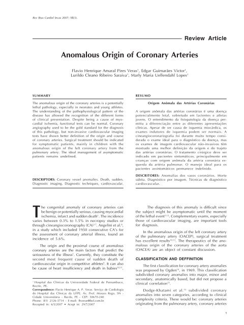
Veras FHAP, et al. <strong>Anomalous</strong> <strong>Origin</strong> <strong>of</strong> <strong>Coronary</strong> <strong>Arteries</strong>. Rev Bras Cardiol Invas 2007; 15(3).with an anomalous origin in the aorta, congenital atresia<strong>of</strong> the left coronary trunk, coronary arteriovenous fistulas,coronary arteries forming myocardial bridges, aneurysms<strong>of</strong> coronary arteries and coronary stenosis.For Angelini et al. 8 , a coronary anomaly should bedefined as any coronary pattern, with a characteristic(number <strong>of</strong> ostios, proximal course, distal bed… ) “rarely”encountered in the general population. The followingdefinitions being favorable: normal, any morphologicalcharacteristic observed in more than 1% <strong>of</strong> a nonselectedpopulation; variable <strong>of</strong> normality, a relativelyuncommon morphological characteristic prevalent inmore than 1% <strong>of</strong> the population; and anomaly, a morphologicalcharacteristic seen in less than 1% <strong>of</strong> thatpopulation. Normal coronary anatomy, universally accepted,is defined in this way 16,17 :• The coronary trunk originates from the left coronarysinus and divides into descendent anterior arteryand circumflex artery;• The anterior descendent artery follows behindthe pulmonary trunk in the intraventricular anteriorgrove;• The circumflex artery follows the posterioratrioventricular grove;• The right coronary artery originates from the rightcoronary sinus and follows along the anterioratrioventricular grove.Any alteration in this pattern must be consideredas an anomalous coronary anatomy 16,17 . The circumflexcoronary artery, originating from the right coronarysinus or from the right coronary artery, with a retroaorticcourse, is the most common congenital anomaly<strong>of</strong> the coronary artery 18, 19 .PHYSIOPATHOLOGY<strong>Coronary</strong> anomalies have been implicated as a causefor thoracic pain, sudden death, heart insufficiency,syncope, dyspnea, ventricular fibrillation and AMI 8 .In the OACEP, it is observed that, during intrauterinelife, no functional alteration takes place, however,after birth, in the measure that arteriolar resistance fallsand blood pressure decreases in the pulmonary artery,the oxi-hemoglobinic saturation <strong>of</strong> the venous blooddiminishes, the arterial channel and the oval foramenclose, progressively diminishing the myocardial bloodperfusion, irrigated by the anomalous artery, leadingto ischemic alterations.The OACEP is generally an isolated defect, butmight be associated, in about 5% <strong>of</strong> cases, to othercardiac defects such as, intraventricular communication,interatrial communication, aortal coarctation, Fallot’sTetralogy and other anomalies 13 . In Fallot’s Tetralogybearers, the incidence <strong>of</strong> coronary anomaly can varyfrom 3% to 36% 20,21 .The congenital atresy <strong>of</strong> the left coronary trunk(LCT) is an extremely rare anomaly and generally hasa benign evolution 22 . In some cases, it can lead tomyocardium ischemia and sudden death 22 . The bearers<strong>of</strong> this anomaly survive thanks to the development <strong>of</strong>collateral circulation for the anterior descendent artery 22 .Collateral circulation can be made up by branches/bunches from the cone artery, also known as VieussensCircle; or anastomosis between the anterior ventricularbranches <strong>of</strong> the right coronary artery and the anteriordescendent artery 22,23 .The explanation for myocardial ischemia in theatresia <strong>of</strong> the TCE, in the absence <strong>of</strong> arterioscleroses orvessel spasm, is not clear and the most logical justificationis the fact that an only coronary artery is incapable <strong>of</strong>supplying, adequately, the myocardial blood demand 22 .The anomalous origin <strong>of</strong> the left coronary artery<strong>of</strong> the right coronary sinus can be related to suddendeath in 59% <strong>of</strong> cases, preceded by physical activityin 81% <strong>of</strong> events 8 . This anomaly can present 4 courses:anterior, to the pulmonary artery; posterior, to the aorta;intra-septal, between the aorta and the pulmonary artery;and intra-arterial, between the aorta and the pulmonary(Figure 1) 24 . All the subtypes <strong>of</strong> this anomaly have beenassociated to sudden death, but the intra-arterial variety,which is the most common pattern, has a strongerrelation with catastrophic events 1,8,24-26 .Potential mechanisms have been erected to explainthe presence <strong>of</strong> myocardial ischemia and sudden deathin patients with OACEA; the formation <strong>of</strong> an acute andcontorted angle at the origin <strong>of</strong> the anomalous coronary<strong>of</strong> the aorta; a narrowing <strong>of</strong> the coronary orifice, secondaryto the anomalous anatomy; compression <strong>of</strong>the anomalous coronary artery during its course betweenthe aorta and the pulmonary trunk during exercise; andspasm <strong>of</strong> the anomalous artery, possibly as a result <strong>of</strong>endothelial injury 8,27,28 .According with current evidence, the coronarysegments with anomalous course are not more susceptibleto obstructive arterosclerotic disease than normalsegments in a same individual 8,9 (Figure 1).CLINICAL OUTLOOKThe OACEP presents two levels: an infantile formand an adult form. The infantile form is characterizedby poor intercoronary collateral circulation, which canresult in AMI, cardiac insufficiency or sudden death 11 .In this type <strong>of</strong> presentation, the symptoms appear in thefirst months <strong>of</strong> life, with constant crying, intense paleness,sudden stop when suckling, weight loss and signs <strong>of</strong>cardiac insufficiency, such as taquipnea tachycardia,tiredness while suckling, sweating, hepathomegalia andperipherical cyanosis. On auscultation, a galloping rhythmcan be heard, muffling <strong>of</strong> the bustle, especially thefirst one, and occasionally, the appearance <strong>of</strong> systolic2
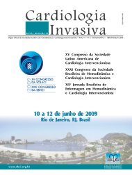
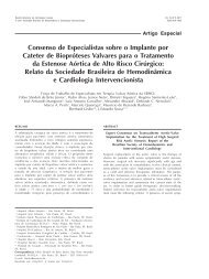
Veras FHAP, et al. <strong>Anomalous</strong> <strong>Origin</strong> <strong>of</strong> <strong>Coronary</strong> <strong>Arteries</strong>. Rev Bras Cardiol Invas 2007; 15(3).Figura 1 - Origem anômala da artéria coronária esquerda no seio de Valsalva direito. Cada painel tem uma representação de um cortecraniocaudal, ao nível das válvulas semilunares, mostrando o trajeto da artéria coronária anômala. Os angiogramas em posição oblíqua anteriordireita e as representações esquemáticas mostram exemplos dos quatro trajetos mais comuns de artéria coronária esquerda anômala originandosedo seio de Valsalva: posterior (retroaórtico), interarterial, anterior e septal. CD: artéria coronária direita, DA: artéria descendente anterior;Cx: artéria circunflexa (adaptado de Popma et al. 27 ).3
Veras FHAP, et al. <strong>Anomalous</strong> <strong>Origin</strong> <strong>of</strong> <strong>Coronary</strong> <strong>Arteries</strong>. Rev Bras Cardiol Invas 2007; 15(3).murmur in the mitral area, as a result <strong>of</strong> secondarymitral insufficiency to the increase <strong>of</strong> the cardiac areadue to its dilatation or due to dysfunction <strong>of</strong> the papillarymuscle 10,11 . Mortality during the first year <strong>of</strong> life isestimated at 90%, even with clinical treatment 10,11 .The adult or mature form can occur in childrenand necessarily in adults in around 10 – 15% <strong>of</strong> cases.It is characterized by an abundance <strong>of</strong> intercoronarycollateral circulation, <strong>of</strong> sufficient magnitude to allowa survival rate until adult age, in some cases, it hasbeen told, until 72 years <strong>of</strong> age.Sometimes, there are no appreciable clinical manifestationsand the diagnosis <strong>of</strong> OACEP is suspecteddue to the presence <strong>of</strong> murmurs: continuous, as aresult <strong>of</strong> the passage <strong>of</strong> blood through the coronarypulmonaryfistula, or systolic, because <strong>of</strong> dysfunction<strong>of</strong> the mitral valve 10 . Some patients evolve towardscardiac insufficiency; others towards angina 10,11 .In the OACEA, some symptoms such as syncope,chest pain and palpitations may be present 12,28 . Butmost bearers are asymptomatic and the only expression<strong>of</strong> this coronary anomaly in young athletes might besudden death 12,28 . The studies suggest that these coronaryanomalies can be lethal only during or immediatelyafter extenuating physical exercise, typically in youngsubjects 8 . (Table 1).COMPLEMENTARY EXAMSElectrocardiogram (ECG) and ergometric test (ET)One study demonstrated that the presence <strong>of</strong> the“Q” wave with a depth over 3 mm, duration higherthan 30 ms and a “QR” pattern in at least one <strong>of</strong> thedeviations – DI, aVL, V5-V7, was present in 100% <strong>of</strong>exams <strong>of</strong> patients with OACEP 29 . The absence <strong>of</strong> the“Q” wave in the lower wall is also a prevalent finding(82% <strong>of</strong> cases) 13,15,29 . The axel deviation towards theleft is less frequent (64% <strong>of</strong> cases) 13 . Atypical electrocardiographicalterations can mask the illness whenassociated to lesions with important pulmonary hip<strong>of</strong>lux.In the OACEA, the ECG and the TE are almostalways normal 8,9,12,28 . In spite <strong>of</strong> the OACEA being thecause <strong>of</strong> myocardial ischemia, this is incidental, with manifestationsunder extenuating clinical conditions 8,9,12,28 .Thorax X-RayIn general, it shows alterations in the cases associatedto cardiac insufficiency. In the OACEP, young agedchildren can present an increase <strong>of</strong> the cardiac area 10 .Lung circulation is normal, however, when cardiacinsufficiency is present, a picture <strong>of</strong> venous pulmonarycongestion can be observed 10 .EchocardiogramThe bidimensional echocardiogram with coloredDoppler is useful in the morph<strong>of</strong>unctional assessment<strong>of</strong> the heart. For the diagnosis <strong>of</strong> anomalous origin <strong>of</strong>the coronary arteries, the studies show a variablesensibility, with better results in children 11,13,15,26 .The transesophageal echocardiogram (TEE) is anexam with a much higher sensibility for the detection<strong>of</strong> the anomalous origin <strong>of</strong> coronary arteries 1,26 . TheTEE, specially with the use <strong>of</strong> a multi-plane probe/catheter, is capable <strong>of</strong> drawing, with precision, theproximal course and the flux pattern in the anomalouscoronary arteries 1,26 .Computer Tomography <strong>of</strong> <strong>Coronary</strong> <strong>Arteries</strong> (CTCA)The studies show a quick evolution <strong>of</strong> this noninvasivemethod in the assessment <strong>of</strong> coronary arteries.Initially, the studies with the Electron Beam ComputedTomography (EBCT), demonstrated excellent accuracyin the assessment <strong>of</strong> the origin and proximal course <strong>of</strong>coronary anomalies 19,25 .With the advent <strong>of</strong> Computer Tomography withMultiple Detectors (16 and 64 acquisitions), a betterassessment <strong>of</strong> the middle and distal segments waspossible, for the detection <strong>of</strong> obstructive coronary arteryillness 16. It uses ionizing radiation and requires theuse <strong>of</strong> medication to diminish heart frequency 25. Themethod requires the administration <strong>of</strong> a contrast meansin the veins, which is potentially allergic and nephrotoxic.The data in the literature show it to be a safe andefficient method with excellent spatial resolution 16,24,25,30 .Comparative studies with coronary angiography haveshown a higher efficacy, suggesting that ComputerTomography with Multiple Detectors is the ideal examfor the detection <strong>of</strong> coronary anomalies 16,19 .TABELA 1Incidência de morte súbita relacionada a anomalias de artérias coronárias (adaptado de Angelini et al. 8 ).Grupo (idade) Número de mortes Mortes relacionadas a anomalias coronarianas,%Indivíduos fazendo exercício (8-66 a) 550 11População geral (< 40 a) 162 0,6Atletas competitivos(idade média de 17 a) 134 23Corredores (30-46 a) 120 1,6Indivíduos fazendo exercício, Maryland - EUA 62 04
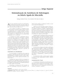
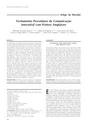
Veras FHAP, et al. <strong>Anomalous</strong> <strong>Origin</strong> <strong>of</strong> <strong>Coronary</strong> <strong>Arteries</strong>. Rev Bras Cardiol Invas 2007; 15(3).Figure 2 illustrates a CTCA where an OACEA withan inter-arterial course is evidenced.Nuclear Magnetic Resonance (NMR)<strong>of</strong> the coronary arteries<strong>Coronary</strong> angiography through MR is a noninvasiveexam, with high accuracy for the anatomicalidentification <strong>of</strong> the origin and proximal course <strong>of</strong> thecoronary arteries. The free choice <strong>of</strong> the image plane,with a trydimensional view, is an important advantageover the limited angle possibilities <strong>of</strong> conventionalcoronary angiography. It requires a non-nephrotoxiccontrast and does not use ionizing radiation 16 .It has the disadvantage <strong>of</strong> the long time <strong>of</strong> theexam (45-50 minutes), the need <strong>of</strong> periods <strong>of</strong> apnea,and the limitation <strong>of</strong> the analysis <strong>of</strong> the distal coronarysegments 19,31-34 .Bunce et al. 34 developed a technique for the detection<strong>of</strong> coronary anomalies were there is no need<strong>of</strong> apnea and with a shorter time <strong>of</strong> exam (average time26 minutes).It is an exam that possibilitates cardiac morph<strong>of</strong>unctionalassessment, being <strong>of</strong> great usefulness in congenitalcardiopathies, since it allows a perfect spatialassessment between the coronary arteries, the greatvessels and all other cardiac structures 19,31-34 .Figure 3 illustrates a NMR diagnosing an athresia<strong>of</strong> the TCE.CineangiocoronarographyIt is traditionally considered the ideal exam for thediagnosis <strong>of</strong> coronary anomalies. It is an invasive exam,which uses nephro-toxic contrast and ionizing radiation.The evolution <strong>of</strong> the RNM and <strong>of</strong> the TCAC, bothwith trydimensional analysis, have shown failures inthe CA for the diagnosis <strong>of</strong> coronary anomalies 19,31-33 .The CA might be incapable <strong>of</strong> differentiating, duringthe course through an anomalous coronary, the highrisk intra-arterial anatomy, from the septal anatomyretro-aortal or <strong>of</strong> the anterior wall, as well as the identification<strong>of</strong> the coronary sinus and the morphology <strong>of</strong>the coronary ostium in the wall <strong>of</strong> the aorta 19,31-33 .The CA is still the best method for the assessment<strong>of</strong> the distal coronary bed and associated arteroscleroticlesions 19,35,36 .TREATMENTOnce diagnosed, the OACEP must be surgicallycorrected immediately after the diagnosis, to avoidcomplications and sequels, characteristic <strong>of</strong> the naturalhistory <strong>of</strong> this illness 10,11,13,37-40 .The chosen surgical technique is the reimplantationwith translocation <strong>of</strong> the left coronary artery <strong>of</strong> thepulmonary trunk to the aorta, which is possible inmost cases 11,13,37-40 .In the case in which this technique is impossible toapply, “tunnelization” or Takeuchi Technique is recommended,which consists <strong>of</strong> creating a tunnel inside thepulmonary artery, to connect the left coronary arterywith the aorta 11,13,37-40 . Other surgical options are simplyto string or thread, to tie the left coronary artery with theaorta 11,13,37-40 . Surgical mortality for all the combinedFigura 2 - Origem anômala da artéria coronária esquerda com curso interarterial visualizada por TCAC. A: corte axial para avaliação da origemdas artérias coronárias. B: reconstrução tridimensional. As setas indicam o trajeto da artéria descendente anterior entre a aorta ascendentee o tronco da artéria pulmonar. A cabeça da seta indica a artéria coronária esquerda. Ao: aorta; AP: artéria pulmonar; AD: átrio direito; AE:átrio esquerdo(adaptado de Ropers et al. 24 ).5
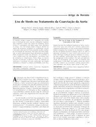
Veras FHAP, et al. <strong>Anomalous</strong> <strong>Origin</strong> <strong>of</strong> <strong>Coronary</strong> <strong>Arteries</strong>. Rev Bras Cardiol Invas 2007; 15(3).techniques varied between 75 and 80% in the 80s 37 .Nowadays, mortality is situated between 0 and 23% 37 .The clinical evolution and functional recovery <strong>of</strong>the left ventricle will depend upon the dysfunctionalventricular degree before the surgery. In most cases,there is a normalization <strong>of</strong> the ventricular function afterthe operation 11,13,37-40 .Bearers <strong>of</strong> OACEA, in general, are asymptomatic 12 .Even in athletes, only a minority is symptomatic 12 . And,in symptomatic individuals, the ECG and ischemia inducingexams, are generally normal 8,12,28 . The presence<strong>of</strong> symptoms suggestive <strong>of</strong> the illness, especially inathletes, must be investigated through imaging exams.In the case in which echocardiograms are unable todiagnose the anomalous origin, TCAC, RNM or CA <strong>of</strong>the coronary arteries must be performed 8,28 .There is no consensus in the literature for the management<strong>of</strong> asymptomatic bearers <strong>of</strong> OACEA. In symptomaticpatients, the surgical option must be chosen 12,28 .Figura 3 - Atresia do TCE em um paciente portador de valva aórtica bicúspide e ducto arterioso patente. A: cineangiocoronariografia mostrandoque a artéria circunflexa(Cx) origina-se da artéria coronária direita(CD). A artéria descendente anterior(DA) enche-se retrogradamente e formauma fístula com a artéria pulmonar(AP). B: no corte axial da RNM, observa-se uma falsa inserção da DA na AP. C: imagem axial da Cxoriginando-se da CD. Ao: aorta; AD: átrio direito; VE: ventrículo esquerdo(adaptado de Taylor et al. 17 )6
Veras FHAP, et al. <strong>Anomalous</strong> <strong>Origin</strong> <strong>of</strong> <strong>Coronary</strong> <strong>Arteries</strong>. Rev Bras Cardiol Invas 2007; 15(3).The surgical options are the implant <strong>of</strong> arterial or venousgraft or, reimplantation <strong>of</strong> the anomalous coronary arteryin the anatomical coronary sinus, plastia <strong>of</strong> the intramuralsegment <strong>of</strong> the coronary artery and translocation <strong>of</strong> thepulmonary artery trunk 12,28 . There is no data which definesthe best surgical technique.CONCLUSIONThe anomalous origin <strong>of</strong> coronary arteries is arare illness, but potentially lethal if not diagnosed andtreated early. Children and young athletes are the riskgroup for the catastrophic sequelae. In these patients,therapeutic surgery brings good results, being this adefinitive treatment. The identification <strong>of</strong> the illness inasymptomatic patients continues being a challenge.And, new studies are necessary, for the definition <strong>of</strong>the ideal treatment in these patients.BIBLIOGRAPHIC REFERENCES1. Dawn B, Talley JD, Prince CR, Hoque A, Morris GT,Xenopoulos NP, et al. Two-dimensional and dopplertransesophageal echocardiographic delineation and flowcharacterization <strong>of</strong> anomalous coronary arteries in adults.J Am Soc Echocardiogr. 2003;16(12):1274-86.2. Hayashi JH, Abreu Fº LM, Sumita MK, Takimura CK, HottaVT, Forte AAC. Incidência de anomalias coronárias emhospital geral. Rev Bras Cardiol Invas. 2002;10 (3):26-32.3. Yamanaka O, Hobbs RE. <strong>Coronary</strong> artery anomalies in126.595 patients undergoing coronary arteriography. CathetCardiovasc Diagn. 1990;21(1):28-40.4. Baltaxe HA, Wixson D. The incidence <strong>of</strong> congenital anomalies<strong>of</strong> the coronary arteries in the adult population. Radiology.1977;122(1):47-52.5. Leberthson RR, Dinsmore RE, Bharati S, Rubenstein JJ, CaulfieldJ, Wheeler BO, et al. Aberrant coronary artery origin fromthe aorta. Diagnosis and clinical significance. Circulation.1974;50(4):774-9.6. Engel HJ, Torres C, Page HL Jr. Major variations in anatomicalorigin <strong>of</strong> the coronary arteries: angiographic observationsin 4.250 patients without associated congenital heartdisease. Cathet Cardiovasc Diagn. 1975;1(2):157-69.7. Chaitman BR, Lesperance J, Saltiel J, Bourassa MG. Clinical,angiographic and hemodynamic findings in patients withanomalous origin <strong>of</strong> the coronary arteries. Circulation. 1976;53(1):122-31.8. Angelini P, Velasco JA, Flamm S. <strong>Coronary</strong> anomalies:incidence, pathophysiology, and clinical relevance. Circulation.2002;105(20):2449-54.9. Maron BJ. Sudden death in young athletes. N Engl J Med.2003;349(11):1064-75.10. Oliveira SA, Snitcowsky R. Origem anômala da artéria coronáriaesquerda do tronco pulmonar. In: Macruz R, Snitcowsky R,eds. Cardiologia pediátrica. 1ª ed. São Paulo:Sarvier;1983.p.508-43.11. Takimura CH, Nakamoto A, Hotta VT, Campos MF, MálamoM, Otsubo R. Origem anômala da artéria coronária esquerdano tronco pulmonar. relato de um caso em adulto. Arq BrasCardiol. 2002;78(3):309-11.12. Erez E, Tam VK, Doublin NA, Stakes J. <strong>Anomalous</strong> coronaryartery with aortic origin and course between the greatarteries: improved diagnosis, anatomic findings, and surgicaltreatment. Ann Thorac Surg. 2006;82(3):973-7.13. Amaral F, Carvalho JS, Granzotti JA, Shinebourne EA. Origemanômala da artéria coronária esquerda do tronco pulmonar:perfil clínico e resultados a médio prazo do tratamentocirúrgico. Arq Bras Cardiol. 1999;72(3):307-20.14. Ogden JA. Congenital anomalies <strong>of</strong> the coronary arteries.Am J Cardiol. 1970;25(4):474-9.15. Dodge-Khatami A, Mavroudis C, Backer CL. CongenitalHeart Surgery Nomenclature and Database Project: anomalies<strong>of</strong> the coronary arteries. Ann Thorac Surg. 2000;69(4 Suppl):S270-97.16. Shi H, Asch<strong>of</strong>f AJ, Brambs HJ, H<strong>of</strong>fmann MH. Multislice CTimaging <strong>of</strong> anomalous coronary arteries. Eur Radiol. 2004;14(12):2172-81.17. Taylor AM, Thorne SA, Rubens MB, Jhooti P, Keegan J,Gatehouse PD, et al. <strong>Coronary</strong> artery imaging in grown upcongenital heart disease: complementary role <strong>of</strong> magneticresonance and X-ray coronary angiography. Circulation.2000;101(14):1670-8.18. Kruskal JB, Hartnell GG. Nonatherosclerotic coronary arterydisease: more than just stenosis. Radiographics. 1995;15(2):383-96.19. Ropers D, Moshage W, Daniel WG, Jessl J, Gottwik M,Achenbach S. Visualization <strong>of</strong> coronary artery anomalies andtheir anatomic course by contrast-enhanced electron beamtomography and three-dimensional reconstruction. Am J Cardiol.2001;87(2):193-7.20. Dabizzi RP, Teodori G, Barletta GA, Caprioli G, BaldrighiG, Baldrighi V. Associated coronary and cardiac anomaliesin the tetralogy <strong>of</strong> Fallot. An angiographic study. Eur HeartJ. 1990;11(8):692-704.21. Carvalho JS, Silva CM, Rigby ML, Shinebourne EA. Angiographicdiagnosis <strong>of</strong> anomalous coronary artery in the tetralogy<strong>of</strong> Fallot. Br Heart J. 1993;70(1):75-8.22. Ruiz CE, Lau FYK. Congenital atresia <strong>of</strong> left main coronaryartery: proposed mechanism for severe disabling angina in apatient with non-atherosclerotic single right coronary artery- a case report. Cathet Cardiovasc Diagn. 1991;23(3):190-3.23. Ghosh PK, Friedman M, Vidne BA. Isolated congenitalatresia <strong>of</strong> the left main coronary artery and atherosclerosis.Ann Thorac Surg. 1993;55(6):1564-5.24. Ropers D, Gehling G, Pohle K, Maeffert R, Regenfus M,Moshage W, et al. <strong>Anomalous</strong> course <strong>of</strong> the left main orleft anterior descending coronary artery originating from theright sinus <strong>of</strong> Valsalva: identification <strong>of</strong> four common variationsby electron beam tomography. Circulation. 2002;105(6):e42-3.25. Memisoglu E, Ropers D, Hobikoglu G, Tepe MS, LabovitzAJ. Usefulness <strong>of</strong> electron beam computed tomography fordiagnosis <strong>of</strong> an anomalous origin <strong>of</strong> a coronary artery fromthe opposite sinus. Am J Cardiol. 2005;96(10):1452-5.26. Kasprzak JD, Kratochwil D, Peruga JZ, Drozdz J, RafalskaK, Religa W, et al. <strong>Coronary</strong> anomalies diagnosed withtransesophageal echocardiography: complementary clinicalvalue in adults. Int J Card Imaging. 1998;14(2):89-95.27. Popma JJ, Bittl J. <strong>Coronary</strong> angiography and intravascularultrasonography. In: Braunwald E, Zipes DP, Libby P, eds.Heart disease. 6 th ed. Philadelphia: W.B. Saunders;2001.p.387-421.28. Basso C, Maron BJ, Corrado D, Thiene G. Clinical pr<strong>of</strong>ile<strong>of</strong> congenital coronary artery anomalies with origin from thewrong aortic sinus leading to sudden death in young competitiveathletes. J Am Coll Cardiol. 2000;35(6):1493-501.29. Johnsrude CL, Perry JC, Cecchin F, Smith EO, Fraley K,Friedman RA, et al. Differentiating anomalous left maincoronary artery originating from the pulmonary artery in7
Veras FHAP, et al. <strong>Anomalous</strong> <strong>Origin</strong> <strong>of</strong> <strong>Coronary</strong> <strong>Arteries</strong>. Rev Bras Cardiol Invas 2007; 15(3).infants from myocarditis and dilated cardiomyopathy byelectrocardiogram. Am J Cardiol. 1995;75(1):71-4.30. Berbarie RF, Dockery WD, Johnson KB, Rosenthal RL, StolerRC, Schussler JM. Use <strong>of</strong> multislice computed tomographiccoronary angiography for the diagnosis <strong>of</strong> anomalous coronaryarteries. Am J Cardiol. 2006;98(3):402-6.31. Post JC, van Rossum AC, Bronzwaer JG, de Cock CC, H<strong>of</strong>manMB, Valk J, et al. Magnetic resonance angiography <strong>of</strong> anomalouscoronary arteries: a new gold standard for delineatingthe proximal course? Circulation. 1995;92(11):3163-71.32. McConnell MV, Ganz P, Selwyn AP, Li W, Edelman RR,Manning WJ. Identification <strong>of</strong> anomalous coronary arteriesand their anatomic course by magnetic resonance coronaryangiography. Circulation. 1995;92(11):3158-62.33. Vliegen HW, Doornbos J, Ross A, Jukema JW, Bekedam MA,van der Wall EE. Value <strong>of</strong> fast gradient echo magnetic resonanceangiography as an adjunct to coronary arteriography indetecting and confirming the course <strong>of</strong> clinically significantcoronary artery anomalies. Am J Cardiol. 1997;79(6):773-6.34. Bunce NH, Lorenz CH, Keegan J, Lesser J, Reyes EM, FirminDN, et al. <strong>Coronary</strong> artery anomalies: assessment with freebreathingthree-dimensional coronary MR angiography. Radiology.2003;227(1):201-8.35. Pinto IMF, Sousa AGMR, Ishikawa W, Sasdelli Neto R, Pivae Mattos LA, Sousa JEMR. Ressonância magnética e tomografiacomputadorizada no diagnóstico de insuficiência coronária.Rev Bras Cardiol Invas. 2006;14(2):168-77.36. Hamon M, Biondi-Zoccai GGL, Malagutti P, Agostini P, MorelloR, Valgimigli M, et al. Diagnostic performance <strong>of</strong> multislicespiral computed tomography <strong>of</strong> coronary arteries as comparedwith conventional invasive coronary angiography: a metaanalysis.J Am Coll Cardiol. 2006;48(9):1896-910.37. Dodge-Khatami A, Mavroudis C, Backer CL. <strong>Anomalous</strong>origin <strong>of</strong> the left coronary artery from the pulmonary artery:collective review <strong>of</strong> surgical therapy. Ann Thorac Surg.2002;74(3):946-55.38. Moraes F, Lincoln C. <strong>Anomalous</strong> origin <strong>of</strong> left coronaryartery: evolution <strong>of</strong> surgical treatment. Eur J CardiothoracSurg. 1996;10(8):603-8.39. Reul RM, Cooley DA, Hallman GL, Reul GJ. Surgical treatment<strong>of</strong> coronary artery anomalies: report <strong>of</strong> 37 ½-yearexperience at the Texas Heart Institute. Tex Heart Inst J.2002;29(4):299-307.40. Selzman CH, Zimmerman MA, Campbell DN. ALCAPA inan adult with preserved left ventricular function. J CardSurg. 2003;18(1):25-8.8