A review on White Spot Disease in penaeid ... - Middle East - OIE
A review on White Spot Disease in penaeid ... - Middle East - OIE
A review on White Spot Disease in penaeid ... - Middle East - OIE

You also want an ePaper? Increase the reach of your titles
YUMPU automatically turns print PDFs into web optimized ePapers that Google loves.
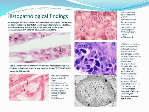
Histopathological f<strong>in</strong>d<strong>in</strong>gsCowdry type A <strong>in</strong>clusi<strong>on</strong> bodies are <strong>in</strong>tranuclear, eos<strong>in</strong>ophilic, amorphousand surrounded by a clear halo beneath the nuclear membrane but later,<strong>in</strong>clusi<strong>on</strong>s become lightly to deeply basophilic and fill the entirenucleus(LightnerD.V. 1996,<strong>OIE</strong> Manual of Aquatic 2009).Left. IntranuclearIBs characteristicof WSSV<strong>in</strong>fecti<strong>on</strong>(arrow)<strong>in</strong>the haemapoeitictissue cells ofL.vannamei <strong>in</strong> Iranshow<strong>in</strong>g signs ofWSSV(H&E 100X).Source:M.Afsharnasab.Above. Intranuclear IBs characteristic of WSSV <strong>in</strong>fecti<strong>on</strong>(arrow)<strong>in</strong> thegill tissue cells of L.vannamei <strong>in</strong> Iran show<strong>in</strong>g signs of WSSV(H&E 100X).Source: M.Afsharnasab.Left. Intranuclear IBscharacteristic ofWSSV<strong>in</strong>fecti<strong>on</strong>(arrow)<strong>in</strong>the heart tissue cellsof L.vannamei <strong>in</strong> Iranshow<strong>in</strong>g signs ofWSSV(H&E 100X).Source:M.Afsharnasab.**Left.Penaeus vannamei.Hypertrophied nucleiwith<strong>in</strong> sub-cuticularepithelium. Notegranular eos<strong>in</strong>ophillicsta<strong>in</strong><strong>in</strong>g (*) with<strong>in</strong> somenuclei and densehaemotoxyl<strong>in</strong>sta<strong>in</strong><strong>in</strong>g <strong>in</strong> others(arrows). H&E sta<strong>in</strong>.Scale = 10μm.And (below left)Penaeus vannamei.WSSV particlesdevelop<strong>in</strong>g with<strong>in</strong>nucleus of <strong>in</strong>fectedcuticular epithelial cell.TEM. Scale = 1μm.Source:EuropeanCommunity ReferenceLaboratory forCrustacean <strong>Disease</strong>sleaflet 2008