GPU-Based Real-Time Imaging Software Suite for Medical Ultrasound
GPU-Based Real-Time Imaging Software Suite for Medical Ultrasound
GPU-Based Real-Time Imaging Software Suite for Medical Ultrasound

Create successful ePaper yourself
Turn your PDF publications into a flip-book with our unique Google optimized e-Paper software.
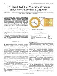
Figure 7. <strong>Real</strong>-time images of a mouse subcutaneous kidney tumor from PA Imager obtained using a 24-element 1-D CMUT array. The vasculature in the tumoris visible in the photoacoustic image.TABLE IIIEXPERIMENTAL CONDITIONS AND IMAGING RATES OF FIGS. 5-7Fig. 5 Fig. 6(a) Fig. 6(b) Fig. 7<strong>Imaging</strong> program General Imager MIP Imager MIP Imager PA ImagerProbe128-element ring CMUT array 64-element ring CMUT array 128-element ring CMUT array 24-element 1-D CMUT array(4.84-mm radius)(1.16-mm radius)(4.84-mm radius)(63-μm pitch)Probe center frequency 6.5 MHz 10 MHz 6.5 MHz 8.5 MHz<strong>Imaging</strong> scheme SPA-HW 1 SPA-HW 1 SPA-HW 1 CPA 2 + Photoacoustic<strong>Imaging</strong> targetTen fluorocarbon fishing wires A metal springA metal spring(150-μm thickness)(6-mm diameter)(13-mm diameter)A mouse subcutaneous tumor<strong>Imaging</strong> depth 25 mm 15 mm 40 mm 15 mm<strong>Imaging</strong> rate100 <strong>for</strong> ultrasound image14 104 11in frames per second10 <strong>for</strong> photoacoustic image 31 SPA-HW: Synthetic phased array imaging with Hadamard coding and aperture weighting (Norton weighting and cosine weighting)2 CPA: Classic phased array imaging3 <strong>Imaging</strong> rate <strong>for</strong> photoacoustic image is limited by the 10-Hz laser repetition rate.obtained using different targets and different probes.Experimental conditions and acquired imaging rates from theseexperiments are summarized in Table III.V. CONCLUSIONWe developed a <strong>GPU</strong>-based ultrasound imaging softwaresuite that is capable of real-time volumetric imaging witharbitrary probe geometries and various imaging schemesincluding non-conventional techniques such as syntheticbeam<strong>for</strong>ming and Hadamard coding. Exploiting massive datalevelparallelism in beam<strong>for</strong>ming operations, this softwaresuccessfully generated volumetric images in real-time <strong>for</strong>various imaging schemes. The real-time imaging wasdemonstrated using our custom CMUT probes with annular,linear, and rectangular shapes.ACKNOWLEDGMENTThis work is funded by the National Institutes of Healthunder Grants HL67647 and CA134720. We also thank Nvidia<strong>for</strong> donating a C2070 graphics card.REFERENCES[1] Ö. Oralkan, A. S. Ergun, J. A. Johnson, U. Demirci, M. Karaman, K.Kaviani, T. H. Lee, and B. T. Khuri-Yakub, “Capacitive micromachinedultrasonic transducers: Next-generation arrays <strong>for</strong> acoustic imaging?,”IEEE Trans. Ultrason., Ferroelect., Freq. Contr., vol. 49, no. 11, pp.1596–1610, Nov. 2002.[2] J. W. Choe, Ö. Oralkan, A. Nikoozadeh, A. Bhuyan, B. C. Lee, M.Gencel, and B. T. Khuri-Yakub, “<strong>Real</strong>-time volumetric imaging system<strong>for</strong> CMUT arrays,” in Proc. IEEE Ultrason. Symp., pp. 1064–1067,2011.[3] J. W. Choe, Ö. Oralkan, A. Nikoozadeh, M. Gencel, D. N. Stephens, M.O’Donnell, D. J. Sahn, and B. T. Khuri-Yakub, "Volumetric real-timeimaging using a CMUT ring array," IEEE Trans. Ultrason., Ferroelect.,Freq. Contr., vol. 59, no. 6, pp. 1201–1211, Jun. 2012.[4] J. Bercoff, G. Montaldo, T. Loupas, D. Savery, F. Mézière, M. Fink, andM. Tanter, “Ultrafast compound Doppler imaging: Providing full bloodflow characterization,” IEEE Trans. Ultrason., Ferroelect., Freq. Contr.,vol. 58, no. 1, pp. 134–147, Jan. 2011.[5] J. W. Choe, A. Nikoozadeh, Ö. Oralkan, and B. T. Khuri-Yakub, "<strong>GPU</strong>basedreal-time volumetric ultrasound image reconstruction <strong>for</strong> a ringarray," IEEE Trans. <strong>Medical</strong> <strong>Imaging</strong>, vol. 32, no. 7, pp. 1258–1264, Jul.2013.[6] A. Nikoozadeh, J. W. Choe, S. Kothapalli, A. Moini, S. S. Sanjani, A.Kamaya, Ö. Oralkan, S. S. Gambhir, and B. T. Khuri-Yakub,“Photoacousitc imaging using a 9F MicroLinear CMUT ICE catheter,”presented at the IEEE Ultrason. Symp., Dresden, Germany, Oct. 7-10,2012.