Etudes sur le mécanisme de remodelage des nucléosomes par ...
Etudes sur le mécanisme de remodelage des nucléosomes par ...
Etudes sur le mécanisme de remodelage des nucléosomes par ...

You also want an ePaper? Increase the reach of your titles
YUMPU automatically turns print PDFs into web optimized ePapers that Google loves.
tel-00413908, version 1 - 7 Sep 2009<br />
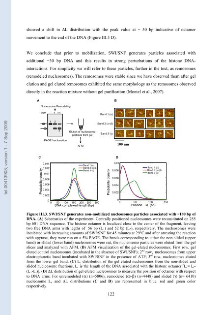
showed a shift in ΔL distribution with the peak value at ~ 50 bp indicative of octamer<br />
movement to the end of the DNA (Figure III.3 D).<br />
We conclu<strong>de</strong> that prior to mobilization, SWI/SNF generates <strong>par</strong>tic<strong>le</strong>s associated with<br />
additional ~30 bp DNA and this results in strong perturbations of the histone DNA-<br />
interactions. For simplicity we will refer to these <strong>par</strong>tic<strong>le</strong>s, further in the text, as remosomes<br />
(remo<strong>de</strong><strong>le</strong>d nuc<strong>le</strong>osomes). The remosomes were stab<strong>le</strong> since we have observed them after gel<br />
elution and gel eluted remosomes exhibited the same morphology as the remosomes observed<br />
directly in the reaction mixture without gel purification (Montel et al., 2007).<br />
A B<br />
Probability <strong>de</strong>nsity<br />
Nuc<strong>le</strong>osome Remo<strong>de</strong>ling<br />
SWI - + ++<br />
1 2<br />
3<br />
PAGE fractionation<br />
0.014<br />
0.012<br />
0.01<br />
0.008<br />
0.006<br />
0.004<br />
0.002<br />
Elution of nuc<strong>le</strong>osome<br />
<strong>par</strong>tic<strong>le</strong>s from gel<br />
AFM<br />
Band 1 (α)<br />
Band 2 (α+β)<br />
Band 3 (γ)<br />
0<br />
0 50 100 150 200 250 300<br />
DNA comp<strong>le</strong>xed <strong>le</strong>ngth (bp)<br />
Band 1 (α)<br />
Band 2 (α+β)<br />
Band 3 (γ)<br />
C D<br />
Probability <strong>de</strong>nsity<br />
Figure III.3. SWI/SNF generates non-mobilized nuc<strong>le</strong>osomes <strong>par</strong>tic<strong>le</strong>s associated with ~180 bp of<br />
DNA. (A) Schematics of the experiment. Centrally positioned nuc<strong>le</strong>osomes were reconstituted on 255<br />
bp 601 DNA sequence. The histone octamer is localized close to the center of the fragment, <strong>le</strong>aving<br />
two free DNA arms with <strong>le</strong>gths of 56 bp (L+) and 52 bp (L-), respectively. The nuc<strong>le</strong>osomes were<br />
incubated with increasing amounts of SWI/SNF for 45 minutes at 29°C and after arresting the reaction<br />
with apyrase, they were run on a 5% PAGE. The bands corresponding to either the non-sli<strong>de</strong>d (upper<br />
band) or sli<strong>de</strong>d (lower band) nuc<strong>le</strong>osomes were cut, the nuc<strong>le</strong>osome <strong>par</strong>tic<strong>le</strong>s were eluted from the gel<br />
slices and analyzed with AFM. (B) AFM visualization of the gel-eluted nuc<strong>le</strong>osomes. First row, gel<br />
eluted control nuc<strong>le</strong>osomes (incubated in the absence of SWI/SNF); 2 nd row, nuc<strong>le</strong>osomes from upper<br />
e<strong>le</strong>ctrophoretic band incubated with SWI/SNF in the presence of ATP; 3 rd row, nuc<strong>le</strong>osomes eluted<br />
from the lower gel band. (C) Lc distribution of the gel eluted nuc<strong>le</strong>osomes from the non-sli<strong>de</strong>d and<br />
sli<strong>de</strong>d nuc<strong>le</strong>osome fractions, Lc is the <strong>le</strong>ngth of the DNA associated with the histone octamer [Lc= Lt-<br />
(L+-L-)]. (D) ΔL distribution of gel eluted nuc<strong>le</strong>osomes to mea<strong>sur</strong>e the position of octamer with respect<br />
to DNA arms. For unremo<strong>de</strong><strong>le</strong>d (α) (n=5806), remo<strong>de</strong><strong>le</strong>d (α+β) (n=4448) and sli<strong>de</strong>d (γ) (n= 6410)<br />
nuc<strong>le</strong>osome Lc and ΔL distributions (C and D) are represented in blue, red and green color<br />
respectively.<br />
122<br />
0.01<br />
0.008<br />
0.006<br />
0.004<br />
0.002<br />
100 nm<br />
Band 1 (α)<br />
Band 2 (α+β)<br />
Band 3 (γ)<br />
0<br />
0 50 100<br />
Position ΔL (bp)<br />
150