Etudes sur le mécanisme de remodelage des nucléosomes par ...
Etudes sur le mécanisme de remodelage des nucléosomes par ...
Etudes sur le mécanisme de remodelage des nucléosomes par ...

You also want an ePaper? Increase the reach of your titles
YUMPU automatically turns print PDFs into web optimized ePapers that Google loves.
tel-00413908, version 1 - 7 Sep 2009<br />
crosslinked to the DNA, and the remo<strong>de</strong>ling was assessed by sensitivity to nuc<strong>le</strong>ases.<br />
Interestingly, hSWI/SNF could still increase the sensitivity towards DNaseI even in the<br />
absence of nuc<strong>le</strong>osome movement. Using a photo affinity labelling and crosslinking<br />
approach Kassabov et al., (2003) have shown that SWI/SNF moves the nuc<strong>le</strong>osomes in<br />
increments of ~50 bp whi<strong>le</strong> for ISWI the step size was ~10 bp. These were interpreted as the<br />
size of the loop or the bulge created by these remo<strong>de</strong><strong>le</strong>rs. Using this information about the<br />
step size authors have tried to explain the observed differences in nuc<strong>le</strong>osome disruption<br />
properties of these two remo<strong>de</strong><strong>le</strong>rs. It must be noted that, however, that new histone DNA<br />
crosslinks generated due to remo<strong>de</strong>ling could represent final products of the remo<strong>de</strong>ling rather<br />
than reaction intermediates. Another support towards formation of bulge by remo<strong>de</strong><strong>le</strong>rs come<br />
from the fact that remo<strong>de</strong><strong>le</strong>rs are ATP <strong>de</strong>pen<strong>de</strong>nt DNA translocases and are ab<strong>le</strong> to pump<br />
DNA insi<strong>de</strong> the nuc<strong>le</strong>osome (Saha et al., 2005; Zofall et al., 2006).<br />
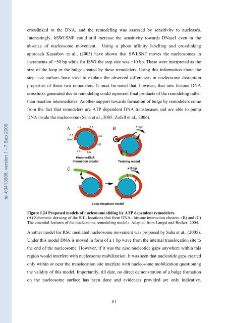
Figure I.24 Proposed mo<strong>de</strong>ls of nuc<strong>le</strong>osome sliding by ATP <strong>de</strong>pen<strong>de</strong>nt remo<strong>de</strong><strong>le</strong>rs.<br />
(A) Schematic drawing of the SHL locations that form DNA– histone interaction clusters. (B) and (C)<br />
The essential features of the nuc<strong>le</strong>osome remo<strong>de</strong>ling mo<strong>de</strong>ls. Adapted from Langst and Becker, 2004.<br />
Another mo<strong>de</strong>l for RSC mediated nuc<strong>le</strong>osome movement was proposed by Saha et at., (2005).<br />
Un<strong>de</strong>r this mo<strong>de</strong>l DNA is moved in form of a 1 bp wave from the internal translocation site to<br />
the end of the nuc<strong>le</strong>osome. However, if it was the case nuc<strong>le</strong>oti<strong>de</strong> gaps anywhere within this<br />
region would interfere with nuc<strong>le</strong>osome mobilization. It was seen that nuc<strong>le</strong>oti<strong>de</strong> gaps created<br />
only within or near the translocation site interfere with nuc<strong>le</strong>osome mobilization questioning<br />
the validity of this mo<strong>de</strong>l. Importantly, till date, no direct <strong>de</strong>monstration of a bulge formation<br />
on the nuc<strong>le</strong>osome <strong>sur</strong>face has been done and evi<strong>de</strong>nces provi<strong>de</strong>d are only indicative.<br />
81