Optimizing visualization and ergonomics. - Academy of Laser Dentistry
Optimizing visualization and ergonomics. - Academy of Laser Dentistry
Optimizing visualization and ergonomics. - Academy of Laser Dentistry

Create successful ePaper yourself
Turn your PDF publications into a flip-book with our unique Google optimized e-Paper software.
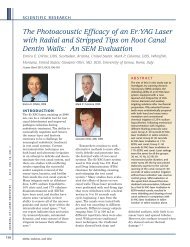
Figure 3: Illustration <strong>of</strong> the convenient<br />
arrangement <strong>of</strong> video camera on the left<br />
<strong>and</strong> a digital, single lens reflex camera<br />
(Nikon D70) on the right <strong>of</strong> the scope<br />
1. Improved Precision <strong>of</strong><br />
Treatment<br />
The visual information provided by<br />
the operating microscope is in fact<br />
not indicative <strong>of</strong> the magnification<br />
that is being employed. The actual<br />
amount <strong>of</strong> visual information is the<br />
area <strong>of</strong> view through the scope <strong>and</strong><br />
is therefore the product <strong>of</strong> the horizontal<br />
times the vertical number <strong>of</strong><br />
pixels. Therefore, the clinician<br />
using the 2X magnification power<br />
<strong>of</strong> entry-level loupes sees approximately<br />
4 times the visual<br />
information <strong>of</strong> a dentist not using<br />
any magnification (unaided eye).<br />
Likewise, 3X loupes provide 9 times<br />
the visual information <strong>of</strong> the<br />
unmagnified view <strong>and</strong> more than<br />
double the view <strong>of</strong> the 2X set. Table<br />
1 summarizes the relative advantages<br />
<strong>of</strong> a variety <strong>of</strong> magnifications.<br />
The author uses his microscope<br />
typically at 10X magnification<br />
which provides 100X the amount <strong>of</strong><br />
visual information compared to the<br />
unaided eye view. This is 25 times<br />
the information from 2X loupes <strong>and</strong><br />
more than 10 times as that seen<br />
with 3X.<br />
Carr 52 reported that the unaided<br />
human eye has the inherent ability<br />
to resolve or distinguish two separate<br />
lines or entities that are at<br />
least 200 µm or 0.2 mm apart. If<br />
the lines are closer together, then<br />
even 20/20 unmagnified vision will<br />
van As<br />
not allow the<br />
clinician to<br />
resolve them as<br />
two separate<br />
entities <strong>and</strong> the<br />
objects will<br />
appear as one.<br />
Thus with<br />
magnification<br />
the resolution <strong>of</strong><br />
the human eye<br />
improves<br />
dramatically<br />
(Table 2).<br />
Baldissara et<br />
al. 53 showed that<br />
the experienced<br />
clinician, when<br />
using a sharp,<br />
new explorer,<br />
can feel<br />
marginal gaps <strong>of</strong><br />
around 36 µm.<br />
Thus, when<br />
COVER FEATURE<br />
Table 1: Comparison <strong>of</strong> Unaided Eye, 2X Loupes, <strong>and</strong> Other Levels <strong>of</strong><br />
Magnification<br />
Magnification Visual Information (VI)<br />
VI Compared<br />
to 2X Loupes<br />
Unaided eye 1X 1/4<br />
2X loupes 4X Even = 1<br />
3X loupes 9X 2.25<br />
4X loupes 16X 4<br />
6X microscope 36X 9<br />
10X microscope 100X 25<br />
20X microscope 400X 100<br />
Table 2: Resolution vs. Assessment Method<br />
Assessment Method Magnification Resolution<br />
(µm)<br />
Resolution<br />
(mm)<br />
Unaided eye 1X 200 0.2<br />
Low-power loupes 2X 100 0.1<br />
Medium-power loupes 4X 50 0.05<br />
Sharp explorer NA 36 0.036<br />
Low-magnification microscope 6X 36 0.036<br />
Medium-magnification microscope 10X 20 0.02<br />
High-magnification microscope 20X 10 0.01<br />
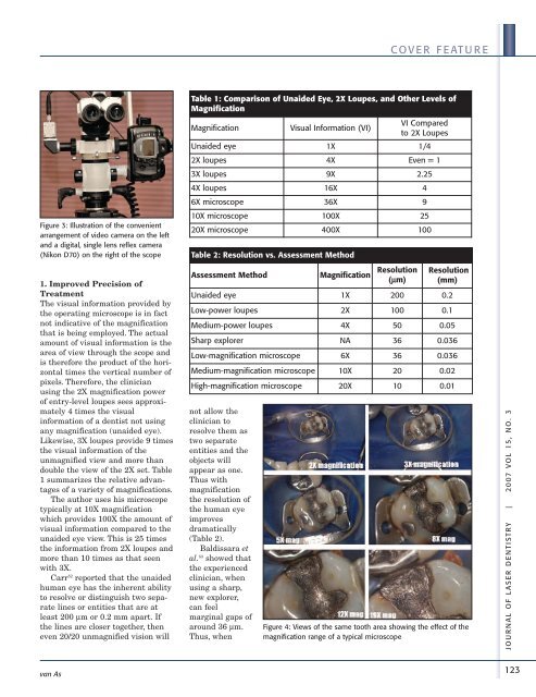
Figure 4: Views <strong>of</strong> the same tooth area showing the effect <strong>of</strong> the<br />
magnification range <strong>of</strong> a typical microscope<br />
JOUR NAL OF LASER DENTIS TRY | 2007 VOL 15, NO. 3<br />
123