Optimizing visualization and ergonomics. - Academy of Laser Dentistry
Optimizing visualization and ergonomics. - Academy of Laser Dentistry
Optimizing visualization and ergonomics. - Academy of Laser Dentistry

You also want an ePaper? Increase the reach of your titles
YUMPU automatically turns print PDFs into web optimized ePapers that Google loves.
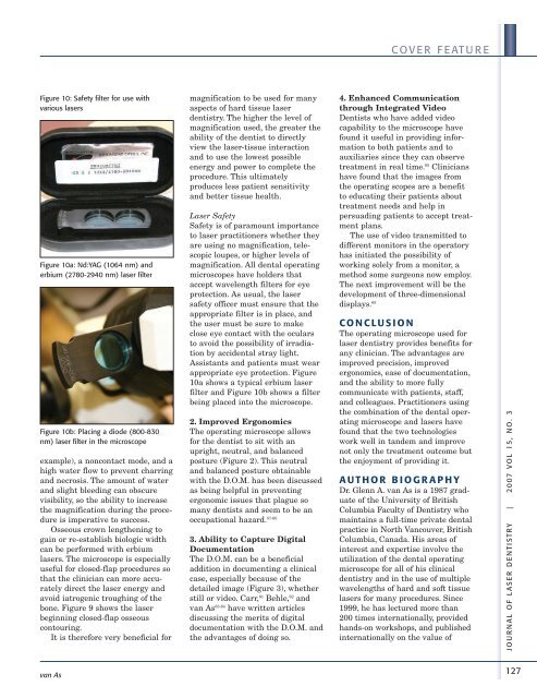
Figure 10: Safety filter for use with<br />
various lasers<br />
Figure 10a: Nd:YAG (1064 nm) <strong>and</strong><br />
erbium (2780-2940 nm) laser filter<br />
Figure 10b: Placing a diode (800-830<br />
nm) laser filter in the microscope<br />
example), a noncontact mode, <strong>and</strong> a<br />
high water flow to prevent charring<br />
<strong>and</strong> necrosis. The amount <strong>of</strong> water<br />
<strong>and</strong> slight bleeding can obscure<br />
visibility, so the ability to increase<br />
the magnification during the procedure<br />
is imperative to success.<br />
Osseous crown lengthening to<br />
gain or re-establish biologic width<br />
can be performed with erbium<br />
lasers. The microscope is especially<br />
useful for closed-flap procedures so<br />
that the clinician can more accurately<br />
direct the laser energy <strong>and</strong><br />
avoid iatrogenic troughing <strong>of</strong> the<br />
bone. Figure 9 shows the laser<br />
beginning closed-flap osseous<br />
contouring.<br />
It is therefore very beneficial for<br />
van As<br />
magnification to be used for many<br />
aspects <strong>of</strong> hard tissue laser<br />
dentistry. The higher the level <strong>of</strong><br />
magnification used, the greater the<br />
ability <strong>of</strong> the dentist to directly<br />
view the laser-tissue interaction<br />
<strong>and</strong> to use the lowest possible<br />
energy <strong>and</strong> power to complete the<br />
procedure. This ultimately<br />
produces less patient sensitivity<br />
<strong>and</strong> better tissue health.<br />
<strong>Laser</strong> Safety<br />
Safety is <strong>of</strong> paramount importance<br />
to laser practitioners whether they<br />
are using no magnification, telescopic<br />
loupes, or higher levels <strong>of</strong><br />
magnification. All dental operating<br />
microscopes have holders that<br />
accept wavelength filters for eye<br />
protection. As usual, the laser<br />
safety <strong>of</strong>ficer must ensure that the<br />
appropriate filter is in place, <strong>and</strong><br />
the user must be sure to make<br />
close eye contact with the oculars<br />
to avoid the possibility <strong>of</strong> irradiation<br />
by accidental stray light.<br />
Assistants <strong>and</strong> patients must wear<br />
appropriate eye protection. Figure<br />
10a shows a typical erbium laser<br />
filter <strong>and</strong> Figure 10b shows a filter<br />
being placed into the microscope.<br />
2. Improved Ergonomics<br />
The operating microscope allows<br />
for the dentist to sit with an<br />
upright, neutral, <strong>and</strong> balanced<br />
posture (Figure 2). This neutral<br />
<strong>and</strong> balanced posture obtainable<br />
with the D.O.M. has been discussed<br />
as being helpful in preventing<br />
ergonomic issues that plague so<br />
many dentists <strong>and</strong> seem to be an<br />
occupational hazard. 57-60<br />
3. Ability to Capture Digital<br />
Documentation<br />
The D.O.M. can be a beneficial<br />
addition in documenting a clinical<br />
case, especially because <strong>of</strong> the<br />
detailed image (Figure 3), whether<br />
still or video. Carr, 61 Behle, 62 <strong>and</strong><br />
van As 63-64 have written articles<br />
discussing the merits <strong>of</strong> digital<br />
documentation with the D.O.M. <strong>and</strong><br />
the advantages <strong>of</strong> doing so.<br />
COVER FEATURE<br />
4. Enhanced Communication<br />
through Integrated Video<br />
Dentists who have added video<br />
capability to the microscope have<br />
found it useful in providing information<br />
to both patients <strong>and</strong> to<br />
auxiliaries since they can observe<br />
treatment in real time. 65 Clinicians<br />
have found that the images from<br />
the operating scopes are a benefit<br />
to educating their patients about<br />
treatment needs <strong>and</strong> help in<br />
persuading patients to accept treatment<br />
plans.<br />
The use <strong>of</strong> video transmitted to<br />
different monitors in the operatory<br />
has initiated the possibility <strong>of</strong><br />
working solely from a monitor, a<br />
method some surgeons now employ.<br />
The next improvement will be the<br />
development <strong>of</strong> three-dimensional<br />
displays. 65<br />
CONCLUSION<br />
The operating microscope used for<br />
laser dentistry provides benefits for<br />
any clinician. The advantages are<br />
improved precision, improved<br />
<strong>ergonomics</strong>, ease <strong>of</strong> documentation,<br />
<strong>and</strong> the ability to more fully<br />
communicate with patients, staff,<br />
<strong>and</strong> colleagues. Practitioners using<br />
the combination <strong>of</strong> the dental operating<br />
microscope <strong>and</strong> lasers have<br />
found that the two technologies<br />
work well in t<strong>and</strong>em <strong>and</strong> improve<br />
not only the treatment outcome but<br />
the enjoyment <strong>of</strong> providing it.<br />
AUTHOR BIOGRAPHY<br />
Dr. Glenn A. van As is a 1987 graduate<br />
<strong>of</strong> the University <strong>of</strong> British<br />
Columbia Faculty <strong>of</strong> <strong>Dentistry</strong> who<br />
maintains a full-time private dental<br />
practice in North Vancouver, British<br />
Columbia, Canada. His areas <strong>of</strong><br />
interest <strong>and</strong> expertise involve the<br />
utilization <strong>of</strong> the dental operating<br />
microscope for all <strong>of</strong> his clinical<br />
dentistry <strong>and</strong> in the use <strong>of</strong> multiple<br />
wavelengths <strong>of</strong> hard <strong>and</strong> s<strong>of</strong>t tissue<br />
lasers for many procedures. Since<br />
1999, he has lectured more than<br />
200 times internationally, provided<br />
h<strong>and</strong>s-on workshops, <strong>and</strong> published<br />
internationally on the value <strong>of</strong><br />
JOUR NAL OF LASER DENTIS TRY | 2007 VOL 15, NO. 3<br />
127