Optimizing visualization and ergonomics. - Academy of Laser Dentistry
Optimizing visualization and ergonomics. - Academy of Laser Dentistry
Optimizing visualization and ergonomics. - Academy of Laser Dentistry

Create successful ePaper yourself
Turn your PDF publications into a flip-book with our unique Google optimized e-Paper software.
Reyhanian et al.<br />
CLINICAL REVIEW AND CASE REPORT<br />
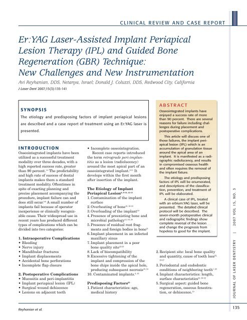
Er:YAG <strong>Laser</strong>-Assisted Implant Periapical<br />
Lesion Therapy (IPL) <strong>and</strong> Guided Bone<br />
Regeneration (GBR) Technique:<br />
New Challenges <strong>and</strong> New Instrumentation<br />
Avi Reyhanian, DDS, Netanya, Israel; Donald J. Coluzzi, DDS, Redwood City, California<br />
J <strong>Laser</strong> Dent 2007;15(3):135-141<br />
SYNOPSIS<br />
The etiology <strong>and</strong> predisposing factors <strong>of</strong> implant periapical lesions<br />
are described <strong>and</strong> a case report <strong>of</strong> treatment using an Er:YAG laser is<br />
presented.<br />
INTRODUCTION<br />
Osseointegrated implants have been<br />
utilized as a successful treatment<br />
modality over three decades, with a<br />
high reported success rate, greater<br />
than 90 percent. 1-4 The predictability<br />
<strong>and</strong> high rate <strong>of</strong> success <strong>of</strong> dental<br />
implants makes them a st<strong>and</strong>ard<br />
treatment modality. Oftentimes in<br />
spite <strong>of</strong> exacting planning <strong>and</strong><br />
precise placement accompanying the<br />
procedure, implant failure can <strong>and</strong><br />
does still occur. 5-8 A small number <strong>of</strong><br />
implants fail because <strong>of</strong> operator<br />
inexperience or clinically recognizable<br />
cause. Their widespread use in<br />
recent years has produced different<br />
types <strong>of</strong> complications which can be<br />
divided into two categories:<br />
1. Intraoperative Complications<br />
• Bleeding<br />
• Nerve injury<br />
• M<strong>and</strong>ibular fractures<br />
• Implant displacements<br />
• Accidental bone perforations<br />
• Incomplete flap closure<br />
2. Postoperative Complications<br />
• Mucositis <strong>and</strong> peri-implantitis<br />
• Implant periapical lesion (IPL)<br />
• Surgical wound dehiscence<br />
• Lesions on adjacent teeth<br />
• Incomplete osseointegration.<br />
Recent case reports introduced<br />
the term retrograde peri-implantitis<br />
as a lesion (radiolucency)<br />
around the most apical part <strong>of</strong> an<br />
osseointegrated implant. 9-11 It<br />
develops within the first month<br />
after insertion <strong>of</strong> the implant.<br />
The Etiology <strong>of</strong> Implant<br />
6, 9-10, 12-15<br />
Periapical Lesion<br />
1. Contamination <strong>of</strong> the implant<br />
surface<br />
10, 16-18<br />
2. Overheating <strong>of</strong> bone<br />
3. Overloading <strong>of</strong> the implant19 4. Presence <strong>of</strong> preexisting bone <strong>and</strong><br />
12, 16, 20<br />
microbial pathology<br />
5. Presence <strong>of</strong> residual root fragments<br />
<strong>and</strong> foreign bodies in bone21 6. Implant placement in an infected<br />
maxillary sinus<br />
7. Implant placement in a poor<br />
bone quality site22-23 8. Lack <strong>of</strong> biocompatibility<br />
9. Excessive tightening <strong>of</strong> the<br />
implant <strong>and</strong> compression <strong>of</strong> the<br />
bone chips inside the apical hole,<br />
16, 24<br />
producing subsequent necrosis<br />
9, 16<br />
10. Contaminated implants.<br />
Predisposing Factors 25<br />
1. Patient characteristics: age,<br />
medical history<br />
ABSTRACT<br />
Osseointegrated implants have<br />
enjoyed a success rate <strong>of</strong> more<br />
than 90 percent. There are several<br />
reasons for failure including challenges<br />
during placement <strong>and</strong><br />
postoperative complications.<br />
This article will discuss one <strong>of</strong><br />
those failures, the implant periapical<br />
lesion (IPL) which is an<br />
accumulation <strong>of</strong> granulation tissue<br />
around the apical area <strong>of</strong> an<br />
implant. It is manifested as a radiographic<br />
radiolucency, <strong>and</strong> results<br />
in compromised osseous health<br />
<strong>and</strong> <strong>of</strong>ten requires the removal <strong>of</strong><br />
the implant fixture.<br />
The etiology <strong>and</strong> predisposing<br />
factors <strong>of</strong> IPL will be enumerated,<br />
<strong>and</strong> descriptions <strong>of</strong> the classification,<br />
prevention, <strong>and</strong> treatment <strong>of</strong><br />
IPL will be elaborated.<br />
A clinical case <strong>of</strong> IPL, treated<br />
with an erbium:YAG laser, will be<br />
presented. The detailed clinical<br />
protocol will be described. The<br />
seven-month postoperative clinical<br />
<strong>and</strong> radiographic findings show<br />
complete reversal <strong>of</strong> the lesion<br />
<strong>and</strong> change the prognosis from<br />
hopeless to good for the implant.<br />
2. Recipient site: local bone quality<br />
<strong>and</strong> quantity, cause <strong>of</strong> tooth loss 22,<br />
26-27<br />
3. Periodontal <strong>and</strong> endodontic<br />
7, 28<br />
conditions <strong>of</strong> neighboring teeth<br />
4. Implant characteristics: length,<br />
22, 29-32<br />
surface characteristics<br />
5. Surgical aspect: guided bone<br />
regeneration, osseous fenestration,<br />
or dehiscence. 10<br />
JOUR NAL OF LASER DENTIS TRY | 2007 VOL 15, NO. 3<br />
135