Optimizing visualization and ergonomics. - Academy of Laser Dentistry
Optimizing visualization and ergonomics. - Academy of Laser Dentistry
Optimizing visualization and ergonomics. - Academy of Laser Dentistry

You also want an ePaper? Increase the reach of your titles
YUMPU automatically turns print PDFs into web optimized ePapers that Google loves.
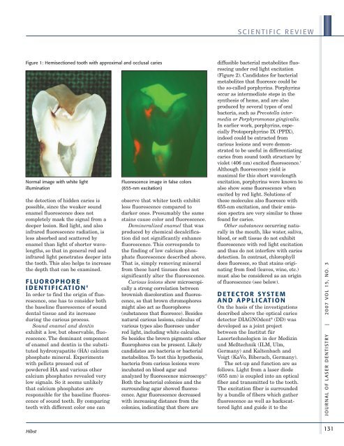
Figure 1: Hemisectioned tooth with approximal <strong>and</strong> occlusal caries<br />
Normal image with white light<br />
illumination<br />
the detection <strong>of</strong> hidden caries is<br />
possible, since the weaker sound<br />
enamel fluorescence does not<br />
completely mask the signal from a<br />
deeper lesion. Red light, <strong>and</strong> also<br />
infrared fluorescence radiation, is<br />
less absorbed <strong>and</strong> scattered by<br />
enamel than light <strong>of</strong> shorter wavelengths,<br />
so that in general red <strong>and</strong><br />
infrared light penetrates deeper into<br />
the tooth. This also helps to increase<br />
the depth that can be examined.<br />
FLUOROPHORE<br />
I DENTIF ICATION 6<br />
In order to find the origin <strong>of</strong> fluorescence,<br />
one has to consider both<br />
the baseline fluorescence <strong>of</strong> sound<br />
dental tissue <strong>and</strong> its increase<br />
during the carious process.<br />
Sound enamel <strong>and</strong> dentin<br />
exhibit a low, but observable, fluorescence.<br />
The dominant component<br />
<strong>of</strong> enamel <strong>and</strong> dentin is the substituted<br />
hydroxyapatite (HA) calcium<br />
phosphate mineral. Experiments<br />
with pellets pressed out <strong>of</strong><br />
powdered HA <strong>and</strong> various other<br />
calcium phosphates revealed very<br />
low signals. So it seems unlikely<br />
that calcium phosphates are<br />
responsible for the baseline fluorescence<br />
<strong>of</strong> sound teeth. By comparing<br />
teeth with different color one can<br />
Hibst<br />
Fluorescence image in false colors<br />
(655-nm excitation)<br />
observe that whiter teeth exhibit<br />
less fluorescence compared to<br />
darker ones. Presumably the same<br />
stains cause color <strong>and</strong> fluorescence.<br />
Demineralized enamel that was<br />
produced by chemical decalcification<br />
did not significantly enhance<br />
fluorescence. This corresponds to<br />
the finding <strong>of</strong> low calcium phosphate<br />
fluorescence described above.<br />
That is, simply removing mineral<br />
from these hard tissues does not<br />
significantly alter the fluorescence.<br />
Carious lesions show microscopically<br />
a strong correlation between<br />
brownish discoloration <strong>and</strong> fluorescence,<br />
so that brown chromophores<br />
might also act as fluorophores<br />
(substances that fluoresce). Besides<br />
natural carious lesions, calculus <strong>of</strong><br />
various types also fluoresce under<br />
red light, including white calculus.<br />
So besides the brown pigments other<br />
fluorophores can be present. Likely<br />
c<strong>and</strong>idates are bacteria or bacterial<br />
metabolites. To test this hypothesis,<br />
bacteria from carious lesions were<br />
incubated on blood agar <strong>and</strong><br />
analyzed by fluorescence microscopy. 6<br />
Both the bacterial colonies <strong>and</strong> the<br />
surrounding agar showed fluorescence.<br />
Agar fluorescence decreased<br />
with increasing distance from the<br />
colonies, indicating that there are<br />
SCIENTIFIC REVIEW<br />
diffusible bacterial metabolites fluorescing<br />
under red light excitation<br />
(Figure 2). C<strong>and</strong>idates for bacterial<br />
metabolites that fluoresce could be<br />
the so-called porphyrins. Porphyrins<br />
occur as intermediate steps in the<br />
synthesis <strong>of</strong> heme, <strong>and</strong> are also<br />
produced by several types <strong>of</strong> oral<br />
bacteria, such as Prevotella intermedia<br />
or Porphyromonas gingivalis.<br />
In earlier work, porphyrins, especially<br />
Protoporphyrine IX (PPIX),<br />
indeed could be extracted from<br />
carious lesions <strong>and</strong> were demonstrated<br />
to be useful in differentiating<br />
caries from sound tooth structure by<br />
violet (406 nm) excited fluorescence. 7<br />
Although fluorescence yield is<br />
maximal for this short wavelength<br />
excitation, porphyrins were known to<br />
also show some fluorescence when<br />
excited by red light. Solutions <strong>of</strong><br />
these molecules also fluoresce with<br />
655-nm excitation, <strong>and</strong> their emission<br />
spectra are very similar to those<br />
found for caries.<br />
Other substances occurring naturally<br />
in the mouth, like water, saliva,<br />
blood, or s<strong>of</strong>t tissue do not exhibit<br />
fluorescence with red light excitation<br />
<strong>and</strong> thus do not interfere with caries<br />
detection. In contrast, chlorophyll<br />
does fluoresce, so that stains originating<br />
from food (leaves, wine, etc.)<br />
must also be considered as an origin<br />
<strong>of</strong> fluorescence (see below).<br />
DETECTOR SY S TEM<br />
AND APPLICATION<br />
On the basis <strong>of</strong> the investigations<br />
described above the optical caries<br />
detector DIAGNOdent ® (DD) was<br />
developed as a joint project<br />
between the Institut für<br />
<strong>Laser</strong>technologien in der Medizin<br />
und Meßtechnik (ILM, Ulm,<br />
Germany) <strong>and</strong> Kaltenbach <strong>and</strong><br />
Voigt (KaVo, Biberach, Germany).<br />
The set-up <strong>and</strong> function are as<br />
follows. Light from a laser diode<br />
(655 nm) is coupled into an optical<br />
fiber <strong>and</strong> transmitted to the tooth.<br />
The excitation fiber is surrounded<br />
by a bundle <strong>of</strong> fibers which gather<br />
fluorescence as well as backscattered<br />
light <strong>and</strong> guide it to the<br />
JOUR NAL OF LASER DENTIS TRY | 2007 VOL 15, NO. 3<br />
131