Optimizing visualization and ergonomics. - Academy of Laser Dentistry
Optimizing visualization and ergonomics. - Academy of Laser Dentistry
Optimizing visualization and ergonomics. - Academy of Laser Dentistry

You also want an ePaper? Increase the reach of your titles
YUMPU automatically turns print PDFs into web optimized ePapers that Google loves.
JOUR NAL OF LASER DENTIS TRY | 2007 VOL 15, NO. 3<br />
152<br />
CLINICAL CASE<br />
4. Hard Tissue Status<br />
All bone levels <strong>and</strong> ridge topography<br />
had historically been within<br />
acceptable limits. The area around<br />
tooth #13 showed no vertical bone<br />
loss. The palatal cusp <strong>of</strong> tooth #13<br />
had fractured below the attachment<br />
level <strong>and</strong> after removal would<br />
show a termination at the osseous<br />
crest. There were signs <strong>of</strong> bruxism<br />
present <strong>and</strong> the patient reported<br />
that he was wearing a nighttime<br />
protective appliance that had been<br />
prescribed many years prior to this<br />
presentation. The tooth tested vital<br />
to air spray stimulation <strong>and</strong> it was<br />
necessary to use injected local<br />
anesthesia to fully evaluate the<br />
extent <strong>of</strong> the fracture.<br />
5. Other Tests<br />
TMJ evaluation showed normal<br />
range <strong>of</strong> motion <strong>and</strong> no joint<br />
sounds were present.<br />
B. Diagnosis <strong>and</strong> Treatment<br />
Plan<br />
1. Provisional Diagnosis<br />
A provisional diagnosis <strong>of</strong> vertical<br />
fracture <strong>of</strong> the palatal cusp <strong>of</strong> tooth<br />
#13 was made. It was thought that<br />
the fracture would extend to the<br />
osseous crest in a limited area,<br />
making impression-taking difficult.<br />
The position <strong>of</strong> the osseous crest<br />
obviated a consideration <strong>of</strong> biologic<br />
width issues. There was no pulpal<br />
exposure evident.<br />
2. Final Diagnosis<br />
A final diagnosis <strong>of</strong> vertical fracture<br />
<strong>of</strong> the palatal cusp <strong>of</strong> tooth #13<br />
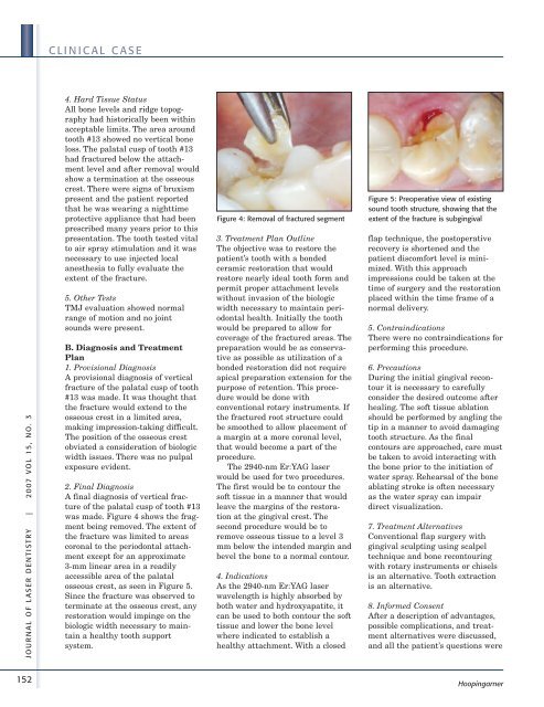
was made. Figure 4 shows the fragment<br />
being removed. The extent <strong>of</strong><br />
the fracture was limited to areas<br />
coronal to the periodontal attachment<br />
except for an approximate<br />
3-mm linear area in a readily<br />
accessible area <strong>of</strong> the palatal<br />
osseous crest, as seen in Figure 5.<br />
Since the fracture was observed to<br />
terminate at the osseous crest, any<br />
restoration would impinge on the<br />
biologic width necessary to maintain<br />
a healthy tooth support<br />
system.<br />
Figure 4: Removal <strong>of</strong> fractured segment<br />
3. Treatment Plan Outline<br />
The objective was to restore the<br />
patient’s tooth with a bonded<br />
ceramic restoration that would<br />
restore nearly ideal tooth form <strong>and</strong><br />
permit proper attachment levels<br />
without invasion <strong>of</strong> the biologic<br />
width necessary to maintain periodontal<br />
health. Initially the tooth<br />
would be prepared to allow for<br />
coverage <strong>of</strong> the fractured areas. The<br />
preparation would be as conservative<br />
as possible as utilization <strong>of</strong> a<br />
bonded restoration did not require<br />
apical preparation extension for the<br />
purpose <strong>of</strong> retention. This procedure<br />
would be done with<br />
conventional rotary instruments. If<br />
the fractured root structure could<br />
be smoothed to allow placement <strong>of</strong><br />
a margin at a more coronal level,<br />
that would become a part <strong>of</strong> the<br />
procedure.<br />
The 2940-nm Er:YAG laser<br />
would be used for two procedures.<br />
The first would be to contour the<br />
s<strong>of</strong>t tissue in a manner that would<br />
leave the margins <strong>of</strong> the restoration<br />
at the gingival crest. The<br />
second procedure would be to<br />
remove osseous tissue to a level 3<br />
mm below the intended margin <strong>and</strong><br />
bevel the bone to a normal contour.<br />
4. Indications<br />
As the 2940-nm Er:YAG laser<br />
wavelength is highly absorbed by<br />
both water <strong>and</strong> hydroxyapatite, it<br />
can be used to both contour the s<strong>of</strong>t<br />
tissue <strong>and</strong> lower the bone level<br />
where indicated to establish a<br />
healthy attachment. With a closed<br />
Figure 5: Preoperative view <strong>of</strong> existing<br />
sound tooth structure, showing that the<br />
extent <strong>of</strong> the fracture is subgingival<br />
flap technique, the postoperative<br />
recovery is shortened <strong>and</strong> the<br />
patient discomfort level is minimized.<br />
With this approach<br />
impressions could be taken at the<br />
time <strong>of</strong> surgery <strong>and</strong> the restoration<br />
placed within the time frame <strong>of</strong> a<br />
normal delivery.<br />
5. Contraindications<br />
There were no contraindications for<br />
performing this procedure.<br />
6. Precautions<br />
During the initial gingival recontour<br />
it is necessary to carefully<br />
consider the desired outcome after<br />
healing. The s<strong>of</strong>t tissue ablation<br />
should be performed by angling the<br />
tip in a manner to avoid damaging<br />
tooth structure. As the final<br />
contours are approached, care must<br />
be taken to avoid interacting with<br />
the bone prior to the initiation <strong>of</strong><br />
water spray. Rehearsal <strong>of</strong> the bone<br />
ablating stroke is <strong>of</strong>ten necessary<br />
as the water spray can impair<br />
direct <strong>visualization</strong>.<br />
7. Treatment Alternatives<br />
Conventional flap surgery with<br />
gingival sculpting using scalpel<br />
technique <strong>and</strong> bone recontouring<br />
with rotary instruments or chisels<br />
is an alternative. Tooth extraction<br />
is an alternative.<br />
8. Informed Consent<br />
After a description <strong>of</strong> advantages,<br />
possible complications, <strong>and</strong> treatment<br />
alternatives were discussed,<br />
<strong>and</strong> all the patient’s questions were<br />
Hoopingarner