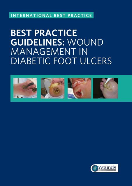
best practice guidelines: wound management in diabetic foot ulcers
best practice guidelines: wound management in diabetic foot ulcers
best practice guidelines: wound management in diabetic foot ulcers
- No tags were found...
You also want an ePaper? Increase the reach of your titles
YUMPU automatically turns print PDFs into web optimized ePapers that Google loves.
INTERNATIONAL BEST PRACTICEBEST PRACTICEGUIDELINES: WOUNDMANAGEMENT INDIABETIC FOOT ULCERS3 BEST PRACTICE GUIDELINES FOR SKIN AND WOUND CARE IN EPIDERMOLYSIS BULLOSA
FOREWORDSupported by an educationalgrant from B BraunThe views presented <strong>in</strong> thisdocument are the work of theauthors and do not necessarilyreflect the op<strong>in</strong>ions of B Braun.Published byWounds InternationalA division of SchofieldHealthcare Media LimitedEnterprise House1–2 HatfieldsLondon SE1 9PG, UKwww.<strong>wound</strong>s<strong>in</strong>ternational.comTo cite this document.International Best PracticeGuidel<strong>in</strong>es: Wound Management<strong>in</strong> Diabetic Foot Ulcers.Wounds International,2013. Available from: www.<strong>wound</strong>s<strong>in</strong>ternational.comThis document focuses on <strong>wound</strong> <strong>management</strong> <strong>best</strong> <strong>practice</strong> for <strong>diabetic</strong><strong>foot</strong> <strong>ulcers</strong> (DFUs). It aims to offer specialists and non-specialists everywherea practical, relevant cl<strong>in</strong>ical guide to appropriate decision mak<strong>in</strong>g and effective<strong>wound</strong> heal<strong>in</strong>g <strong>in</strong> people present<strong>in</strong>g with a DFU.In recognition of the gap <strong>in</strong> the literature <strong>in</strong> the field of <strong>wound</strong> <strong>management</strong>,this document concentrates on the importance of <strong>wound</strong> assessment,debridement and cleans<strong>in</strong>g, recognition and treatment of <strong>in</strong>fection andappropriate dress<strong>in</strong>g selection to achieve optimal heal<strong>in</strong>g for patients. However,it acknowledges that heal<strong>in</strong>g of the ulcer is only one aspect of <strong>management</strong>and the role of <strong>diabetic</strong> control, offload<strong>in</strong>g strategies and an <strong>in</strong>tegrated<strong>wound</strong> care approach to DFU <strong>management</strong> (which are all covered extensivelyelsewhere) are also addressed. Prevention of DFUs is not discussed <strong>in</strong>this document.The scope of the many local and <strong>in</strong>ternational <strong>guidel<strong>in</strong>es</strong> on manag<strong>in</strong>g DFUsis limited by the lack of high-quality research. This document aims to gofurther than exist<strong>in</strong>g guidance by draw<strong>in</strong>g, <strong>in</strong> addition, from the wide-rang<strong>in</strong>gexperience of an extensive <strong>in</strong>ternational panel of expert practitioners. However,it is not <strong>in</strong>tended to represent a consensus, but rather a <strong>best</strong> <strong>practice</strong>guide that can be tailored to the <strong>in</strong>dividual needs and limitations of differenthealthcare systems and to suit regional <strong>practice</strong>.EXPERT WORKING GROUPDevelopment groupPaul Chadwick, Pr<strong>in</strong>cipal Podiatrist, Salford Royal Foundation Trust, UKMichael Edmonds, Professor of Diabetes and Endocr<strong>in</strong>ology, Diabetic Foot Cl<strong>in</strong>ic, K<strong>in</strong>g's CollegeHospital, London, UKJoanne McCardle, Advanced Cl<strong>in</strong>ical and Research Diabetes Podiatrist, NHS Lothian UniversityHospital, Ed<strong>in</strong>burgh, UKDavid Armstrong, Professor of Surgery and Director, Southern Arizona Limb Salvage Alliance (SALSA),University of Arizona College of Medic<strong>in</strong>e, Arizona, USAReview groupJan Apelqvist, Senior Consultant, Department of Endocr<strong>in</strong>ology, Skåne University Hospital, Malmo,SwedenMariam Botros, Director, Diabetic Foot Canada, Canadian Wound Care Association and Cl<strong>in</strong>icalCoord<strong>in</strong>ator, Women's College Wound Heal<strong>in</strong>g Cl<strong>in</strong>ic, Toronto, CanadaGiacomo Clerici, Chief Diabetic Foot Cl<strong>in</strong>ic, IRCC Casa di Cura Multimedica, Milan, ItalyJill Cundell, Lecturer/Practitioner, University of Ulster, Belfast Health and Social Care Trust, NorthernIrelandSolange Ehrler, Functional Rehabilitation Department, IUR Clémenceau (Institut Universitaire deRéadaptation Clémenceau), Strasbourg, FranceMichael Hummel, MD, Diabetes Center Rosenheim & Institute of Diabetes Research, HelmholtzZentrum München, GermanyBenjam<strong>in</strong> A Lipsky, Emeritus Professor of Medic<strong>in</strong>e, University of Wash<strong>in</strong>gton, USA; Visit<strong>in</strong>g Professor,Infectious Diseases, University of Geneva, Switzerland; Teach<strong>in</strong>g Associate, University of Oxford andDeputy Director, Graduate Entry Course, University of Oxford Medical School, UKJosé Luis Lázaro Mart<strong>in</strong>ez, Full Time Professor, Diabetic Foot Unit, Complutense University, Madrid,Spa<strong>in</strong>Rosalyn Thomas, Deputy Head of Podiatry, Abertawe Bro Morgannwg University Health Board,Swansea, WalesSusan Tulley, Senior Podiatrist, Mafraq Hospital, Abu Dhabi, United Arab Emirates3C BEST PRACTICEBEST PRACTICEGUIDELINESGUIDELINES:FOR SKINWOUNDAND WOUNDMANAGEMENTCARE ININEPIDERMOLYSISDIABETIC FOOTBULLOSAULCERS
INTRODUCTIONIntroductionDFUs are complex, chronic <strong>wound</strong>s, whichhave a major long-term impact on themorbidity, mortality and quality of patients’lives 1,2 . Individuals who develop a DFU are atgreater risk of premature death, myocardial<strong>in</strong>farction and fatal stroke than those withouta history of DFU 3 . Unlike other chronic<strong>wound</strong>s, the development and progression ofa DFU is often complicated by wide-rang<strong>in</strong>g<strong>diabetic</strong> changes, such as neuropathy andvascular disease. These, along with thealtered neutrophil function, dim<strong>in</strong>ished tissueperfusion and defective prote<strong>in</strong> synthesisthat frequently accompany diabetes, presentpractitioners with specific and unique <strong>management</strong>challenges 1 .DFUs are relatively common — <strong>in</strong> the UK,5–7% of people with diabetes currently haveor have had a DFU 4,5 . Furthermore, around25% of people with diabetes will develop aDFU dur<strong>in</strong>g their lifetime 6 . Globally, around370 million people have diabetes and thisnumber is <strong>in</strong>creas<strong>in</strong>g <strong>in</strong> every country 7 . DiabetesUK estimates that by 2030 some 552million people worldwide will have diabetes 8 .DFUs have a major economic impact. A USstudy <strong>in</strong> 1999 estimated the average outpatientcost of treat<strong>in</strong>g one DFU episode as$28,000 USD over a two–year period 9 . Average<strong>in</strong>patient costs for lower limb complications<strong>in</strong> 1997 were reported as $16,580 USDfor DFUs, $25,241 USD for toe or toe plusother distal amputations and $31,436 USDfor major amputations 10,11 .such as the effect on physical, psychologicaland social wellbe<strong>in</strong>g and the fact that manypatients are unable to work long term as aresult of their <strong>wound</strong>s 6 .A DFU is a pivotal event <strong>in</strong> the life of aperson with diabetes and a marker of seriousdisease and comorbidities. Without earlyand optimal <strong>in</strong>tervention, the <strong>wound</strong> canrapidly deteriorate, lead<strong>in</strong>g to amputation ofthe affected limb 5,13 .It has been estimated that every 20 secondsa lower limb is amputated due to complicationsof diabetes 14 .In Europe, the annual amputation rate forpeople with diabetes has been cited as 0.5-0.8% 1,15 , and <strong>in</strong> the US it has been reportedthat around 85% of lower-extremityamputations due to diabetes beg<strong>in</strong> with <strong>foot</strong>ulceration 16,17 .Mortality follow<strong>in</strong>g amputation <strong>in</strong>creaseswith level of amputation 18 and ranges from50–68% at five years, which is comparableor worse than for most malignancies 13,19(Figure 1).The statistics need not make for such grimread<strong>in</strong>g. With appropriate and careful<strong>management</strong> it is possible to delay or avoidmost serious complications of DFUs 1 .FIGURE 1: Relative five-year mortality (%) (adapted from 19 )The EURODIALE study exam<strong>in</strong>ed total directand <strong>in</strong>direct costs for one year across severalEuropean countries. Average total costsbased on 821 patients were approximately10,000 euros, with hospitalisation represent<strong>in</strong>gthe highest direct cost. Based on prevalencedata for Europe, they estimated thatcosts associated with treatment of DFUsmay be as high as 10 billion euros per year 12 .In England, <strong>foot</strong> complications account for20% of the total National Health Servicespend on diabetes care, which equates toaround £650 million per year (or £1 <strong>in</strong> every£150) 5 . Of course, these figures do not takeaccount of the <strong>in</strong>direct costs to patients,Prostate cancerBreast cancerHodgk<strong>in</strong>'s lymphomaNeuropathic DFUAmputationColon cancerIschaemic DFUPeripheral arterial diseaseLung cancerPancreatic cancerBEST PRACTICE GUIDELINES: WOUND MANAGEMENT IN DIABETIC FOOT ULCERS 1
INTRODUCTIONIt has been suggested that up to 85% ofamputations can be avoided when an effectivecare plan is adopted 20 . Unfortunately,<strong>in</strong>sufficient tra<strong>in</strong><strong>in</strong>g, suboptimal assessmentand treatment methods, failure to referpatients appropriately and poor access to specialist<strong>foot</strong>care teams h<strong>in</strong>der the prospects ofachiev<strong>in</strong>g optimal outcomes 21,22 .Successful diagnosis and treatment ofpatients with DFUs <strong>in</strong>volves a holisticapproach that <strong>in</strong>cludes: Optimal diabetes control Effective local <strong>wound</strong> care Infection control Pressure reliev<strong>in</strong>g strategies Restor<strong>in</strong>g pulsatile blood flow.Many studies have shown that planned <strong>in</strong>terventionaimed at heal<strong>in</strong>g of DFUs is mosteffective <strong>in</strong> the context of a multidiscipl<strong>in</strong>aryteam with the patient at the centre of thiscare.One of the key tenets underp<strong>in</strong>n<strong>in</strong>g thisdocument is that <strong>in</strong>fection is a major threatto DFUs — much more so than to <strong>wound</strong>sof other aetiologies not subject to <strong>diabetic</strong>changes. A European-wide study found that58% of patients attend<strong>in</strong>g a <strong>foot</strong> cl<strong>in</strong>ic with anew ulcer had a cl<strong>in</strong>ically <strong>in</strong>fected <strong>wound</strong> 23 .Similarly a s<strong>in</strong>gle-centre US study found thatabout 56% of DFUs were cl<strong>in</strong>ically <strong>in</strong>fected 24 .This study also showed the risk of hospitalisationand lower-extremity amputation to be56–155 times greater for diabetes patientswith a <strong>foot</strong> <strong>in</strong>fection than those without 24 .Recognis<strong>in</strong>g the importance of start<strong>in</strong>g treatmentearly may allow practitioners to preventprogression to severe and limb-threaten<strong>in</strong>g<strong>in</strong>fection and potentially halt the <strong>in</strong>evitablepathway to amputation 25 .This document offers a global <strong>wound</strong> careplan for practitioners (page 20), which<strong>in</strong>cludes a series of steps for prevent<strong>in</strong>gcomplications through active <strong>management</strong>— namely prompt and appropriate treatmentof <strong>in</strong>fection, referral to a vascular specialist tomanage ischaemia and optimal <strong>wound</strong> care.This should be comb<strong>in</strong>ed with appropriatepatient education and an <strong>in</strong>tegrated approachto care.32 BEST PRACTICEBEST PRACTICEGUIDELINESGUIDELINES:FOR SKINWOUNDAND WOUNDMANAGEMENTCARE ININEPIDERMOLYSISDIABETIC FOOTBULLOSAULCERS
AETIOLOGY OFDFUsAetiology of DFUsThe underly<strong>in</strong>g cause(s) of DFUs will have a significant bear<strong>in</strong>g on the cl<strong>in</strong>ical<strong>management</strong> and must be determ<strong>in</strong>ed before a care plan is put <strong>in</strong>to placeIn most patients, peripheral neuropathy andperipheral arterial disease (PAD) (or both)play a central role and DFUs are thereforecommonly classified as (Table 1) 26 : Neuropathic Ischaemic Neuroischaemic (Figures 2–4).Neuroischaemia is the comb<strong>in</strong>ed effectof <strong>diabetic</strong> neuropathy and ischaemia,whereby macrovascular disease and, <strong>in</strong>some <strong>in</strong>stances, microvascular dysfunctionimpair perfusion <strong>in</strong> a <strong>diabetic</strong> <strong>foot</strong> 26,27 .is <strong>in</strong>creas<strong>in</strong>g and it is reported to be a contributoryfactor <strong>in</strong> the development of DFUs<strong>in</strong> up to 50% of patients 14,28,33 .It is important to remember that even <strong>in</strong> theabsence of a poor arterial supply, microangiopathy(small vessel dysfunction)contributes to poor ulcer heal<strong>in</strong>g <strong>in</strong> neuroischaemicDFUs 34 . Decreased perfusion <strong>in</strong>the <strong>diabetic</strong> <strong>foot</strong> is a complex scenario andis characterised by various factors relat<strong>in</strong>gto microvascular dysfunction <strong>in</strong> addition toPAD 34 .FIGURE 2: Neuropathic DFUPERIPHERAL NEUROPATHYPeripheral neuropathy may predispose the<strong>foot</strong> to ulceration through its effects on thesensory, motor and autonomic nerves: The loss of protective sensation experiencedby patients with sensory neuropathyrenders them vulnerable to physical,chemical and thermal trauma Motor neuropathy can cause <strong>foot</strong>deformities (such as hammer toes andclaw <strong>foot</strong>), which may result <strong>in</strong> abnormalpressures over bony prom<strong>in</strong>ences Autonomic neuropathy is typicallyassociated with dry sk<strong>in</strong>, which can result<strong>in</strong> fissures, crack<strong>in</strong>g and callus. Anotherfeature is bound<strong>in</strong>g pulses, which isoften mis<strong>in</strong>terpreted as <strong>in</strong>dicat<strong>in</strong>g a goodcirculation 28 .Loss of protective sensation is a majorcomponent of nearly all DFUs 29,30 . It is associatedwith a seven–fold <strong>in</strong>crease <strong>in</strong> riskof ulceration 6 .DFUs usually result from two or more riskfactors occurr<strong>in</strong>g together. Intr<strong>in</strong>sic elementssuch as neuropathy, PAD and <strong>foot</strong> deformity(result<strong>in</strong>g, for example, from neuropathicstructural changes), accompanied by anexternal trauma such as poorly fitt<strong>in</strong>g <strong>foot</strong>wearor an <strong>in</strong>jury to the <strong>foot</strong> can, over time,lead to a DFU 7 .TABLE 1: Typical features of DFUs accord<strong>in</strong>g to aetiologyFeature Neuropathic Ischaemic NeuroischaemicSensation Sensory loss Pa<strong>in</strong>ful Degree of sensorylossCallus/necrosisWound bedFoot temperatureand pulsesCallus present andoften thickP<strong>in</strong>k and granulat<strong>in</strong>g,surrounded bycallusWarm with bound<strong>in</strong>gpulsesNecrosis commonPale and sloughywith poorgranulationCool with absentpulsesFIGURE 3: Ischaemic DFUFIGURE 4: NeuroischaemicDFUM<strong>in</strong>imal callusProne to necrosisPoor granulationCool with absentpulsesPatients with a loss of sensation will havedecreased awareness of pa<strong>in</strong> and othersymptoms of ulceration and <strong>in</strong>fection 31 .PERIPHERAL ARTERIAL DISEASEPeople with diabetes are twice as likely tohave PAD as those without diabetes 32 . Itis also a key risk factor for lower extremityamputation 30 . The proportion of patientswith an ischaemic component to their DFUOtherTypical locationPrevalence(based on 35 )Dry sk<strong>in</strong> andfissur<strong>in</strong>gWeight-bear<strong>in</strong>gareas of the <strong>foot</strong>,such as metatarsalheads, the heel andover the dorsum ofclawed toesDelayed heal<strong>in</strong>gTips of toes, nailedges and betweenthe toes and lateralborders of the <strong>foot</strong>35% 15% 50%High risk of<strong>in</strong>fectionMarg<strong>in</strong>s of the<strong>foot</strong> and toesBEST PRACTICE GUIDELINES: WOUND MANAGEMENT IN DIABETIC FOOT ULCERS 3
ASSESSING DFUsAssess<strong>in</strong>g DFUsPatients with a DFU need to be assessed holistically and <strong>in</strong>tr<strong>in</strong>sic and extr<strong>in</strong>sicfactors consideredFor the non-specialist practitioner, the key skillrequired is know<strong>in</strong>g when and how to refer apatient with a DFU to the multidiscipl<strong>in</strong>ary <strong>foot</strong>careteam (MDFT; see page 19). Patients witha DFU should be assessed by the team with<strong>in</strong>one work<strong>in</strong>g day of presentation — or sooner<strong>in</strong> the presence of severe <strong>in</strong>fection 22,36,37 . Inmany places, however, MDFTs do not exist andpractitioners <strong>in</strong>stead work as <strong>in</strong>dividuals. Inthese situations, the patient’s prognosis oftendepends on a particular practitioner’s knowledgeand <strong>in</strong>terest <strong>in</strong> the <strong>diabetic</strong> <strong>foot</strong>.Patients with a DFU need to be assessed holisticallyto identify <strong>in</strong>tr<strong>in</strong>sic and extr<strong>in</strong>sic factors.This should encompass a full patient history<strong>in</strong>clud<strong>in</strong>g medication, comorbidities and diabetesstatus 38 . It should also take <strong>in</strong>to considerationthe history of the <strong>wound</strong>, previous DFUs oramputations and any symptoms suggestive ofneuropathy or PAD 28 .EXAMINATION OF THE ULCERA physical exam<strong>in</strong>ation should determ<strong>in</strong>e: Is the <strong>wound</strong> predom<strong>in</strong>antly neuropathic,ischaemic or neuroischaemic? If ischaemic, is there critical limb ischaemia? Are there any musculoskeletal deformities? What is the size/depth/location of the<strong>wound</strong>? What is the colour/status of the <strong>wound</strong>bed?— Black (necrosis)— Yellow, red, p<strong>in</strong>k Is there any exposed bone? Is there any necrosis or gangrene? Is the <strong>wound</strong> <strong>in</strong>fected? If so, are theresystemic signs and symptoms of <strong>in</strong>fection(such as fevers, chills, rigors, metabolic<strong>in</strong>stability and confusion)? Is there any malodour? Is there local pa<strong>in</strong>? Is there any exudate? What is the level ofproduction (high, moderate, low, none),colour and consistency of exudate, and is itpurulent? What is the status of the <strong>wound</strong> edge(callus, maceration, erythema, oedema,underm<strong>in</strong><strong>in</strong>g)?Document<strong>in</strong>g ulcer characteristicsRecord<strong>in</strong>g the size, depth, appearance and locationof the DFU will help to establish a basel<strong>in</strong>efor treatment, develop a treatment plan andmonitor any response to <strong>in</strong>terventions. It isimportant also to assess the area around the<strong>wound</strong>: erythema and maceration <strong>in</strong>dicateadditional complications that may h<strong>in</strong>der<strong>wound</strong> heal<strong>in</strong>g 38 .Digitally photograph<strong>in</strong>g DFUs at the firstconsultation and periodically thereafterto document progress is helpful 39 . This isparticularly useful for ensur<strong>in</strong>g consistencyof care among healthcare practitioners,facilitat<strong>in</strong>g telehealth <strong>in</strong> remote areas andillustrat<strong>in</strong>g improvement to the patient.TESTING FOR LOSS OF SENSATIONTwo simple and effective tests for peripheralneuropathy are commonly used: 10g (Semmes-We<strong>in</strong>ste<strong>in</strong>) monofilament Standard 128Hz tun<strong>in</strong>g fork.The 10g monofilament is the most frequentlyused screen<strong>in</strong>g tool to determ<strong>in</strong>e the presenceof neuropathy <strong>in</strong> patients with diabetes 28 . Itshould be applied at various sites along theplantar aspect of the <strong>foot</strong>. Guidel<strong>in</strong>es vary <strong>in</strong> thenumber of sites advocated, but the <strong>in</strong>ternationalconsensus is to test at three sites (see Figure5) 7 . A positive result is the <strong>in</strong>ability to feel themonofilament when it is pressed aga<strong>in</strong>st the<strong>foot</strong> with enough force to bend it 40 .Neuropathy is also demonstrated by an <strong>in</strong>abilityto sense vibration from a standard tun<strong>in</strong>g fork.Other tests are available, such as the biothesiometerand neurothesiometer, which are morecomplex handheld devices for assess<strong>in</strong>g theperception of vibration.Do not test for neuropathy <strong>in</strong> areas of callusas this can mask feel<strong>in</strong>g from any of theneuropathy test<strong>in</strong>g devices and may give afalse-positive result.Be aware that patients with small nerve fibredamage and <strong>in</strong>tact sensory nerves may have34 BEST PRACTICEBEST PRACTICEGUIDELINESGUIDELINES:FOR SKINWOUNDAND WOUNDMANAGEMENTCARE ININEPIDERMOLYSISDIABETIC FOOTBULLOSAULCERS
ASSESSING DFUsFIGURE 5: Procedure for carry<strong>in</strong>g out the monofilament test (adapted from 7 )The International Work<strong>in</strong>g Group on the Diabetic Foot (IWGDF) recommends the follow<strong>in</strong>g procedure for carry<strong>in</strong>g out themonofilament test. The sensory exam<strong>in</strong>ation should be carried out <strong>in</strong> a quiet and relaxed sett<strong>in</strong>g The patient should close their eyes so as not to see whether or where the exam<strong>in</strong>erapplies the monofilament The patient should sit sup<strong>in</strong>e with both feet level First apply the monofilament on the patient’s hands or on the <strong>in</strong>side of the arm sothey know what to expect Apply the monofilament perpendicular to the sk<strong>in</strong> surface with sufficient force tobend or buckle the monofilament Ask the patient:— Whether they feel the pressure applied (yes/no)— Where they feel the pressure (left <strong>foot</strong>/right <strong>foot</strong>) Apply the monofilament along the perimeter of (not on) the ulcer site Do not allow the monofilament to slide across the sk<strong>in</strong> or make repetitive contact atthe test site The total duration of the approach (sk<strong>in</strong> contact and removal of the monofilament)should be around 2 seconds Apply the monofilament to each site three times, <strong>in</strong>clud<strong>in</strong>g at least one additional‘mock’ application <strong>in</strong> which no filament is applied Encourage the patient dur<strong>in</strong>g test<strong>in</strong>g by giv<strong>in</strong>g positive feedback— Protective sensation is present at each site if the patient correctly answers twoout of three applications— Protective sensation is absent with two out of three <strong>in</strong>correct answersNote: The monofilament should not be used on more than 10 patients without arecovery period of 24 hoursUs<strong>in</strong>g a monofilament to test for neuropathya pa<strong>in</strong>ful neuropathy. They may describesharp, stabb<strong>in</strong>g, burn<strong>in</strong>g, shoot<strong>in</strong>g or electricshock type pa<strong>in</strong>, which may be worse at nightand can disrupt sleep 41 . The absence of coldwarmdiscrim<strong>in</strong>ation may help to identifypatients with small nerve fibre damage.TESTING FOR VASCULAR STATUSPalpation of peripheral pulses should be arout<strong>in</strong>e component of the physical exam<strong>in</strong>ationand <strong>in</strong>clude assessment of the femoral,popliteal and pedal (dorsalis pedis andposterior tibial) pulses. Assessment of pulsesis a learned skill and has a high degree of<strong>in</strong>ter-observer variability, with high falsepositiveand false-negative rates. The dorsalispedis pulse is reported to be absent <strong>in</strong> 8.1%of healthy <strong>in</strong>dividuals, and the posterior tibialpulse is absent <strong>in</strong> 2.0%. Nevertheless, theabsence of both pedal pulses, when assessedby an experienced cl<strong>in</strong>ician, strongly suggeststhe presence of pedal vascular disease 42 . Ifthere is any doubt regard<strong>in</strong>g diagnosis of PAD,it is important to refer to a specialist for a fullvascular assessment.Where available, Doppler ultrasound, anklebrachialpressure <strong>in</strong>dex (ABPI) and Dopplerwaveform may be used as adjuncts tothe cl<strong>in</strong>ical f<strong>in</strong>d<strong>in</strong>gs when carried out by acompetent practitioner. Toe pressures, and<strong>in</strong> some <strong>in</strong>stances, transcutaneous oxygenmeasurement (where equipment is available),may be useful for measur<strong>in</strong>g localtissue perfusion.An ischaemic <strong>foot</strong> may appear p<strong>in</strong>k and relativelywarm even with impaired perfusion dueto arteriovenous shunt<strong>in</strong>g. Delayed discolouration(rubor) or venous refill<strong>in</strong>g greater thanfive seconds on dependency may <strong>in</strong>dicatepoor arterial perfusion 43 .Other signs suggestive of ischaemia <strong>in</strong>clude 40 : Claudication: pa<strong>in</strong> <strong>in</strong> the leg muscles andCOMMON TERMS EXPLAINEDCritical limb ischaemia: this isa chronic manifestation of PADwhere the arteries of the lowerextremities are severely blocked.This results <strong>in</strong> ischaemic pa<strong>in</strong><strong>in</strong> the feet or toes even at rest.Complications of poor circulation<strong>in</strong>clude sk<strong>in</strong> <strong>ulcers</strong> or gangrene.If left untreated it will result <strong>in</strong>amputation of the affected limb.Acute limb ischaemia: thisoccurs when there is a suddenlack of blood flow to a limb andis due to either an embolism orthrombosis. Without surgicalrevascularisation, completeacute ischaemia leads to extensivetissue necrosis with<strong>in</strong> sixhours.BEST PRACTICE GUIDELINES: WOUND MANAGEMENT IN DIABETIC FOOT ULCERS 5
ASSESSING DFUsusually exercise-<strong>in</strong>duced (although this isoften absent <strong>in</strong> people with diabetes) A temperature difference between the feet.If you suspect severe ischaemia <strong>in</strong> a patientwith a DFU you should refer as quickly aspossible to a MDFT with access to a vascularsurgeon. If the patient has critical limbischaemia this should be done urgently. Apatient with acute limb ischaemia characterisedby the six ‘Ps’ (pulselessness, pa<strong>in</strong>,pallor [mottled colouration], perish<strong>in</strong>g cold,paraesthesia and paralysis) poses a cl<strong>in</strong>icalemergency and may be at great risk if notmanaged <strong>in</strong> a timely and effective way 44 .IDENTIFYING INFECTIONRecognis<strong>in</strong>g <strong>in</strong>fection <strong>in</strong> patients with DFUscan be challeng<strong>in</strong>g, but it is one of the mostimportant steps <strong>in</strong> the assessment. It is at thiscrucial early stage that practitioners have thepotential to curb what is often progressionfrom simple (mild) <strong>in</strong>fection to a more severeproblem, with necrosis, gangrene and oftenamputation 45 . Around 56% of DFUs become<strong>in</strong>fected and overall about 20% of patientswith an <strong>in</strong>fected <strong>foot</strong> <strong>wound</strong> will undergo alower extremity amputation 30 .Risk factors for <strong>in</strong>fectionPractitioners should be aware of the factors that<strong>in</strong>crease the likelihood of <strong>in</strong>fection 46 :TABLE 2: Classification and severity of <strong>diabetic</strong> <strong>foot</strong> <strong>in</strong>fections (adapted from 46 )Cl<strong>in</strong>ical criteriaNo cl<strong>in</strong>ical signs of <strong>in</strong>fectionSuperficial tissue lesion with at least two of the follow<strong>in</strong>gsigns:— Local warmth— Erythema >0.5–2cm around the ulcer— Local tenderness/pa<strong>in</strong>— Local swell<strong>in</strong>g/<strong>in</strong>duration— Purulent dischargeOther causes of <strong>in</strong>flammation of the sk<strong>in</strong> must be excludedErythema >2cm and one of the f<strong>in</strong>d<strong>in</strong>gs above or:— Infection <strong>in</strong>volv<strong>in</strong>g structures beneath the sk<strong>in</strong>/subcutaneous tissues (eg deep abscess, lymphangitis,osteomyelitis, septic arthritis or fascitis)— No systemic <strong>in</strong>flammatory response (see Grade 4)Presence of systemic signs with at least two of the follow<strong>in</strong>g:— Temperature >39°C or 90bpm— Respiratory rate >20/m<strong>in</strong>— PaCO 2
ASSESSING DFUsBOX 1: Signs of spread<strong>in</strong>g<strong>in</strong>fection (adapted from 49 ) Spread<strong>in</strong>g, <strong>in</strong>tenseerythema Increas<strong>in</strong>g <strong>in</strong>duration Lymphangitis Regional lymphadenitis Hypotension, tachypnoea,tachycardia RigorsFIGURE 6: Necrotic toe whichhas been allowed to autoamputateRISK OF AMPUTATIONArmstrong et al 52 found thatpatients were 11 times morelikely to receive a mid<strong>foot</strong>or higher level amputationif their <strong>wound</strong> had apositive probe-to-bone test.Furthermore, patients with<strong>in</strong>fection and ischaemiawere nearly 90 times morelikely to receive a mid<strong>foot</strong>or higher amputation thanpatients with less advancedDFUs. There may also be apossible correlation betweenlocation of osteomyelitisand major amputation, witha higher rate of transtibialamputation reported whenosteomyelitis <strong>in</strong>volved theheel <strong>in</strong>stead of the mid<strong>foot</strong>or fore<strong>foot</strong> <strong>in</strong> <strong>diabetic</strong>patients 53 .Cultures should not be taken from cl<strong>in</strong>icallynon-<strong>in</strong>fected <strong>wound</strong>s as all <strong>ulcers</strong> will be contam<strong>in</strong>ated;microbiological sampl<strong>in</strong>g cannotdiscrim<strong>in</strong>ate colonisation from <strong>in</strong>fection.Extensive <strong>in</strong>flammation, crepitus, bullae, necrosisor gangrene are signs suggestive of severe<strong>foot</strong> <strong>in</strong>fections 50 . Refer patients immediatelyto an MDFT if you suspect a deep or limbthreaten<strong>in</strong>g<strong>in</strong>fection. Where there is no MDFT,the referral should be to the most appropriatepractitioner, notably the person(s) champion<strong>in</strong>gthe cause of the <strong>diabetic</strong> <strong>foot</strong>, for example anexperienced <strong>foot</strong> surgeon.Refer patients urgently to a member of thespecialist <strong>foot</strong> care team for urgent surgicaltreatment and prompt revascularisation if thereis acute spread<strong>in</strong>g <strong>in</strong>fection (Box 1), critical limbischaemia, wet gangrene or an unexpla<strong>in</strong>ed hot,red, swollen <strong>foot</strong> with or without the presenceof pa<strong>in</strong> 37,51 . These cl<strong>in</strong>ical signs and symptomsare potentially limb- and even life-threaten<strong>in</strong>g.Where necrosis occurs on the distal part of thelimb due to ischaemia and <strong>in</strong> the absence of<strong>in</strong>fection (dry gangrene), mummification of thetoes and auto-amputation may occur. In most ofthese situations, surgery is not recommended.However, if the necrosis is more superficial thenthe toe can be removed with a scalpel (Figure 6).Assess<strong>in</strong>g bone <strong>in</strong>volvementOsteomyelitis may frequently be present <strong>in</strong>patients with moderate to severe <strong>diabetic</strong> <strong>foot</strong><strong>in</strong>fection. If any underly<strong>in</strong>g osteomyelitis is notidentified and treated appropriately, the <strong>wound</strong>is unlikely to heal 17 .Osteomyelitis can be difficult to diagnose <strong>in</strong>the early stages. Wounds that are chronic,large, deep or overlie a bony prom<strong>in</strong>ence areat high risk for underly<strong>in</strong>g bone <strong>in</strong>fection, whilethe presence of a 'sausage toe' or visible boneis suggestive of osteomyelitis. A simple cl<strong>in</strong>icaltest for bone <strong>in</strong>fection is detect<strong>in</strong>g bone by itshard, gritty feel when gently <strong>in</strong>sert<strong>in</strong>g a sterileblunt metal probe <strong>in</strong>to the ulcer 54,55 . This canhelp to diagnose bone <strong>in</strong>fection (when thelikelihood is high) or exclude (when the likelihoodis low) 46 .Pla<strong>in</strong> x-rays can help to confirm the diagnosis,but they have a relatively low sensitivity (early <strong>in</strong>the <strong>in</strong>fection) and specificity (late <strong>in</strong> the courseof <strong>in</strong>fection) for osteomyelitis 46,56 .The National Institute for Health and CareExcellence (NICE) <strong>in</strong> the UK and IDSArecommend that if <strong>in</strong>itial x-rays do not confirmthe presence of osteomyelitis and suspicionrema<strong>in</strong>s high, the next advanced imag<strong>in</strong>g testto consider is magnetic resonance imag<strong>in</strong>g(MRI) 1,46 . If MRI is contra<strong>in</strong>dicated or unavailable,white blood cell scann<strong>in</strong>g comb<strong>in</strong>ed witha radionuclide bone scan may be performed<strong>in</strong>stead 46 . The most def<strong>in</strong>itive way to diagnoseosteomyelitis is by the comb<strong>in</strong>ed f<strong>in</strong>d<strong>in</strong>gs ofculture and histology from a bone specimen.Bone may be obta<strong>in</strong>ed dur<strong>in</strong>g deep debridementor by biopsy 46 .INSPECTING FEET FORDEFORMITIESExcessive or abnormal plantar pressure, result<strong>in</strong>gfrom limited jo<strong>in</strong>t mobility, often comb<strong>in</strong>edwith <strong>foot</strong> deformities, is a common underly<strong>in</strong>gcause of DFUs <strong>in</strong> <strong>in</strong>dividuals with neuropathy 6 .These patients may also develop atypicalwalk<strong>in</strong>g patterns (Figure 7). The result<strong>in</strong>galtered biomechanical load<strong>in</strong>g of the <strong>foot</strong> canresult <strong>in</strong> callus, which <strong>in</strong>creases the abnormalpressure and can cause subcutaneous haemorrhage7 . Because there is commonly loss ofsensation, the patient cont<strong>in</strong>ues to walk on the<strong>foot</strong>, <strong>in</strong>creas<strong>in</strong>g the risk of further problems.Typical presentations result<strong>in</strong>g <strong>in</strong> high plantarpressure areas <strong>in</strong> patients with motor neuropathyare 7 : A high-arch <strong>foot</strong> Clawed lesser toes Visible muscle wast<strong>in</strong>g <strong>in</strong> the plantar archand on the dorsum between the metatarsalshafts (a ‘hollowed-out’ appearance) Gait changes, such as the <strong>foot</strong> ‘slapp<strong>in</strong>g’ onthe ground Hallux valgus, hallux rigidus and fatty paddepletion.In people with diabetes, even m<strong>in</strong>or traumacan precipitate a chronic ulcer 7 . This mightbe caused by wear<strong>in</strong>g poorly fitt<strong>in</strong>g <strong>foot</strong>wearor walk<strong>in</strong>g bare<strong>foot</strong>, or from an acute <strong>in</strong>jury.In some cultures the frequent adoption of theprayer position and/or sitt<strong>in</strong>g cross-legged willcause ulcerations on the lateral malleoli, andto a lesser extent the dorsum of the <strong>foot</strong>, <strong>in</strong>the mid-tarsal area. The dorsal, plantar andposterior surfaces of both feet and betweenthe toes should be checked thoroughly forbreaks <strong>in</strong> the sk<strong>in</strong> or newly established DFUs.BEST PRACTICE GUIDELINES: WOUND MANAGEMENT IN DIABETIC FOOT ULCERS 7
ASSESSING DFUsFIGURE 7: Areas at risk for DFU (adaptedfrom 7 )Corrective <strong>foot</strong> surgery to offload pressureareas may be considered where structuraldeformities cannot be accommodated bytherapeutic <strong>foot</strong>wear.CLASSIFICATION OF DFUsClassification systems grade <strong>ulcers</strong> accord<strong>in</strong>gto the presence and extent of various physicalcharacteristics, such as size, depth, appearanceand location. They can help <strong>in</strong> the plann<strong>in</strong>gand monitor<strong>in</strong>g of treatment and <strong>in</strong> predict<strong>in</strong>goutcome 17,58 , and also for research and audit.Classification systems should be used consistentlyacross the healthcare team and berecorded appropriately <strong>in</strong> the patient’s records.However, it is the assessment of the <strong>wound</strong>that <strong>in</strong>forms <strong>management</strong>.Table 3 summarises the key features of thesystems most commonly used for DFUs.FIGURE 8: Charcot <strong>foot</strong>.Top — Charcot <strong>foot</strong> with plantarulcer. Middle — Charcot <strong>foot</strong>with sepsis. Bottom — ChronicCharcot <strong>foot</strong>Charcot jo<strong>in</strong>t is a form of neuroarthropathythat occurs most often <strong>in</strong> the <strong>foot</strong> and <strong>in</strong> peoplewith diabetes 57 . Nerve damage from diabetescauses decreased sensation, muscle atrophyand subsequent jo<strong>in</strong>t <strong>in</strong>stability, which is madeworse by walk<strong>in</strong>g on an <strong>in</strong>sensitive jo<strong>in</strong>t. In theacute stage there is <strong>in</strong>flammation and bonereabsorption, which weakens the bone. In laterstages, the arch falls and the <strong>foot</strong> may developa ‘rocker bottom’ appearance (Figure 8). Earlytreatment, particularly offload<strong>in</strong>g pressure,can help stop bone destruction and promoteheal<strong>in</strong>g.TABLE 3: Key features of common <strong>wound</strong> classification systems for DFUsClassificationsystemWagnerUniversity ofTexas(Armstrong)PEDISSINBADKey po<strong>in</strong>ts Pros/cons ReferencesAssesses ulcer depth along with presenceof gangrene and loss of perfusion us<strong>in</strong>g sixgrades (0-5)Assesses ulcer depth, presence of <strong>in</strong>fectionand presence of signs of lower-extremityischaemia us<strong>in</strong>g a matrix of four gradescomb<strong>in</strong>ed with four stagesAssesses Perfusion, Extent (size), Depth(tissue loss), Infection and Sensation (neuropathy)us<strong>in</strong>g four grades (1-4)Assesses Site, Ischaemia, Neuropathy, Bacterial<strong>in</strong>fection and DepthUses a scor<strong>in</strong>g system to help predictoutcomes and enable comparisons betweendifferent sett<strong>in</strong>gs and countriesWell established 58Does not fully address <strong>in</strong>fection and ischaemiaWell established 58Describes the presence of <strong>in</strong>fection and ischaemiabetter than Wagner and may help <strong>in</strong> predict<strong>in</strong>g theoutcome of the DFUDeveloped by IWGDFUser-friendly (clear def<strong>in</strong>itions, few categories) forpractitioners with a lower level of experience with<strong>diabetic</strong> <strong>foot</strong> <strong>management</strong>Wagner 1981 59Lavery et al 1996 60Armstrong et al1998 52Lipsky et al 2012 46Simplified version of the S(AD)SAD classification Ince et al 2008 63system 61Includes ulcer site as data suggests this might bean important determ<strong>in</strong>ant of outcome 6238 BEST PRACTICEBEST PRACTICEGUIDELINESGUIDELINES:FOR SKINWOUNDAND WOUNDMANAGEMENTCARE ININEPIDERMOLYSISDIABETIC FOOTBULLOSAULCERS
DFU WOUNDMANAGEMENTDFU <strong>wound</strong> <strong>management</strong>Practitioners must strive to prevent DFUs develop<strong>in</strong>g elsewhere on the <strong>foot</strong> or onthe contralateral limb and to achieve limb preservation 64The pr<strong>in</strong>ciple aim of DFU <strong>management</strong> is<strong>wound</strong> closure 17 . More specifically, the <strong>in</strong>tentionshould be to treat the DFU at an earlystage to allow prompt heal<strong>in</strong>g 65 .The essential components of <strong>management</strong>are: Treat<strong>in</strong>g underly<strong>in</strong>g disease processes Ensur<strong>in</strong>g adequate blood supply Local <strong>wound</strong> care, <strong>in</strong>clud<strong>in</strong>g <strong>in</strong>fectioncontrol Pressure offload<strong>in</strong>g.Effective <strong>foot</strong> care should be a partnershipbetween patients, carers and healthcareprofessionals 1,66 . This means provid<strong>in</strong>gappropriate <strong>in</strong>formation to enable patientsand carers to participate <strong>in</strong> decision mak<strong>in</strong>gand understand the rationale beh<strong>in</strong>d someof the cl<strong>in</strong>ical decisions as well as support<strong>in</strong>ggood self-care.TREATING THE UNDERLYINGDISEASE PROCESSESPractitioners should identify the underly<strong>in</strong>gcause of the DFU dur<strong>in</strong>g the patient assessmentand, where possible, correct orelim<strong>in</strong>ate it. Treat<strong>in</strong>g any severe ischaemia is criticalto <strong>wound</strong> heal<strong>in</strong>g, regardless of other<strong>in</strong>terventions 17 . It is recommended thatall patients with critical limb ischaemia,<strong>in</strong>clud<strong>in</strong>g rest pa<strong>in</strong>, ulceration and tissueloss, should be referred for considerationof arterial reconstruction 31 . Achiev<strong>in</strong>g optimal <strong>diabetic</strong> control. Thisshould <strong>in</strong>volve tight glycaemic control andmanag<strong>in</strong>g risk factors such as high bloodpressure, hyperlipidaemia and smok<strong>in</strong>g 67 .Nutritional deficiencies should also bemanaged 7 . Address<strong>in</strong>g the physical cause of thetrauma. As well as exam<strong>in</strong><strong>in</strong>g the <strong>foot</strong>,practitioners should exam<strong>in</strong>e the patient's<strong>foot</strong>wear for proper fit, wear and tear andthe presence of any foreign bodies (suchas small stones, glass fragments, draw<strong>in</strong>gp<strong>in</strong>s, pet hairs) that may traumatisethe <strong>foot</strong> 1 . When possible and appropriate,practitioners should check other <strong>foot</strong>wearworn at home and at work (eg slippersand work boots).ENSURING ADEQUATE BLOODSUPPLYA patient with acute limb ischaemia (seepage 5) is a cl<strong>in</strong>ical emergency and may beat great risk if not managed <strong>in</strong> a timely andeffective way.It is important to appreciate that, aside fromcritical limb ischaemia, decreased perfusionor impaired circulation may be an <strong>in</strong>dicatorfor revascularisation <strong>in</strong> order to achieveand ma<strong>in</strong>ta<strong>in</strong> heal<strong>in</strong>g and to avoid or delay afuture amputation 34 .OPTIMISING LOCAL WOUND CAREThe European Wound Management Association(EWMA) states that the emphasis <strong>in</strong><strong>wound</strong> care for DFUs should be on radical andrepeated debridement, frequent <strong>in</strong>spectionand bacterial control and careful moisturebalance to prevent maceration 49 . Its positiondocument on <strong>wound</strong> bed preparationsuggests the follow<strong>in</strong>g TIME framework formanag<strong>in</strong>g DFUs (see also Box 2): Tissue debridement Inflammation and <strong>in</strong>fection control Moisture balance (optimal dress<strong>in</strong>gselection) Epithelial edge advancement.Tissue debridementThere are many methods of debridementused <strong>in</strong> the <strong>management</strong> of DFUs <strong>in</strong>clud<strong>in</strong>gsurgical/sharp, larval, autolytic and, morerecently, hydrosurgery and ultrasonic 68,69 .Debridement may be a one-off procedure orit may need to be ongo<strong>in</strong>g for ma<strong>in</strong>tenanceof the <strong>wound</strong> bed 69 . The requirement forfurther debridement should be determ<strong>in</strong>edat each dress<strong>in</strong>g change. If the <strong>wound</strong> is notprogress<strong>in</strong>g, practitioners should reviewthe current treatment plan and look for anunderly<strong>in</strong>g cause of delayed heal<strong>in</strong>g (suchBOX 2: Wound bed preparationand TIME framework(adapted from 49 )Wound bed preparationis not a static concept,but a dynamic and rapidlychang<strong>in</strong>g oneThere are fourcomponents to <strong>wound</strong>bed preparation, whichaddress the differentpathophysiologicalabnormalities underly<strong>in</strong>gchronic <strong>wound</strong>s The TIME framework canbe used to apply <strong>wound</strong>bed preparation to<strong>practice</strong>BEST PRACTICE GUIDELINES: WOUND MANAGEMENT IN DIABETIC FOOT ULCERS 9
DFU WOUNDMANAGEMENTFIGURE 9: Neuropathic ulcerpre- (top) and post- (bottom)debridementFIGURE 10: Neuroischaemic ulcerpre- (top) and post- (bottom)debridementas ischaemia, <strong>in</strong>fection or <strong>in</strong>flammation)and consider patient concordance withrecommended treatment regimens (such asnot wear<strong>in</strong>g offload<strong>in</strong>g devices or not tak<strong>in</strong>ganti<strong>diabetic</strong> medication) 69 .Sharp debridementNo one debridement method has beenshown to be more effective <strong>in</strong> achiev<strong>in</strong>gcomplete ulcer heal<strong>in</strong>g 70 . However, <strong>in</strong><strong>practice</strong>, the gold standard technique fortissue <strong>management</strong> <strong>in</strong> DFUs is regular, local,sharp debridement us<strong>in</strong>g a scalpel, scissorsand/or forceps 1,7,27,37,71, . The benefits ofdebridement <strong>in</strong>clude 72 : Removes necrotic/sloughy tissue andcallus Reduces pressure Allows full <strong>in</strong>spection of the underly<strong>in</strong>gtissues Helps dra<strong>in</strong>age of secretions or pus Helps optimise the effectiveness of topicalpreparations Stimulates heal<strong>in</strong>g.Sharp debridement should be carried outby experienced practitioners (eg a specialistpodiatrist or nurse) with specialisttra<strong>in</strong><strong>in</strong>g 22,69 .Practitioners must be able to dist<strong>in</strong>guishtissue types and understand anatomy toavoid damage to blood vessels, nerves andtendons 69 . They should also demonstratehigh-level cl<strong>in</strong>ical decision-mak<strong>in</strong>g skills <strong>in</strong>assess<strong>in</strong>g a level of debridement that is safeand effective. The procedure may be carriedout <strong>in</strong> the cl<strong>in</strong>ic or at the bedside.Ulcers may be obscured by the presenceof callus. After discuss<strong>in</strong>g the plan andexpected outcome with the patient <strong>in</strong>advance, debridement should remove alldevitalised tissue, callus and foreign bodiesdown to the level of viable bleed<strong>in</strong>g tissue38,69 (Figures 9 and 10). It is importantto debride the <strong>wound</strong> marg<strong>in</strong>s as well asthe <strong>wound</strong> base to prevent the ‘edge effect’,whereby epithelium fails to migrate across afirm, level granulation base 73,74 .Sharp debridement is an <strong>in</strong>vasive procedureand can be quite radical. Practitioners mustexpla<strong>in</strong> fully to patients the risks and benefitsof debridement <strong>in</strong> order to ga<strong>in</strong> their<strong>in</strong>formed consent. One small study pilot<strong>in</strong>gan <strong>in</strong>formation leaflet showed that manypatients did not understand the proceduredespite hav<strong>in</strong>g undergone debridement onseveral previous occasions 68 .Vascular status must always be determ<strong>in</strong>edprior to sharp debridement. Patients need<strong>in</strong>grevascularisation should not undergoextensive sharp debridement because ofthe risk of trauma to vascularly compromisedtissues. However, the ‘toothpick’approach may be suitable for <strong>wound</strong>srequir<strong>in</strong>g removal of loose callus 45 . Seekadvice from a specialist if <strong>in</strong> doubt about apatient’s suitability.Other debridement methodsWhile sharp debridement is the goldstandard technique, other methods may beappropriate <strong>in</strong> certa<strong>in</strong> situations: As an <strong>in</strong>terim measure (eg by practitionerswithout the necessary skill sets tocarry out sharp debridement; methods<strong>in</strong>clude the use of a monofilament pad orlarval therapy) For patients for whom sharp debridementis contra<strong>in</strong>dicated or unacceptablypa<strong>in</strong>fulWhen the cl<strong>in</strong>ical decision is that anotherdebridement technique may bemore beneficial for the patient For patients who have expressed anotherpreference.Larval therapy The larvae of the greenbottlefly can achieve relatively rapid, atraumaticremoval of moist, slimy slough, and can<strong>in</strong>gest pathogenic organisms present <strong>in</strong> the<strong>wound</strong> 69 . The decision to use larval debridementmust be taken by an appropriatespecialist practitioner, but the techniqueitself may then be carried out by generalistor specialist practitioners with m<strong>in</strong>imaltra<strong>in</strong><strong>in</strong>g 69 .Larval therapy has been shown to be safeand effective <strong>in</strong> the treatment of DFUs 75 .However, it is not recommended as the solemethod of debridement for neuropathicDFUs as the larvae cannot remove callus 76 .A recent review of debridement methodsfound some evidence to suggest that larvaltherapy may improve outcomes whencompared to autolytic debridement with ahydrogel 72 .310 BEST PRACTICEBEST PRACTICEGUIDELINESGUIDELINES:FOR SKINWOUNDAND WOUNDMANAGEMENTCARE ININEPIDERMOLYSISDIABETIC FOOTBULLOSAULCERS
DFU WOUNDMANAGEMENTHydrosurgical debridement This is an alternativemethod of <strong>wound</strong> debridement, whichforces water or sal<strong>in</strong>e <strong>in</strong>to a nozzle to createa high-energy cutt<strong>in</strong>g beam. This enablesprecise visualisation and removal of devitalisedtissue <strong>in</strong> the <strong>wound</strong> bed 77 .Autolytic debridement This is a naturalprocess that uses a moist <strong>wound</strong> dress<strong>in</strong>gto soften and remove devitalised tissue.Care must be taken not to use a moisturedonat<strong>in</strong>gdress<strong>in</strong>g as this can predispose tomaceration. In addition, the application ofmoisture-retentive dress<strong>in</strong>gs <strong>in</strong> the presenceof ischaemia and/or dry gangrene is notrecommended 38,76 .Not debrid<strong>in</strong>g a <strong>wound</strong>, not referr<strong>in</strong>g apatient to specialist staff for debridement, orchoos<strong>in</strong>g the wrong method of debridement,can cause rapid deterioration with potentiallydevastat<strong>in</strong>g consequences.Inflammation and <strong>in</strong>fection controlThe high morbidity and mortality associatedwith <strong>in</strong>fection <strong>in</strong> DFUs means that earlyand aggressive treatment — <strong>in</strong> the presenceof even subtle signs of <strong>in</strong>fection — is moreappropriate than for <strong>wound</strong>s of otheraetiologies (with the exception of immunocompromisedpatients) (Table 4, page12) 38 . In one study, nearly half of patientsadmitted to a specialised <strong>foot</strong> cl<strong>in</strong>ic <strong>in</strong>France with a <strong>diabetic</strong> <strong>foot</strong> <strong>in</strong>fection wenton to have a lower-limb amputation 78 .Both the IDSA 46 and the InternationalDiabetes Federation (IDF) recommendclassify<strong>in</strong>g <strong>in</strong>fected DFUs by severity andus<strong>in</strong>g this to direct appropriate antibiotictherapy 27 . Cl<strong>in</strong>ically un<strong>in</strong>fected <strong>wound</strong>sshould not be treated with systemic antibiotictherapy. However, virtually all <strong>in</strong>fected<strong>wound</strong>s require antibiotic therapy 46 .Superficial DFUs with sk<strong>in</strong> <strong>in</strong>fection (mild<strong>in</strong>fection)For mild <strong>in</strong>fections <strong>in</strong> patients who have notrecently received antibiotic treatment 7,46 : Start empiric oral antibiotic therapy targetedat Staphylococcus aureus andß-haemolytic Streptococcus Change to an alternate antibiotic if theculture results <strong>in</strong>dicate a more appropriateantibiotic Obta<strong>in</strong> another optimum specimen forculture if the <strong>wound</strong> does not respond totreatment.Role of topical antimicrobials The <strong>in</strong>creas<strong>in</strong>gprevalence of antimicrobial resistance(eg meticill<strong>in</strong>-resistant S. aureus [MRSA]) orother complications (eg Clostridium difficile<strong>in</strong>fection) has led to a rise <strong>in</strong> the use oftopical antimicrobial treatments for<strong>in</strong>creased <strong>wound</strong> bioburden 79 (Box 3).Antimicrobial agents that are used topicallyhave the advantage of not driv<strong>in</strong>g resistance.Such agents provide high local concentrations,but do not penetrate <strong>in</strong>tact sk<strong>in</strong> or <strong>in</strong>todeeper soft tissue 80 .Topical antimicrobials may be beneficial <strong>in</strong>certa<strong>in</strong> situations 79 : Where there are concerns regard<strong>in</strong>greduced antibiotic tissue penetration —for example, where the patient has a poorvascular supply In non-heal<strong>in</strong>g <strong>wound</strong>s where the classicsigns and symptoms of <strong>in</strong>fection are absent,but where there is a cl<strong>in</strong>ical suspicionof <strong>in</strong>creased bacterial bioburden.In these situations topical antimicrobials(either alone or as an adjunctive therapyto systemic therapy) have the potential toreduce bacterial load and may protect the<strong>wound</strong> from further contam<strong>in</strong>ation 79 . In addition,treatment at an early stage may preventspread of <strong>in</strong>fection to deeper tissues 82 .An <strong>in</strong>itial two-week period with regularreview is recommended for the use of topicalantimicrobials <strong>in</strong> <strong>wound</strong>s that are mildly<strong>in</strong>fected or heavily colonised. A recentconsensus offers recommendations on appropriateuse of silver dress<strong>in</strong>gs 83 . If aftertwo weeks: There is improvement <strong>in</strong> the <strong>wound</strong>, butcont<strong>in</strong>u<strong>in</strong>g signs of <strong>in</strong>fection, it may becl<strong>in</strong>ically justifiable to cont<strong>in</strong>ue the chosentreatment with further regular reviews The <strong>wound</strong> has improved and the signsand symptoms of <strong>wound</strong> <strong>in</strong>fection are nolonger present, the antimicrobial shouldbe discont<strong>in</strong>ued and a non-antimicrobialdress<strong>in</strong>g applied to cover the open <strong>wound</strong> There is no improvement, consider discont<strong>in</strong>u<strong>in</strong>gthe antimicrobial treatmentand re-cultur<strong>in</strong>g the <strong>wound</strong> and reassess<strong>in</strong>gthe need for surgical therapy orrevascularisation.BOX 3: Common topicalantimicrobial agents thatmay be considered for useas an adjunctive therapy for<strong>diabetic</strong> <strong>foot</strong> <strong>in</strong>fections* Silver — dress<strong>in</strong>gs conta<strong>in</strong><strong>in</strong>gsilver (elemental,<strong>in</strong>organic compound ororganic complex) or silversulphadiaz<strong>in</strong>e cream/dress<strong>in</strong>gs Polyhexamethylenebiguanide (PHMB) —solution, gel or impregnateddress<strong>in</strong>gs Iod<strong>in</strong>e — povidone iod<strong>in</strong>e(impregnated dress<strong>in</strong>g) orcadexomer iod<strong>in</strong>e (o<strong>in</strong>tment,beads or impregnateddress<strong>in</strong>gs) Medical-grade honey —gel, o<strong>in</strong>tment or impregnateddress<strong>in</strong>gs*NB: Topical antimicrobialagents should not be usedalone <strong>in</strong> those with cl<strong>in</strong>icalsigns of <strong>in</strong>fectionBEST PRACTICE GUIDELINES: WOUND MANAGEMENT IN DIABETIC FOOT ULCERS 11
DFU WOUNDMANAGEMENTTABLE 4: General pr<strong>in</strong>ciples of bacterial <strong>management</strong> (adapted from 49 ) At <strong>in</strong>itial presentation of <strong>in</strong>fection it is important to assess its severity, take appropriatecultures and consider need for surgical procedures Optimal specimens for culture should be taken after <strong>in</strong>itial cleans<strong>in</strong>g and debridementof necrotic material Patients with severe <strong>in</strong>fection require empiric broad-spectrum antibiotic therapy,pend<strong>in</strong>g culture results. Those with mild (and many with moderate) <strong>in</strong>fection can betreated with a more focused and narrow-spectrum antibioticPatients with diabetes have immunological disturbances; therefore even bacteria regardedas sk<strong>in</strong> commensals can cause severe tissue damage and should be regardedas pathogens when isolated from correctly obta<strong>in</strong>ed tissue specimens Gram-negative bacteria, especially when isolated from an ulcer swab, are oftencolonis<strong>in</strong>g organisms that do not require targeted therapy unless the person is at riskfor <strong>in</strong>fection with those organisms Blood cultures should be sent if fever and systemic toxicity are present Even with appropriate treatment, the <strong>wound</strong> should be <strong>in</strong>spected regularly for earlysigns of <strong>in</strong>fection or spread<strong>in</strong>g <strong>in</strong>fection Cl<strong>in</strong>ical microbiologists/<strong>in</strong>fectious diseases specialists have a crucial role; laboratoryresults should be used <strong>in</strong> comb<strong>in</strong>ation with the cl<strong>in</strong>ical presentation and history toguide antibiotic selection Timely surgical <strong>in</strong>tervention is crucial for deep abscesses, necrotic tissue and forsome bone <strong>in</strong>fectionsBOX 4: Guidel<strong>in</strong>es for the useof systemic antibiotic therapyAntibiotics should be prescribedus<strong>in</strong>g local protocolsand, <strong>in</strong> complex cases, theadvice of a cl<strong>in</strong>ical microbiologistor <strong>in</strong>fectious diseasesspecialist. Avoid prescrib<strong>in</strong>gantibiotics for un<strong>in</strong>fectedulcerations. IDSA 46 offersevidence-based suggestions,which can be adapted to localneeds.http://www.idsociety.org/uploadedFiles/IDSA/Guidel<strong>in</strong>es-Patient_Care/PDF_Library/2012%20Diabetic%20Foot%20Infections%20Guidel<strong>in</strong>e.pdfIf there are cl<strong>in</strong>ical signs of <strong>in</strong>fection atdress<strong>in</strong>g change, systemic antibiotic therapyshould be started. Topical antimicrobialsare not <strong>in</strong>dicated as the only anti-<strong>in</strong>fectivetreatment for moderate or severe <strong>in</strong>fectionof deep tissue or bone 38,46 .Patients may also require debridement toremove <strong>in</strong>fected material. In addition, <strong>in</strong>fected<strong>wound</strong>s should be cleansed at eachdress<strong>in</strong>g change with sal<strong>in</strong>e or an appropriateantiseptic <strong>wound</strong> cleans<strong>in</strong>g agent.Deep tissue <strong>in</strong>fection (moderate to severe<strong>in</strong>fection)For treat<strong>in</strong>g deep tissue <strong>in</strong>fection (cellulitis,lymphangitis, septic arthritis, fasciitis): Start patients quickly on broad-spectrumantibiotics, commensurate with the cl<strong>in</strong>icalhistory and accord<strong>in</strong>g to local protocolswhere possible 37 Take deep tissue specimens or aspiratesof purulent secretions for cultures at thestart of treatment to identify specificorganisms <strong>in</strong> the <strong>wound</strong>, but do not waitfor results before <strong>in</strong>itiat<strong>in</strong>g therapy 1,37 Change to an alternate antibiotic if:— <strong>in</strong>dicated by microbiology results 46— the signs of <strong>in</strong>flammation are notimprov<strong>in</strong>g 84 Adm<strong>in</strong>ister antibiotics parenterally forall severe and some moderate <strong>in</strong>fections,and switch to the oral route when thepatient is systemically well and cultureresults are available 46 Cont<strong>in</strong>ue antibiotic therapy until the <strong>in</strong>fectionresolves, but not through to completeheal<strong>in</strong>g 46 . In most cases 1–3 weeks oftherapy is sufficient for soft tissue <strong>in</strong>fections Consider giv<strong>in</strong>g empiric therapy directedaga<strong>in</strong>st MRSA 46 :— <strong>in</strong> patients with a prior history of MRSA<strong>in</strong>fection— when the local prevalence of MRSAcolonisation or <strong>in</strong>fection is high— if the <strong>in</strong>fection is cl<strong>in</strong>ically severe.Note that the optimal duration of antibiotictreatment is not clearly def<strong>in</strong>ed andwill depend on the severity of <strong>in</strong>fection andresponse to treatment 84 .Infection <strong>in</strong> a neuroischaemic <strong>foot</strong> is oftenmore serious than <strong>in</strong> a neuropathic <strong>foot</strong>(which has a good blood supply), and thisshould <strong>in</strong>fluence antibiotic policy 49 . Antibiotictherapy should not be given as a preventivemeasure <strong>in</strong> the absence of signs of <strong>in</strong>fection(see Box 4). This is likely to cause <strong>in</strong>fectionwith more resistant pathogens.Obta<strong>in</strong> an urgent consultation with experts(eg <strong>foot</strong> surgeon) for patients who havea rapidly deteriorat<strong>in</strong>g <strong>wound</strong> that is notrespond<strong>in</strong>g to antibiotic therapy. Infectionsaccompanied by a deep abscess, extensivebone or jo<strong>in</strong>t <strong>in</strong>volvement, crepitus, substantialnecrosis or gangrene, or necrotis<strong>in</strong>gfasciitis, need prompt surgical <strong>in</strong>terventionalong with appropriate antibiotic therapy, toreduce the risk of major amputation 51,85 .Biofilms and chronic persistent <strong>in</strong>fectionPolymicrobial <strong>in</strong>fections predom<strong>in</strong>ate <strong>in</strong>severe <strong>diabetic</strong> <strong>foot</strong> <strong>in</strong>fections and thisdiversity of bacterial populations <strong>in</strong> chronic<strong>wound</strong>s, such as DFUs, may be an importantcontributor to chronicity 86,87 . Biofilms arecomplex polymicrobial communities thatdevelop on the surface of chronic <strong>wound</strong>s,which may lack the overt cl<strong>in</strong>ical signs of <strong>in</strong>fection34 . They are not visible to the naked eye andcannot be detected by rout<strong>in</strong>e cultures 88 .The microbes produce an extra-polymericsubstance that contributes to the structure ofthe biofilm. This matrix acts as a thick, slimyprotective barrier, mak<strong>in</strong>g it very difficult for312 BEST PRACTICEBEST PRACTICEGUIDELINESGUIDELINES:FOR SKINWOUNDAND WOUNDMANAGEMENTCARE ININEPIDERMOLYSISDIABETIC FOOTBULLOSAULCERS
DFU WOUNDMANAGEMENTantimicrobial agents to penetrate it 89 . Theimpact of biofilms may depend on which speciesare present rather than the bioburden 34 .Treatment should aim to 88 : Disrupt the biofilm burden through regular,repeated debridement and vigorous <strong>wound</strong>cleans<strong>in</strong>g Prevent reformation and attachment of thebiofilm by us<strong>in</strong>g antimicrobial dress<strong>in</strong>gs.Appropriate <strong>wound</strong> bed preparation rema<strong>in</strong>sthe gold standard for biofilm removal 90 .Moisture balance: optimal dress<strong>in</strong>gselectionMost dress<strong>in</strong>gs are designed to create a moist<strong>wound</strong> environment and support progressiontowards <strong>wound</strong> heal<strong>in</strong>g. They are not asubstitute for sharp debridement, manag<strong>in</strong>gsystemic <strong>in</strong>fection, offload<strong>in</strong>g devices and<strong>diabetic</strong> control.Moist <strong>wound</strong> heal<strong>in</strong>g has the potential toaddress multiple factors that affect <strong>wound</strong>heal<strong>in</strong>g. It <strong>in</strong>volves ma<strong>in</strong>ta<strong>in</strong><strong>in</strong>g a balanced<strong>wound</strong> environment that is not too moist ortoo dry. Dress<strong>in</strong>gs that can help to manage<strong>wound</strong> exudate optimally and promote abalanced environment are key to improv<strong>in</strong>goutcomes 91 . However, a dress<strong>in</strong>g that may beideal for <strong>wound</strong>s of other aetiologies may beentirely <strong>in</strong>appropriate for certa<strong>in</strong> DFUs. Thedress<strong>in</strong>g selected may have a considerableeffect on outcome and, due to the vary<strong>in</strong>gcomplexities of DFUs, there is no s<strong>in</strong>gledress<strong>in</strong>g to suit all scenarios.Many practitioners are confused by the greatrange of dress<strong>in</strong>gs available. Impressiveclaims are rarely supported by scientificstudies and there is often a lack of highqualityevidence to support decision mak<strong>in</strong>g.One <strong>in</strong>herent problem is whether thecharacteristics of each <strong>wound</strong> randomised toa specific dress<strong>in</strong>g <strong>in</strong> a trial correspond to thecharacteristics that the dress<strong>in</strong>g was designedto manage 92 . Many dress<strong>in</strong>gs are designedfor non-<strong>foot</strong> areas of the body and may bedifficult to apply between or over the toes orplantar surface. In addition, most practitionershave historically had little specific, practicalguidance on select<strong>in</strong>g dress<strong>in</strong>gs.In the absence of strong evidence of cl<strong>in</strong>icalor cost effectiveness, healthcare professionalsshould use <strong>wound</strong> dress<strong>in</strong>gs that <strong>best</strong> matchthe cl<strong>in</strong>ical appearance and site of the <strong>wound</strong>,as well as patient preferences 1 . Dress<strong>in</strong>gchoice must beg<strong>in</strong> with a thorough patientand <strong>wound</strong> assessment. Factors to consider<strong>in</strong>clude: Location of the <strong>wound</strong> Extent (size/depth) of the <strong>wound</strong> Amount and type of exudate The predom<strong>in</strong>ant tissue type on the <strong>wound</strong>surface Condition of the peri<strong>wound</strong> sk<strong>in</strong> Compatibility with other therapies (egcontact casts) Wound bioburden and risk of <strong>in</strong>fection Avoidance of pa<strong>in</strong> and trauma at dress<strong>in</strong>gchanges Quality of life and patient wellbe<strong>in</strong>g.The status of the <strong>diabetic</strong> <strong>foot</strong> can changevery quickly, especially if <strong>in</strong>fection has notbeen appropriately addressed. The need forregular <strong>in</strong>spection and assessment meansthat dress<strong>in</strong>gs designed to be left <strong>in</strong> situ formore than five days are not usually appropriatefor DFU <strong>management</strong>.Practitioners should also consider the follow<strong>in</strong>gquestions 93 .Does the dress<strong>in</strong>g: Stay <strong>in</strong>tact and rema<strong>in</strong> <strong>in</strong> place throughoutwear time? Prevent leakage between dress<strong>in</strong>gchanges? Cause maceration/allergy or sensitivity? Reduce pa<strong>in</strong>? Reduce odour? Reta<strong>in</strong> fluid? Trap exudate components?Is the dress<strong>in</strong>g: Comfortable, conformable, flexible and of abulk/weight that can be accommodated <strong>in</strong>an offload<strong>in</strong>g device/<strong>foot</strong>wear? Suitable for leav<strong>in</strong>g <strong>in</strong> place for the requiredduration? Easy to remove (does not traumatise thesurround<strong>in</strong>g sk<strong>in</strong> or <strong>wound</strong> bed)? Easy to apply? Cost effective? Likely to cause iatrogenic lesions?Tables 5 and 6 (pages 14-15) provide adviceon type of dress<strong>in</strong>g and how to select accord<strong>in</strong>gto tissue type (see also Figures 11–14).FIGURE 11: Dry necrotic <strong>wound</strong>.Select dress<strong>in</strong>g to rehydrateand soften the escharFIGURE 12: Sloughy <strong>wound</strong> bedwith areas of necrosis. Selectdress<strong>in</strong>g to control moistureand promote debridement ofdevitalised tissueFIGURE 13: Infected <strong>wound</strong>with evidence of swell<strong>in</strong>g andexudate. Start empiric antibiotictherapy and take cultures.Consider select<strong>in</strong>g an antimicrobialdress<strong>in</strong>g to reduce<strong>wound</strong> bioburden and manageexudateFIGURE 14: A newly epithelialis<strong>in</strong>gDFU. It is important toprotect new tissue growthBEST PRACTICE GUIDELINES: WOUND MANAGEMENT IN DIABETIC FOOT ULCERS 13
DFU WOUNDMANAGEMENTTABLE 5: Types of <strong>wound</strong> dress<strong>in</strong>gs availableType Actions Indications/use Precautions/contra<strong>in</strong>dicationsAlg<strong>in</strong>ates/CMC*FoamsHoneyHydrocolloidsHydrogelsAbsorb fluidPromote autolyticdebridementMoisture controlConformability to <strong>wound</strong> bedAbsorb fluidMoisture controlConformability to <strong>wound</strong> bedRehydrate <strong>wound</strong> bedPromote autolyticdebridementAntimicrobial actionAbsorb fluidPromote autolyticdebridementRehydrate <strong>wound</strong> bedMoisture controlPromote autolytic debridementCool<strong>in</strong>gModerate to high exud<strong>in</strong>g <strong>wound</strong>sSpecial cavity presentations <strong>in</strong> the form of ropeor ribbonComb<strong>in</strong>ed presentation with silver forantimicrobial activityModerate to high exud<strong>in</strong>g <strong>wound</strong>sSpecial cavity presentations <strong>in</strong> the form ofstrips or ribbonLow adherent versions available for patientswith fragile sk<strong>in</strong>Comb<strong>in</strong>ed presentation with silver or PHMB forantimicrobial activitySloughy, low to moderate exud<strong>in</strong>g <strong>wound</strong>sCritically colonised <strong>wound</strong>s or cl<strong>in</strong>ical signs of<strong>in</strong>fectionClean, low to moderate exud<strong>in</strong>g <strong>wound</strong>sComb<strong>in</strong>ed presentation with silver forantimicrobial activityDry/low to moderate exud<strong>in</strong>g <strong>wound</strong>sComb<strong>in</strong>ed presentation with silver forantimicrobial activityIod<strong>in</strong>e Antimicrobial action Critically colonised <strong>wound</strong>s or cl<strong>in</strong>ical signs of<strong>in</strong>fectionLow to high exud<strong>in</strong>g <strong>wound</strong>sLow-adherent<strong>wound</strong> contactlayer (silicone)Protect new tissue growthAtraumatic to peri<strong>wound</strong> sk<strong>in</strong>Conformable to body contoursLow to high exud<strong>in</strong>g <strong>wound</strong>sUse as contact layer on superficial low exud<strong>in</strong>g<strong>wound</strong>sDo not use on dry/necrotic <strong>wound</strong>sUse with caution on friable tissue (maycause bleed<strong>in</strong>g)Do not pack cavity <strong>wound</strong>s tightlyDo not use on dry/necrotic <strong>wound</strong>s orthose with m<strong>in</strong>imal exudateMay cause 'draw<strong>in</strong>g' pa<strong>in</strong> (osmoticeffect)Known sensitivityDo not use on dry/necrotic <strong>wound</strong>s orhigh exud<strong>in</strong>g <strong>wound</strong>sMay encourage overgranulationMay cause macerationDo not use on highly exud<strong>in</strong>g <strong>wound</strong>sor where anaerobic <strong>in</strong>fection is suspectedMay cause macerationDo not use on dry necrotictissueKnown sensitivity to iod<strong>in</strong>eShort-term use recommended (risk ofsystemic absorption)May dry out if left <strong>in</strong> place for too longKnown sensitivity to siliconePHMB Antimicrobial action Low to high exud<strong>in</strong>g <strong>wound</strong>sCritically colonised <strong>wound</strong>s or cl<strong>in</strong>ical signs of<strong>in</strong>fectionMay require secondary dress<strong>in</strong>gDo not use on dry/necrotic <strong>wound</strong>sKnown sensitivityOdour control(eg activatedcharcoal)Proteasemodulat<strong>in</strong>gOdour absorptionActive or passive control of<strong>wound</strong> protease levelsMalodorous <strong>wound</strong>s (due to excess exudate)May require antimicrobial if due to <strong>in</strong>creasedbioburdenClean <strong>wound</strong>s that are not progress<strong>in</strong>g despitecorrection of underly<strong>in</strong>g causes, exclusion of<strong>in</strong>fection and optimal <strong>wound</strong> careSilver Antimicrobial action Critically colonised <strong>wound</strong>s or cl<strong>in</strong>ical signs of<strong>in</strong>fectionLow to high exud<strong>in</strong>g <strong>wound</strong>sComb<strong>in</strong>ed presentation with foam and alg<strong>in</strong>ates/CMC for <strong>in</strong>creased absorbency. Also <strong>in</strong> paste formPolyurethane filmMoisture controlBreathable bacterial barrierTransparent (allowvisualisation of <strong>wound</strong>)Primary dress<strong>in</strong>g over superficial low exud<strong>in</strong>g<strong>wound</strong>sSecondary dress<strong>in</strong>g over alg<strong>in</strong>ate or hydrogelfor rehydration of <strong>wound</strong> bedDo not use on dry <strong>wound</strong>sDo not use on dry <strong>wound</strong>s or those withleathery escharSome may cause discolourationKnown sensitivityDiscont<strong>in</strong>ue after 2 weeks if noimprovement and re-evaluateDo not use on patients with fragile/compromised peri<strong>wound</strong> sk<strong>in</strong>Do not use on moderate to high exud<strong>in</strong>g<strong>wound</strong>sOther more advanced dress<strong>in</strong>gs (eg collagen and bioeng<strong>in</strong>eered tissue products) may be considered for <strong>wound</strong>s that are hard to heal 94 .*Wound dress<strong>in</strong>gs may conta<strong>in</strong> alg<strong>in</strong>ates or CMC only; alg<strong>in</strong>ates may also be comb<strong>in</strong>ed with CMC.314 BEST PRACTICEBEST PRACTICEGUIDELINESGUIDELINES:FOR SKINWOUNDAND WOUNDMANAGEMENTCARE ININEPIDERMOLYSISDIABETIC FOOTBULLOSAULCERS
DFU WOUNDMANAGEMENTTABLE 6: Wound <strong>management</strong> dress<strong>in</strong>g guideType of tissue <strong>in</strong> the<strong>wound</strong>Necrotic, black,drySloughy,yellow, brown,black or greyDry to lowexudateSloughy,yellow, brown,black or greyModerate to highexudateGranulat<strong>in</strong>g,clean, redDry to lowexudateGranulat<strong>in</strong>g,clean, redModerate to highexudateEpithelialis<strong>in</strong>g,red, p<strong>in</strong>kNo to lowexudateInfectedLow to highexudateTherapeutic goal Role of dress<strong>in</strong>g Treatment optionsRemove devitalisedtissueDo not attemptdebridement if vascular<strong>in</strong>sufficiency suspectedKeep dry and refer forvascular assessmentRemove sloughProvide clean <strong>wound</strong>bed for granulationtissueRemove sloughProvide clean <strong>wound</strong>bed for granulationtissueExudate <strong>management</strong>Promote granulationProvide healthy <strong>wound</strong>bed for epithelialisationExudate <strong>management</strong>Provide healthy <strong>wound</strong>bed for epithelialisationPromote epithelialisationand <strong>wound</strong> maturation(contraction)Reduce bacterial loadExudate <strong>management</strong>Odour controlHydration of <strong>wound</strong>bedPromote autolyticdebridementRehydrate <strong>wound</strong>bedControl moisturebalancePromote autolyticdebridementAbsorb excess fluidProtect peri<strong>wound</strong>sk<strong>in</strong> to preventmacerationPromote autolyticdebridementMa<strong>in</strong>ta<strong>in</strong> moisturebalanceProtect newtissue growthMa<strong>in</strong>ta<strong>in</strong> moisturebalanceProtect newtissue growthProtect new tissuegrowthAntimicrobial actionMoist <strong>wound</strong> heal<strong>in</strong>gOdour absorptionWound bedpreparationSurgical or mechnicaldebridementSurgical or mechanicaldebridement ifappropriateWound cleans<strong>in</strong>g(consider antiseptic<strong>wound</strong> cleans<strong>in</strong>gsolution)Surgical ormechanical debridementif appropriateWound cleans<strong>in</strong>g(consider antiseptic<strong>wound</strong> cleans<strong>in</strong>gsolution)Considerbarrier productsWound cleans<strong>in</strong>gWound cleans<strong>in</strong>gConsiderbarrier productsWound cleans<strong>in</strong>g(consider antiseptic<strong>wound</strong> cleans<strong>in</strong>gsolution)Consider barrierproductsPrimarydress<strong>in</strong>gHydrogelHoneyHydrogelHoneyAbsorbent dress<strong>in</strong>g(alg<strong>in</strong>ate/CMC/foam)For deep <strong>wound</strong>s, usecavity strips, rope orribbon versionsHydrogelLow adherent (silicone)dress<strong>in</strong>gFor deep <strong>wound</strong>s usecavity strips, rope orribbon versionsAbsorbent dress<strong>in</strong>g(alg<strong>in</strong>ate/CMC/foam)Low adherent (silicone)dress<strong>in</strong>gFor deep <strong>wound</strong>s, usecavity strips, rope orribbon versionsHydrocolloid (th<strong>in</strong>)Polyurethane filmdress<strong>in</strong>gLow adherent (silicone)dress<strong>in</strong>gAntimicrobial dress<strong>in</strong>g(see Table 5 for comb<strong>in</strong>edpresentations)Secondarydress<strong>in</strong>gPolyurethane filmdress<strong>in</strong>gPolyurethane filmdress<strong>in</strong>gLow adherent(silicone)dress<strong>in</strong>gRetention bandageor polyurethanefilm dress<strong>in</strong>gPad and/orretention bandage.Avoid bandagesthat may causeocclusion andmaceration. Tapesshould be usedwith caution dueto allergy potentialand secondarycomplicationsThe purpose of this table is to provide guidance about appropriate dress<strong>in</strong>gs and should be used <strong>in</strong> conjunction with cl<strong>in</strong>ical judgement andlocal protocols. Where <strong>wound</strong>s conta<strong>in</strong> mixed tissue types, it is important to consider the predom<strong>in</strong>ant factors affect<strong>in</strong>g heal<strong>in</strong>g and addressaccord<strong>in</strong>gly. Where <strong>in</strong>fection is suspected it is important to regularly <strong>in</strong>spect the <strong>wound</strong> and to change the dress<strong>in</strong>g frequently.Wound dress<strong>in</strong>gs should be used <strong>in</strong> comb<strong>in</strong>ation with appropriate <strong>wound</strong> bed preparation, systemic antibiotic therapy, pressure offload<strong>in</strong>gand <strong>diabetic</strong> controlBEST PRACTICE GUIDELINES: WOUND MANAGEMENT IN DIABETIC FOOT ULCERS 15
DFU WOUNDMANAGEMENTBOX 5: The use of advanced therapiesDress<strong>in</strong>g application and <strong>wound</strong> monitor<strong>in</strong>gRegularly review<strong>in</strong>g a patient's <strong>wound</strong> anddress<strong>in</strong>g is vital. For <strong>in</strong>fected or highly exud<strong>in</strong>g<strong>wound</strong>s, a healthcare professional should<strong>in</strong>spect the <strong>wound</strong> and change the dress<strong>in</strong>gdaily, and then every two or three days once the<strong>in</strong>fection is stable. A different type of dress<strong>in</strong>gmay be needed as the status of the <strong>wound</strong>changes.Some patients, especially those with mobilityissues or work commitments may prefer tochange their dress<strong>in</strong>gs themselves, or have arelative or carer to do it. These patients shouldbe advised about us<strong>in</strong>g aseptic technique andthe <strong>wound</strong> should cont<strong>in</strong>ue to be reviewedat regular <strong>in</strong>tervals by the MDFT or otherhealthcare team members. Patients should beencouraged to look out for signs of deterioration,such as <strong>in</strong>creased pa<strong>in</strong>, swell<strong>in</strong>g, odour,purulence or septic symptoms. In some cases(eg <strong>in</strong> the first few days of antibiotic therapy) itis a good idea to mark the extent of any cellulitiswith an <strong>in</strong>delible marker and tell the patientto contact the <strong>foot</strong>care team immediately if theredness moves substantially beyond the l<strong>in</strong>e.When apply<strong>in</strong>g dress<strong>in</strong>gs: Avoid bandag<strong>in</strong>g over toes as this maycause a tourniquet effect (<strong>in</strong>stead, layergauze over the toes and secure with a bandagefrom the metatarsal heads to a suitablepo<strong>in</strong>t on <strong>foot</strong>) Use appropriate techniques (eg avoid<strong>in</strong>gcreases and be<strong>in</strong>g too bulky) and take carewhen dress<strong>in</strong>g weight-bear<strong>in</strong>g areas Avoid strong adhesive tapes on fragilesk<strong>in</strong>Adjunctive treatments such as negative pressure <strong>wound</strong> therapy (NPWT), biologicaldress<strong>in</strong>gs, bioeng<strong>in</strong>eered sk<strong>in</strong> equivalents, hyperbaric oxygen therapy, plateletrich plasma and growth factors may be considered, if appropriate and where availablefor DFUs that are not progress<strong>in</strong>g 95 . These techniques require advanced cl<strong>in</strong>icaldecision mak<strong>in</strong>g and should be carried out only by practitioners with appropriateskills and anatomical knowledge 22 .However, such therapies represent considerable greater product cost than standardtherapy. These costs may be justified if they result <strong>in</strong> improved ulcer heal<strong>in</strong>g,reduced morbidity, fewer lower-extremity amputations and improved patientfunctional status 95 . There is a good level of evidence for some biological sk<strong>in</strong>equivalents 95 as well as for the use of NPWT <strong>in</strong> DFU patients without significant<strong>in</strong>fection 96 . More recently, NPWT with <strong>in</strong>stillation therapy (NPWTi) us<strong>in</strong>g antisepticagents (eg PHMB) has become available. Although there are limited dataon its benefits, it could be considered when there is a need for <strong>wound</strong> cleans<strong>in</strong>g ortreatment with topical antimicrobials 97 . Avoid tight bandag<strong>in</strong>g at the fifth toe andthe fifth metatarsal head (trim the bandageback) Ensure <strong>wound</strong> dead space is elim<strong>in</strong>ated (eguse a dress<strong>in</strong>g that conforms to the contoursof the <strong>wound</strong> bed) Remember that <strong>foot</strong>wear needs to accommodateany dress<strong>in</strong>g.Wounds should be cleansed at each dress<strong>in</strong>gchange and after debridement with a <strong>wound</strong>cleans<strong>in</strong>g solution or sal<strong>in</strong>e. Cleans<strong>in</strong>g canhelp remove devitalised tissue, re-balance thebioburden and reduce exudate to help preparethe <strong>wound</strong> bed for heal<strong>in</strong>g 98 . It may also helpto remove biofilms 88 .Manag<strong>in</strong>g pa<strong>in</strong> at dress<strong>in</strong>g changesIt is now acknowledged that many patients —even those with neuropathy or neuroischaemia— can feel pa<strong>in</strong> due to their <strong>wound</strong> or a procedure99 . It is important to <strong>in</strong>corporate strategiesto prevent trauma and m<strong>in</strong>imise <strong>wound</strong>-relatedpa<strong>in</strong> dur<strong>in</strong>g dress<strong>in</strong>g changes 100 . This may<strong>in</strong>clude the use of soft silicone dress<strong>in</strong>gs andavoid<strong>in</strong>g unnecessary manipulation of the<strong>wound</strong> 99 . Remember also that patients whohave lost the protective pa<strong>in</strong> sensation are atgreater risk of trauma at dress<strong>in</strong>g change 99 .When appropriate, use low- or non-adherentdress<strong>in</strong>gs 99 . If a dress<strong>in</strong>g becomes encrustedor is difficult to remove, it is important to soakthe dress<strong>in</strong>g with sal<strong>in</strong>e or a <strong>wound</strong> irrigationsolution and check the <strong>wound</strong> and surround<strong>in</strong>gsk<strong>in</strong> for evidence of trauma and <strong>in</strong>fection ondress<strong>in</strong>g removal 99 .Epithelial edge advancementIt is important to debride the edges of the ulcerto remove potential physical barriers to thegrowth of the epithelium across the ulcer bed 74 .The demarcation l<strong>in</strong>e between any necrotictissue or gangrene and healthy tissue maybecome a site of <strong>in</strong>fection 48 . Similar problemscan be seen when a gangrenous toe touches ahealthy toe 50 .Conversely, ‘die-back’ is an abnormal responseto over-aggressive sharp debridement. It<strong>in</strong>volves necrosis at the <strong>wound</strong> edge andextends through previously healthy tissue 50 .If the <strong>wound</strong> does not respond to standard<strong>wound</strong> <strong>management</strong> <strong>in</strong>terventions despitetreatment of the underly<strong>in</strong>g cause and316 BEST PRACTICEBEST PRACTICEGUIDELINESGUIDELINES:FOR SKINWOUNDAND WOUNDMANAGEMENTCARE ININEPIDERMOLYSISDIABETIC FOOTBULLOSAULCERS
DFU WOUNDMANAGEMENTexclusion of <strong>in</strong>fection, adjunctive therapiesmay be considered (Box 5).e underly<strong>in</strong>g cause and exclusion of <strong>in</strong>fection,Pressure offload<strong>in</strong>gIn patients with peripheral neuropathy, it isimportant to offload at-risk areas of the <strong>foot</strong><strong>in</strong> order to redistribute pressures evenly 101 .Inadequate offload<strong>in</strong>g leads to tissue damageand ulceration. The gold standard is thetotal contact cast (TCC). This is a wellmoulded,m<strong>in</strong>imally padded <strong>foot</strong> and lowerleg cast that distributes pressures evenlyover the entire plantar surface of the <strong>foot</strong>. Itensures compliance because it is not easyfor the patient to remove 74 . Us<strong>in</strong>g a TCC<strong>in</strong> patients with a unilateral uncomplicatedplantar ulcer can reduce heal<strong>in</strong>g time byaround six weeks 37 .Disadvantages of TCCs <strong>in</strong>clude 74 : Must be applied by fully tra<strong>in</strong>ed andexperienced practitioners May cause sk<strong>in</strong> irritation and further<strong>ulcers</strong> if applied <strong>in</strong>appropriately Prevents daily <strong>in</strong>spection (signs ofspread<strong>in</strong>g <strong>in</strong>fection may go unnoticed) May disturb sleep Makes bath<strong>in</strong>g difficult Patient may not tolerate it (especially <strong>in</strong>warm climates) May prevent patient's ability to work Relatively high cost/low availability.In patients with ischaemic or neuroischaemic<strong>ulcers</strong>, the priority is to protect the marg<strong>in</strong>s ofthe <strong>foot</strong> (eg us<strong>in</strong>g Scotchcast boots or heal<strong>in</strong>gsandals).TCCs are contra<strong>in</strong>dicated <strong>in</strong> patients withischaemiabecause of the risk of <strong>in</strong>duc<strong>in</strong>g furtherDFUs 102 . They are also not appropriate forpatients with <strong>in</strong>fected DFUs or osteomyelitisbecause, unlike removable devices,they do not allow <strong>wound</strong> <strong>in</strong>spection 74 .Removable devices (such as removable castwalkers, Scotchcast boots (Figures 15 and16), heal<strong>in</strong>g sandals and crutches, walkersand wheelchairs) should be selected <strong>in</strong>these patients (see Table 7).Removable devices may also be more pragmaticchoices for less motivated patientsbecause they allow patients to bathe andsleep more comfortably. However, us<strong>in</strong>g removabledevices is complicated by patientsnot wear<strong>in</strong>g the device as prescribed. Thismay account for their lower efficacy. Onestudy found that patients wore their removableoffload<strong>in</strong>g device dur<strong>in</strong>g less than 30%of their total daily activity 103 .Exam<strong>in</strong>e <strong>foot</strong>wear thoroughly <strong>in</strong> all patients atevery cl<strong>in</strong>ic visit. The aim should be to providea pressure-reliev<strong>in</strong>g device or to adapt exist<strong>in</strong>g<strong>foot</strong>wear to accommodate pressure.FIGURE 15: Removable castwalkerFIGURE 16: Scotchcast bootTABLE 7: Offload<strong>in</strong>g devices — alternatives to TCCs (adapted from 73 )TypeRemovable cast walkersScotchcast bootsHeal<strong>in</strong>g sandalsCrutches, walkers andwheelchairsKey po<strong>in</strong>ts— Similar pressure reduction to TCCs— More acceptable to patients, but reduced heal<strong>in</strong>g rate compared with TCCs (Armstrong 2001)— Can be used on <strong>in</strong>fected and ischaemic <strong>wound</strong>s— Easy to remove— Lighter and stronger alternative to plaster-of-Paris casts— Padded cast cover<strong>in</strong>g the <strong>foot</strong> to the ankle— Extensive <strong>practice</strong> experience, but no comparative data with the TCC— Can be made non-removable— Designed to limit dorsiflexion of the metatarsophalangeal jo<strong>in</strong>ts— Improved distribution of metatarsal head pressures— Lightweight, stable, reusable— Can <strong>in</strong>crease the risk of fall<strong>in</strong>g for patients with poor balance— Requires time and expertise to produce and modify— Provide complete offload<strong>in</strong>g of the <strong>foot</strong>— Patients need good upper body strength— Patients who do not perceive any limitation <strong>in</strong> function of the affected limb must understand the purpose ofthese devices and be motivated to use them— Wheelchairs may be difficult to use <strong>in</strong> unmodified homesIn many countries some of the items listed are unavailable, but one can f<strong>in</strong>d <strong>in</strong>spired <strong>in</strong>dividuals adapt<strong>in</strong>g local resources to assist patients 104BEST PRACTICE GUIDELINES: WOUND MANAGEMENT IN DIABETIC FOOT ULCERS 17
DFU WOUNDMANAGEMENTRecommendations from the IWGDF 26 on theuse of offload<strong>in</strong>g <strong>in</strong>terventions <strong>in</strong> treat<strong>in</strong>g uncomplicatedneuropathic <strong>foot</strong> <strong>ulcers</strong> are: Pressure relief should always be part of thetreatment plan for an exist<strong>in</strong>g ulcer TCCs and non-removable walkers are thepreferred <strong>in</strong>terventions Fore<strong>foot</strong> offload<strong>in</strong>g shoes or cast shoesmay be used when above ankle devices arecontra<strong>in</strong>dicated Conventional or standard therapeutic <strong>foot</strong>wearshould not be used 101 .However, <strong>in</strong> many countries, recommendeddevices are not available and all that can be offeredis cushion<strong>in</strong>g constructed from items fromlocal shops (eg, kitchen sponges, upholsteryfoams etc). In many regions of the world, walk<strong>in</strong>gbare<strong>foot</strong> or with poorly protective sandals isnormal. Replac<strong>in</strong>g these by advis<strong>in</strong>g shoe wearmay be culturally unacceptable or create other<strong>foot</strong> problems 105 . The use of tra<strong>in</strong>ers or sportsshoes is recommended by some cl<strong>in</strong>icians,which may provide another option to custombuilt<strong>foot</strong>wear where this is not accessible 106 .Patients should also be advised to limit stand<strong>in</strong>gand walk<strong>in</strong>g and to rest with the <strong>foot</strong> elevated 7 .The <strong>in</strong>troduction of medical <strong>in</strong>surance schemesthat do not pay for preventative care has beena significant factor <strong>in</strong> lack of care <strong>in</strong> patientswith diabetes <strong>in</strong> recent years. These schemesalso limit what equipment can be offered to apatient.The hallmark of an appropriately offloaded<strong>wound</strong> is a noticeable lack of underm<strong>in</strong><strong>in</strong>g atthe <strong>wound</strong>’s edge at follow up 74 .Amputation and post-amputationcareLower-extremity amputation often results <strong>in</strong> disability and a loss of <strong>in</strong>dependence;amputation is often more costly than limb salvage 25Accord<strong>in</strong>g to the IDF guidel<strong>in</strong>e, amputationshould not be considered unless a detailedvascular assessment has been performed byvascular staff 27 .Amputation may be <strong>in</strong>dicated <strong>in</strong> the follow<strong>in</strong>gcircumstances 27 : Ischaemic rest pa<strong>in</strong> that cannot be managedby analgesia or revascularisation A life-threaten<strong>in</strong>g <strong>foot</strong> <strong>in</strong>fection that cannotbe managed by other measures A non-heal<strong>in</strong>g ulcer that is accompanied bya higher burden of disease than would resultfrom amputation. In some cases, for example,complications <strong>in</strong> a <strong>diabetic</strong> <strong>foot</strong> renderit functionally useless and a well performedamputation is a better alternative for thepatient.Around half of patients who undergo an amputationwill develop a further DFU on the contralaterallimb with<strong>in</strong> 18 months of amputation. Thethree–year mortality rate after a first amputationis 20–50% 107 . In a six-year follow-up study,almost 50% of patients developed critical limbischaemia <strong>in</strong> the contralateral limb, but theseverity of the DFU and amputation level wassignificantly lower than <strong>in</strong> the unilateral limb. Thismay have been due to prompt <strong>in</strong>tervention madepossible by <strong>in</strong>creased patient awareness 108 .Patients at high risk for ulceration (such aspatients who have undergone an amputation fora DFU) should be reviewed 1–3 monthly by a <strong>foot</strong>protection team 1 . At each review patients' feetshould be <strong>in</strong>spected and the need for vascularassessment reviewed. Provision should be madefor <strong>in</strong>tensified <strong>foot</strong>care education, specialist <strong>foot</strong>wearand <strong>in</strong>soles, and sk<strong>in</strong> and nail care. Specialarrangements should be made for people withdisabilities or immobility 1 . The Scottish IntercollegiateGuidel<strong>in</strong>es Network (SIGN) recommendsspecialist diabetes podiatrist <strong>in</strong>put for patientswith a history of amputation and ulceration 37 .Although amputation <strong>in</strong>cidence may notreflect the quality of local healthcare delivery,there is a need for more consistent deliveryof diabetes care 70 , with the <strong>in</strong>volvement of anMDFT and patient education.318 BEST PRACTICEBEST PRACTICEGUIDELINESGUIDELINES:FOR SKINWOUNDAND WOUNDMANAGEMENTCARE ININEPIDERMOLYSISDIABETIC FOOTBULLOSAULCERS
Integrated care approachINTEGRATEDCARE APPROACHDFUs are a multifaceted condition and no one <strong>in</strong>dividual or cl<strong>in</strong>ical specialty shouldbe expected (or should attempt) to address all aspects of <strong>management</strong> <strong>in</strong> isolationMULTIDISCIPLINARY FOOTCARETEAMEvidence consistently highlights the benefits ofMDFTs <strong>in</strong> the outcomes of DFUs. Over 11 years,one study found total amputations fell by 70%follow<strong>in</strong>g improvements <strong>in</strong> <strong>foot</strong>care services,<strong>in</strong>clud<strong>in</strong>g multidiscipl<strong>in</strong>ary team work 109 .However, <strong>in</strong> England around one-fifth ofhospitals provid<strong>in</strong>g <strong>in</strong>patient care for peoplewith diabetes have no MDFT 5 . Furthermore, <strong>in</strong>many areas of the country there are no clearpathways for referr<strong>in</strong>g patients at <strong>in</strong>creasedrisk or high risk of develop<strong>in</strong>g DFUs, as recommendedby NICE 5 .All the major <strong>guidel<strong>in</strong>es</strong> recommend thatpatients identified with new DFUs should bereferred to a dedicated MDFT 1,4,7,26,27,37,110 .There are many different considered op<strong>in</strong>ionsabout which discipl<strong>in</strong>es should be <strong>in</strong>corporated<strong>in</strong> an MDFT.The IDF recommends that a specialist <strong>foot</strong>careteam will <strong>in</strong>clude doctors with a special <strong>in</strong>terest<strong>in</strong> diabetes, people with educational skillsand people with formal tra<strong>in</strong><strong>in</strong>g <strong>in</strong> <strong>foot</strong> care(usually diabetes podiatrists and tra<strong>in</strong>ed nurses).For comprehensive care, this team wouldbe enhanced by vascular surgeons, orthopaedicsurgeons, <strong>in</strong>fection specialists, orthotists,social workers and psychologists (Box 6).Guidel<strong>in</strong>es aside, it will be local resources thatdictate the skill mix and scope of any <strong>foot</strong>careteam. In the UK there is a move towards hav<strong>in</strong>ga core team of specialist diabetes podiatrists,medical specialty consultants, orthotistsand surgeons, which works with additionalrelevant discipl<strong>in</strong>es (such as nurses and generalpractitioners) almost <strong>in</strong> a virtual manner.The key is the ability to ga<strong>in</strong> immediate accessto relevant healthcare professionals (such as avascular surgeon) as needed.In many countries it is not only specialistequipment that may be unavailable, but alsothe specialist practitioners themselves, suchas podiatrists, vascular surgeons or plastertechnicians and so on. While the MDFT willbe manag<strong>in</strong>g the ongo<strong>in</strong>g challenges of DFUcare, non-specialist practitioners can play akey role <strong>in</strong> the early detection of problemsand prompt referral to the team.PATIENT FOOTCARE EDUCATIONPatient education should be an <strong>in</strong>tegral partof <strong>management</strong> and prevention. Treatmentoutcomes will be directly <strong>in</strong>fluenced bypatients’ knowledge of their own medicalstatus, their ability to care for their <strong>wound</strong>and concordance with their treatment 13,38 .It is vital that patients should know who tocontact if a DFU develops or recurs, <strong>in</strong>clud<strong>in</strong>gemergency numbers for the MDFT and outof-hourscontact details 37 .The development of an ulcer is a major eventand a sign of progressive disease. It is importantto discuss the impact of the ulcer on lifeexpectancy with the patient. Education shouldbe offered on ways <strong>in</strong> which patients canhelp to improve outcomes by mak<strong>in</strong>g lifestylechanges (eg smok<strong>in</strong>g cessation) and work<strong>in</strong>gwith practitioners to reduce the risk of recurrenceand life-threaten<strong>in</strong>g complications 13 .A Cochrane systematic review found thateducat<strong>in</strong>g people with diabetes about theneed to look after their feet improves their<strong>foot</strong>care knowledge and behaviour <strong>in</strong> theshort term. There was <strong>in</strong>sufficient evidencethat education alone, without any additionalpreventive measures, effectively reduces theoccurrence of <strong>ulcers</strong> and amputations 111 .Accord<strong>in</strong>g to the IWGDF, patient educationshould be provided <strong>in</strong> several sessions us<strong>in</strong>ga variety of methods based on standardeffective communication techniques. It isessential to evaluate whether the patient hasunderstood the messages, is motivated to actand has sufficient self-care skills 7 . Rememberthat elderly and disabled patients may needhome or special care 45 .Practitioners should ensure patients understandthe aims of treatment, how to recogniseand report the signs and symptoms of(worsen<strong>in</strong>g) <strong>in</strong>fection and the need for prompttreatment of new <strong>wound</strong>s 7,17 .BOX 6: Recommended levelsof <strong>foot</strong> care <strong>in</strong> acute and communitysett<strong>in</strong>gs 71. General practitioner, diabetespodiatrist and <strong>diabetic</strong>nurse2. Diabetologist, surgeon(general and/or vascular,plastic and/or orthopaedic),<strong>in</strong>fectious dieases/microbiologyspecialist, diabetespodiatrist and <strong>diabetic</strong>nurse3. Specialised <strong>foot</strong> centre withmultiple discipl<strong>in</strong>es specialised<strong>in</strong> <strong>foot</strong> careBEST PRACTICE GUIDELINES: WOUND MANAGEMENT IN DIABETIC FOOT ULCERS 19
GLOBAL WOUNDCARE PLANSteps to avoid amputation: implement<strong>in</strong>g a global <strong>wound</strong> care planA Diagnosis of diabetes (+/_ peripheral sensory neuropathy)AIM: Prevent the development of a DFU1. Implement DFU prevention care plan that <strong>in</strong>cludes treatment of co-morbidities, good glycaemiccontrol and pressure offload<strong>in</strong>g2. Annually perform general <strong>foot</strong> exam<strong>in</strong>ation:— Use 10g monofilament to assess sensory status— Inspection of the feet for deformities— Inspection of <strong>foot</strong>wear for wear and tear and foreign objects that may traumatise <strong>foot</strong>— Ma<strong>in</strong>ta<strong>in</strong> sk<strong>in</strong> hydration (consider emollient therapy) for sk<strong>in</strong> health— Offer patient education on check<strong>in</strong>g feet for trauma3. Ensure regular review and provide patient educationBCDevelopment of DFUAIM: Treat the ulcer and prevent <strong>in</strong>fection1. Determ<strong>in</strong>e cause of ulcer2. Agree treatment aims with patient and implement <strong>wound</strong> care plan:— Debride and regularly cleanse the <strong>wound</strong>— Take appropriate tissue samples for culture if <strong>in</strong>fection is suspected— Select dress<strong>in</strong>gs to ma<strong>in</strong>ta<strong>in</strong> moist <strong>wound</strong> environment and manage exudate effectively3. Initiate antibiotic treatment if <strong>in</strong>fection suspected and consider topical antimicrobial therapyif <strong>in</strong>creased bioburden is suspected4. Review offload<strong>in</strong>g device and ensure <strong>foot</strong>wear accommodates dress<strong>in</strong>g5. Optimise glycaemic control for diabetes <strong>management</strong>6. Refer for vascular assessment if cl<strong>in</strong>ically significant limb ischaemia is suspected7. Offer patient education on how to self-manage and when to raise concernsDevelopment of vascular diseaseAIM: Prevent complications associated with ischaemia1. Ensure early referral to vascular specialist for arterial reconstruction to improve blood flow <strong>in</strong>patients with an ischaemic or neuroischaemic ulcer2. Optimise diabetes controlD Ulcer becomes <strong>in</strong>fectedAIM: Prevent life- or limb-threaten<strong>in</strong>g complications1. For superficial (mild) <strong>in</strong>fections — treat with systemic antibiotics and consider topicalantimicrobials <strong>in</strong> selected cases2. For deep (moderate or severe) <strong>in</strong>fections — treat with appropriately selected empiric systemicantibiotics, modified by the results of culture and sensitivity reports3. Offload pressure correctly and optimise glycaemic control for diabetes <strong>management</strong>4. Consider therapy directed at biofilm <strong>in</strong> <strong>wound</strong>s that are slow to healACTIVE MANAGEMENT OF THE ULCER AND CO-MORBIDITIES SHOULD AIM TO PREVENT AMPUTATIONWhere amputation is not avoidable:1. Implement sk<strong>in</strong> and <strong>wound</strong> care plan to manage surgical <strong>wound</strong> and optimise heal<strong>in</strong>g2. Review regularly and implement prevention care plan to reduce risk of recurrence or further DFUon contralateral limb320 BEST PRACTICEBEST PRACTICEGUIDELINESGUIDELINES:FOR SKINWOUNDAND WOUNDMANAGEMENTCARE ININEPIDERMOLYSISDIABETIC FOOTBULLOSAULCERS
REFERENCES1. National Institute for Health and Cl<strong>in</strong>ical Excellence. Diabetic <strong>foot</strong>problems: <strong>in</strong>patient <strong>management</strong> of <strong>diabetic</strong> <strong>foot</strong> problems. Cl<strong>in</strong>ical guidel<strong>in</strong>e119. London: NICE, 2011. Available at: http://publications.nice.org.uk/<strong>diabetic</strong>-<strong>foot</strong>-problems-cg119. Accessed March 20132. Abetz L, Sutton M, Brady L, et al. The <strong>diabetic</strong> <strong>foot</strong> ulcer scale: aquality of life <strong>in</strong>strument for use <strong>in</strong> cl<strong>in</strong>ical trials. Pract Diab Int 2002; 19:167-75.3. Brownrigg JR, Davey J, Holt et al. The association of ulceration of the<strong>foot</strong> with cardiovascular and all-cause mortality <strong>in</strong> patients with diabetes:a meta-analysis. Diabetologia 2012; 55(11): 2906-12.4. Diabetes UK. Putt<strong>in</strong>g feet first: national m<strong>in</strong>imum skills framework. Jo<strong>in</strong>t<strong>in</strong>itiative from the Diabetes UK, Foot <strong>in</strong> Diabetes UK, NHS Diabetes,the Association of British Cl<strong>in</strong>ical Diabetologists, the Primary CareDiabetes Society, the Society of Chiropodists and Podiatrists. London:Diabetes UK, 2011. Available at: http://diabetes.org.uk/putt<strong>in</strong>g-feetfirst.Accessed March 2013.5. Kerr M. Foot care for people with diabetes: the economic case for change.NHS Diabetes, Newcastle-upon-Tyne, 2012. Available at: http://bit.ly/xjY7FS. Accessed March 2013.6. S<strong>in</strong>gh N, Armstrong DA, Lipsky BA. Prevent<strong>in</strong>g <strong>foot</strong> <strong>ulcers</strong> <strong>in</strong> patientswith diabetes. JAMA 2005; 293: 217-28.7. Bakker K, Apelqvist J, Schaper NC on behalf of the International Work<strong>in</strong>gGroup on the Diabetic Foot Editorial Board. Practical <strong>guidel<strong>in</strong>es</strong> on the<strong>management</strong> and prevention of the <strong>diabetic</strong> <strong>foot</strong> 2011. Diabetes Metab ResRev 2012; 28(Suppl 1): 225-31.8. Diabetes UK. State of the nation 2012 – England. London: Diabetes UK,2012. Available at: http://bit.ly/Kcg0TU. Accessed March 2013.9. Ramsay SD, Newton K, Blough D, et al. Incidence, outcomes, andcost of <strong>foot</strong> <strong>ulcers</strong> <strong>in</strong> patients with diabetes. Diabetes Care 1999; 22:382-87.10. Assal JP, Mehnert H, Tritschler HS, et al. ‘On your feet’ workshop on the<strong>diabetic</strong> <strong>foot</strong>. J Diabet Comp 2002; 16: 183-94.11. Rathur HM, Boulton AJM. The <strong>diabetic</strong> <strong>foot</strong>. Cl<strong>in</strong> Dermatol 2007; 25:109-20.12. Prompers L, Huijberts M, Schaper N, et al. Resource utilisation andcosts associated with the treatment of <strong>diabetic</strong> <strong>foot</strong> <strong>ulcers</strong>. Prospectivedata from the EURODIALE Study. Diabetologia 2008; 51: 1826-34.13. Young MJ, McCardle JE, Randall LE, et al. Improved survival of<strong>diabetic</strong> <strong>foot</strong> ulcer patients 1995-2008: possible impact of aggressivecardiovascular risk <strong>management</strong>. Diabetes Care 2008; 31: 2143-47.14. H<strong>in</strong>chcliffe RJ, Andros G, Apelqvist J, et al. A systematic review ofthe effectiveness of revascularisation of the ulcerated <strong>foot</strong> <strong>in</strong> patientswith diabetes and peripheral arterial disease. Diabetes Metab Res Rev2012; 28(Suppl 1): 179-217.15. Muller IS, Bartel<strong>in</strong>k ML, Wim JC, et al. Foot ulceration and lower limbamputation <strong>in</strong> type 2 <strong>diabetic</strong> patients <strong>in</strong> Dutch Primary Health Care.Diabetes Care 2002; 25(3): 570-74.16. Boulton AJ, Vileikyte L, Ragnarson-Tennvall G, et al. The global burdenof <strong>diabetic</strong> <strong>foot</strong> disease. Lancet 2005; 366: 1719-1724.17. Frykberg RG. Diabetic <strong>foot</strong> <strong>ulcers</strong>: pathogenesis and <strong>management</strong>. AmFam Physician 2002; 66(9): 1655-62.18. Berthel M, Ehrler S. Aspects épidémiologiques de l'amputation demembre <strong>in</strong>férieur en france. K<strong>in</strong>esitherapie Scientifique 2010; 7(512): 5-8.19. Armstrong DG, Wrobel J, Robb<strong>in</strong>s JM. Guest editorial: are diabetesrelated<strong>wound</strong>s and amputations worse than cancer? Int Wound J 2007;4: 286-87.20. Pecoraro RE, Reiber GE, Burgess EM. Pathways to <strong>diabetic</strong> limb amputation.Basis for prevention. Diabetes Care 1990; 13(5): 513-21.21. Chadwick P, Jeffcoate W, McIntosh C. How can we improve the care ofthe <strong>diabetic</strong> <strong>foot</strong>? Wounds UK 2008; 4(4): 144-48.22. TRIEPodD-UK. Podiatry competency framework for <strong>in</strong>tegrated <strong>diabetic</strong><strong>foot</strong> care — a user’s guide. London: TRIEpodD-UK, 2012.23. Prompers L, Schaper N, Apelqvist J, et al. Prediction of outcome <strong>in</strong><strong>in</strong>dividuals with <strong>diabetic</strong> <strong>foot</strong> <strong>ulcers</strong>: focus on the differences between<strong>in</strong>dividuals with and without peripheral arterial disease. The EURODI-ALE Study. Diabetologia 2008; 51(5): 747-55.24. Lavery LA, Armstrong DA, Wunderlich RP, et al. Risk factors for <strong>foot</strong><strong>in</strong>fections <strong>in</strong> <strong>in</strong>dividuals with diabetes. Diabetes Care 2006; 29(6):1288-93.25. Rogers LC. Prevent<strong>in</strong>g amputation <strong>in</strong> patients with diabetes. PodiatryToday 2008; 21(3): 44-50.26. International Work<strong>in</strong>g Group on the Diabetic Foot. International consensuson the <strong>diabetic</strong> <strong>foot</strong> and practical <strong>guidel<strong>in</strong>es</strong> on the <strong>management</strong> and theprevention of the <strong>diabetic</strong> <strong>foot</strong>. Amsterdam, the Netherlands, 2011.27. International Diabetes Federation Cl<strong>in</strong>ical Guidel<strong>in</strong>es Taskforce. Globalguidel<strong>in</strong>e for type 2 diabetes. Brussels: IDF, 2012. Available at: http://www.idf.org. Accessed March 2013.28. Boulton AJ, Armstrong DG, Albert SF, et al. Comprehensive <strong>foot</strong> exam<strong>in</strong>ationand risk assessment. Diabetes Care 2008; 31: 1679-85.29. Reiber GE, Vileikyte L, Boyko EJ, et al. Causal pathways for <strong>in</strong>cidentlower-extremity <strong>ulcers</strong> <strong>in</strong> patients with diabetes from two sett<strong>in</strong>gs.Diabetes Care 1999; 22: 157–62.30. Wu S, Driver VR, Wrobel JS, et al. Foot <strong>ulcers</strong> <strong>in</strong> the <strong>diabetic</strong> patient,prevention and treatment. Vasc Health Risk Manag 2007; 3(1): 65–76.31. Boulton AJM. What you can’t feel can hurt you. J Am Pod Med Assoc2010; 100(5): 349-52.32. Gregg EW, Sorlie P, Paulose-Ram R, et al. Prevalence of lower-extremitydisease <strong>in</strong> the US adult population ≥40 years of age with and withoutdiabetes: 1999-2000 national health and nutrition exam<strong>in</strong>ation survey.Diabetes Care 2004; 27: 1591–97.33. Huijberts MS, Schaper NC, Schalkwijk CG. Advanced glycation endproducts and <strong>diabetic</strong> <strong>foot</strong> disease. Diabetes Metab Res Rev 2008;24(Suppl 1): S19-S24.34. Apelqvist J. Diagnostics and treatment of the <strong>diabetic</strong> <strong>foot</strong>. Endocr<strong>in</strong>e2012; 41(3): 384-97.35. Armstrong DG, Cohen K, Courric S, et al. Diabetic <strong>foot</strong> <strong>ulcers</strong> and vascular<strong>in</strong>sufficiency: our population has changed, but our methods havenot. J Diabetes Sci Technol 2011; 5(6): 1591-95.36. AWMF [National cl<strong>in</strong>ical <strong>practice</strong> guidel<strong>in</strong>e Type 2 diabetes: prevention andtreatment strategies for <strong>foot</strong> complications] Guidel<strong>in</strong>e <strong>in</strong> German. AWMFonl<strong>in</strong>e 2011. Available from: www.awmf.org/leitl<strong>in</strong>ien/detail/ll/nvl-001c.html Accessed April 2013.37. Scottish Intercollegiate Guidel<strong>in</strong>es Network. Management of diabetes.A national cl<strong>in</strong>ical guidel<strong>in</strong>e. Guidel<strong>in</strong>e no 116. Ed<strong>in</strong>burgh: SIGN, 2010.Available at: http:// http://www.sign.ac.uk/<strong>guidel<strong>in</strong>es</strong>/fulltext/116/<strong>in</strong>dex.html. Accessed March 2013.38. Mulder G, Armstrong D, Seaman S. Standard, appropriate, and advancedcare and medical-legal considerations: part one — <strong>diabetic</strong> <strong>foot</strong>ulcerations. Wounds 2003; 15(4): 92-106.39. Ousey K, Cook L. Wound assessment Made Easy. Wounds UK 2012;8(2). Available at: http://www.<strong>wound</strong>s-uk.com/made-easy/<strong>wound</strong>assessment-made-easy.Accessed April 2013.40. Clayton W, Elasy TA. A review of the pathophysiology, classification,and treatment of <strong>foot</strong> <strong>ulcers</strong> <strong>in</strong> <strong>diabetic</strong> patients. Cl<strong>in</strong> Diabetes 2009;27(2): 52-58.41. Malik R, Baker N, Bartlett K, et al. Diabetic Foot J 2010; 13(4): S1-S7.42. Armstrong DW, Tob<strong>in</strong> C, Matangi MF. The accuracy of the physicalexam<strong>in</strong>ation for the detection of lower extremity peripheral arterialdisease. Can J Cardiol 2010; 26(10): e346-50.43. LoGerfo FW, Coffman JD. Vascular and microvascular disease of the<strong>foot</strong> <strong>in</strong> diabetes. N Engl J Med 1984; 311: 1615-19.44. Hirsch AT, Haskal ZJ, Hertzer NR, et al. ACC/AHA <strong>guidel<strong>in</strong>es</strong> forthe <strong>management</strong> of patients with peripheral arterial disease (lowerextremity, renal, mesenteric, and abdom<strong>in</strong>al aortic): a collaborativereport from the American Association for Vascular Surgery/Society forVascular Surgery, Society for Cardiovascular Angiography and Interventions,Society of Interventional Radiology, Society for Vascular Medic<strong>in</strong>eand Biology, and the American College of Cardiology/American HeartAssociation Task Force on Practice Guidel<strong>in</strong>es. J Am Coll Cardiol 2006;47(6): 1239-1312.45. Edmonds ME, Foster AVM. Manag<strong>in</strong>g the <strong>diabetic</strong> <strong>foot</strong>. Oxford: BlackwellScience, 2005.BEST PRACTICE GUIDELINES: WOUND MANAGEMENT IN DIABETIC FOOT ULCERS 21
REFERENCES46. Lipsky B, Berendt A, Cornia PB. Infectious Diseases Society of Americacl<strong>in</strong>ical <strong>practice</strong> guidel<strong>in</strong>e for the diagnosis and treatment of <strong>diabetic</strong><strong>foot</strong> <strong>in</strong>fections. IDSA <strong>guidel<strong>in</strong>es</strong>. Cl<strong>in</strong> Infect Dis 2012; 54(12): 132-73.47. Edmonds M, Foster AVM, Vowden P. Wound bed preparation for<strong>diabetic</strong> <strong>foot</strong> <strong>ulcers</strong>. In: EWMA Position Document. Wound bed preparation<strong>in</strong> <strong>practice</strong>. London: MEP Ltd, 2004. Available at: http://www.<strong>wound</strong>s<strong>in</strong>ternational.com Accessed April 2013.48. O’Meara S, Nelson EA, Golder S, et al. Diabetic Med 2006; 23(4): 341-47.49. European Wound Management Association (EWMA). Position document:Wound bed preparation <strong>in</strong> <strong>practice</strong>. London: MEP Ltd, 2004. Availableat http://<strong>wound</strong>s<strong>in</strong>ternational.com Accessed March 201350. Lipsky BA. Medical treatment of <strong>diabetic</strong> <strong>foot</strong> <strong>in</strong>fections. Cl<strong>in</strong> Infect Dis2004; 39: S104-S114.51. Faglia E, Clerici G, Cam<strong>in</strong>iti M, et al. The role of early surgical debridementand revascularization <strong>in</strong> patients with diabetes and deep <strong>foot</strong>space abscess: retrospective review of 106 patients with diabetes. J FootAnkle Surg 2006; 45(4): 220-26.52. Armstrong DG, Lavery LA, Harkless LB. Validation of a <strong>diabetic</strong> <strong>wound</strong>classification system. The contribution of depth, <strong>in</strong>fection, and ischemiato risk of amputation. Diabetes Care 1998; 21(5): 855-9.53. Faglia E, Clerici G, Cam<strong>in</strong>iti M. Influence of osteomyelitis location <strong>in</strong>the <strong>foot</strong> of <strong>diabetic</strong> patients with transtibial amputation. Foot Ankle Int2013; 34(2): 222-27. Epub 2013 Jan 10.54. Grayson ML, Gibbons GW, Balogh K, et al. Prob<strong>in</strong>g to bone <strong>in</strong> <strong>in</strong>fectedpedal <strong>ulcers</strong>: a cl<strong>in</strong>ical sign of underly<strong>in</strong>g osteomyelitis <strong>in</strong> <strong>diabetic</strong>patients. JAMA 1995; 273: 721-23.55. Lozano RM, Fernandes ML, Hernandez D, et al. Validat<strong>in</strong>g the probe tobone test and other tests for diagnos<strong>in</strong>g chronic osteomyelitis <strong>in</strong> the<strong>diabetic</strong> <strong>foot</strong>. Diabetes Care 2010; 33(10): 2140-45.56. Aragón-Sánchez J, Lipsky BA, Lázaro-Martínez J. Diagnos<strong>in</strong>g <strong>diabetic</strong><strong>foot</strong> osteomyelitis: is the comb<strong>in</strong>ation of probe-to-bone test and pla<strong>in</strong>radiography sufficient for high-risk <strong>in</strong>patients? Diabet Med 2011; 28:191–94.57. Frykberg RG, Belczyk R. Epidemiology of the Charcot <strong>foot</strong>. Cl<strong>in</strong> PodiatrMed Surg 2008; 25(1): 17-28.58. Oyibo SO, Jude EB, Tarawneh I, et al. A comparison of two <strong>diabetic</strong><strong>foot</strong> ulcer classification systems. Diabetes Care 2001; 24(1): 84-88.59. Wagner FW. The dysvascular <strong>foot</strong>: a system of diagnosis and treatment.Foot Ankle 1981; 2: 64-122.60. Lavery LA, Armstrong DG, Harkless LB. Classification of <strong>diabetic</strong> <strong>foot</strong><strong>wound</strong>s. J Foot Ankle Surg 1996; 35: 528-31.61. Treece KA, Macfarlane RM, Pound P, et al. Validation of a system of <strong>foot</strong>ulcer classification <strong>in</strong> diabetes mellitus. Diabet Med 2004; 21: 987–91.62. Ince P, Kendrick D, Game F, Jeffcoate W. The association between basel<strong>in</strong>echaracteristics and the outcome of <strong>foot</strong> lesions <strong>in</strong> a UK populationwith diabetes. Diabet Med 2007; 24: 977–81.63. Ince P, Abbas ZG, Lutale JK, et al. Use of the SINBAD classificationsystem and score <strong>in</strong> compar<strong>in</strong>g outcome of <strong>foot</strong> ulcer <strong>management</strong> onthree cont<strong>in</strong>ents. Diabetes Care 2008; 31(5): 964-67.64. Jeffcoate WJ, Hard<strong>in</strong>g KG. Diabetic <strong>foot</strong> <strong>ulcers</strong>. Lancet 2003; 361: 1545-51.65. Vuorisalo S, Venermo M, Lepantälo M. Treatment of <strong>diabetic</strong> <strong>foot</strong><strong>ulcers</strong>. J Cardiovasc Surg 2009; 50(3): 275-91.66. Graffy J, Eaton S, Sturt J, Chadwick P. Personalized care plann<strong>in</strong>g fordiabetes: policy lessons from systematic reviews of consultation andself-<strong>management</strong> <strong>in</strong>terventions. Primary Health Care Res Dev 2009;10(3): 210-22.67. United K<strong>in</strong>gdom Prospective Diabetes Study Group. Tight blood pressurecontrol and risk of macrovascular and microvascular complications<strong>in</strong> type 2 diabetes. BMJ 1997; 317: 703-13.68. Haycocks S, Chadwick P. Sharp debridement of <strong>diabetic</strong> <strong>foot</strong> <strong>ulcers</strong> andthe importance of mean<strong>in</strong>gful <strong>in</strong>formed consent. Wounds UK 2008;4(1): 51-56.69. Wounds UK. Effective debridement <strong>in</strong> a chang<strong>in</strong>g NHS: a UK consensus.London: Wounds UK, 2013. Available from: www.<strong>wound</strong>s-uk.com.Accessed March 2013.70. National Institute for Health and Care Excellence. NHS Evidence.Diabetic <strong>foot</strong> problems: evidence update March 2013. Available at: http://www. evidence.nhs.uk. Accessed April 2013.71. Steed DL, Donohoe D, Webster MW, et al. Effect of extensive debridementand treatment on heal<strong>in</strong>g of <strong>diabetic</strong> <strong>foot</strong> <strong>ulcers</strong>. J Am Coll Surg1996; 183: 61-64.72. Edwards J, Stapley S. Debridement of <strong>diabetic</strong> <strong>foot</strong> <strong>ulcers</strong>. CochraneDatabase Syst Rev 2010; 1: CD003556. doi:10.1002/14651858.73. Armstrong DG, Athanasiou KA. The edge effect: how and why <strong>wound</strong>sgrow <strong>in</strong> size and depth. Cl<strong>in</strong> Podiatr Med Surg 1998; 15(1): 105-08.74. Armstrong DG, Lavery LA, Nixon BP, et al. It’s not what you put on, butwhat you take off: techniques for debrid<strong>in</strong>g and off-load<strong>in</strong>g the <strong>diabetic</strong><strong>foot</strong> <strong>wound</strong>. Cl<strong>in</strong> Infect Dis 2004; 39(Suppl 2): S92-S99.75. Gottrup F, Jorgensen B. Maggot debridement: an alternative methodfor debridement. Eplasty 2011; 11: e33.76. Game F. The advantages and disadvantages of non-surgical <strong>management</strong>of the <strong>diabetic</strong> <strong>foot</strong>. Diabetes Metab Res Rev 2008; 24(Suppl 1);S72-S75.77. Haycock S, Chadwick P. Debridement of <strong>diabetic</strong> <strong>foot</strong> <strong>wound</strong>s. Nurs<strong>in</strong>gStandard 2012; 26, 24, 51-58.78. Richards JL, Lavigne JP, Got I, et al. Management of patients hospitalizedfor <strong>diabetic</strong> <strong>foot</strong> <strong>in</strong>fection: results of the French OPIDIA study.Diabetes Metab 2011; 37(3): 208-15.79. Chadwick P. International case series: us<strong>in</strong>g Ask<strong>in</strong>a® Calgitrol® Paste <strong>in</strong>the treatment of <strong>diabetic</strong> <strong>foot</strong> <strong>in</strong>fection: case studies. London: WoundsInternational, 2013. Available at: http://www.<strong>wound</strong>s<strong>in</strong>ternational.com.Accessed March 2013.80. Lipsky BA, Holroyd KJ, Zasloff M. Topical antimicrobial therapy fortreat<strong>in</strong>g chronic <strong>wound</strong>s. Cl<strong>in</strong> Infect Dis 2009; 49(10): 1541-49.81. Chadwick P. International case series: us<strong>in</strong>g Ask<strong>in</strong>a® Calgitrol® Paste <strong>in</strong>the treatment of <strong>diabetic</strong> <strong>foot</strong> <strong>in</strong>fection: case studies. London: WoundsInternational, 2013. Available at: http://www.<strong>wound</strong>s<strong>in</strong>ternational.com.Accessed March 2013.82. World Union of Wound Heal<strong>in</strong>g Societies (WUWHS). Wound <strong>in</strong>fection<strong>in</strong> cl<strong>in</strong>ical <strong>practice</strong>. An <strong>in</strong>ternational consensus. London: MEP Ltd, 2008.Available at http://<strong>wound</strong>s<strong>in</strong>ternational.com Accessed March 201383. International Consensus. Appropriate use of silver dress<strong>in</strong>gs <strong>in</strong> <strong>wound</strong>s.An expert work<strong>in</strong>g group review. Wounds International 2012. Availableat: http://www.<strong>wound</strong>s<strong>in</strong>ternational.com Accessed March 2013.84. Richards JL, Sotto A, Lavigne JP. New <strong>in</strong>sights <strong>in</strong> <strong>diabetic</strong> <strong>foot</strong> <strong>in</strong>fection.World J Diabetes 2011; 2(2): 24-32.85. Lepantalo M, Apelqvist J, Stacci C et al. Diabetic Foot. Eur J Vasc EndoSurg 2011; 42(S2): S60-74.86. James GA, Swogger E, Wolcott R, et al. Biofilms <strong>in</strong> chronic <strong>wound</strong>s.Wound Repair Regen 2008; 16(1): 37-44.87. Neut D, Tijdens-Creusen EJA, Bulstra SK, et al. Biofilms <strong>in</strong> chronic<strong>diabetic</strong> <strong>foot</strong> <strong>ulcers</strong> — a study of two cases. Acta Orthop 2011; 82(3):383-85.88. Phillips PL, Wolcott RD, Fletcher J, et al. Biofilms Made Easy. WoundsInternational 2010; 1(3): Available at: http://www.<strong>wound</strong>s<strong>in</strong>ternational.com. Accessed March 2013.89. Davis SC, Mart<strong>in</strong>ez L, Kirsner R. The <strong>diabetic</strong> <strong>foot</strong>: the importance ofbiofilms and <strong>wound</strong> bed preparation. Curr Diab Rep 2006; 6(6): 439-45.90. Kim S, Rahman M, Seol SY, et al. Pseudomonas aerug<strong>in</strong>osa bacteriophagePA1Ø requires type-IV pili for <strong>in</strong>fection and shows broadbacterial and biofilm-removal activity. Appl Environ Microbiol 2012;78(17): 6380-85.91. Bishop SM, Walker M, Rogers AA, Chen WYJ. Importance of moisturebalance at the <strong>wound</strong>-dress<strong>in</strong>g <strong>in</strong>terface. J Wound Care 2993; 12(4):125-28.92. Timmons J, Chadwick P. Right product, right <strong>wound</strong>, right time? DiabeticFoot J 2010; 13(2): 62-66.93. World Union of Wound Heal<strong>in</strong>g Societies (WUWHS). Pr<strong>in</strong>ciples of <strong>best</strong><strong>practice</strong>: <strong>wound</strong> exudate and the role of dress<strong>in</strong>gs. A consensus document.London: MEP Ltd, 2007. Available at http://<strong>wound</strong>s<strong>in</strong>ternational.com.Accessed March 2013.322 BEST PRACTICEBEST PRACTICEGUIDELINESGUIDELINES:FOR SKINWOUNDAND WOUNDMANAGEMENTCARE ININEPIDERMOLYSISDIABETIC FOOTBULLOSAULCERS
REFERENCES94. International Consensus. Acellular matrices for the treatment of <strong>wound</strong>s.An expert work<strong>in</strong>g group review. Wounds International 2010. Availableat http://<strong>wound</strong>s<strong>in</strong>ternational.com Accessed March 201395. Greer N, Foman N, Dorrian J, et al. Advanced <strong>wound</strong> care therapies fornon-heal<strong>in</strong>g <strong>diabetic</strong>, venous, and arterial <strong>ulcers</strong>: A systematic review.Wash<strong>in</strong>gton (DC): Department of Veterans Affairs, 2012.96. Game FL, H<strong>in</strong>chliffe RJ, Apelqvist J et al. (2012) A systematic reviewof <strong>in</strong>terventions to enhance the heal<strong>in</strong>g of chronic <strong>ulcers</strong> of the <strong>foot</strong><strong>in</strong> diabetes. Diabetes/Metabolism Research and Reviews 28(Suppl 1):119–41.97. Rycerz A, Vowden K, Warner V, et al. V.A.C.Ulta® NPWT SystemMade Easy. Wounds International 2012; 3(3). Available at http://<strong>wound</strong>s<strong>in</strong>ternational.com. Accessed March 2013.98. Wolcott RD, Kennedy JP, Dowd SE. Regular debridement is the ma<strong>in</strong>tool for ma<strong>in</strong>ta<strong>in</strong><strong>in</strong>g a healthy <strong>wound</strong> bed <strong>in</strong> most chronic <strong>wound</strong>s. JWound Care 2009; 18(2): 54-56.99. Baker N. Implications of dress<strong>in</strong>g-related trauma and pa<strong>in</strong> <strong>in</strong> patientswith diabetes. Diabetic Foot J 2012; 15(Suppl): S1-S8.100. World Union of Wound Heal<strong>in</strong>g Societies (WUWHS). M<strong>in</strong>imis<strong>in</strong>g pa<strong>in</strong>at dress<strong>in</strong>g-related procedures. Implementation of pa<strong>in</strong> reliev<strong>in</strong>g strategies.WoundPedia Inc, 2007.101. Cavanagh PR, Bus SA. Offload<strong>in</strong>g the <strong>diabetic</strong> <strong>foot</strong> for ulcer preventionand heal<strong>in</strong>g. J Vasc Surg 2010; 52: 37S-43S.102. National Institute for Health and Cl<strong>in</strong>ical Excellence. Type 2 diabetesprevention and <strong>management</strong> of <strong>foot</strong> problems. Cl<strong>in</strong>ical guidel<strong>in</strong>e 10.London: NICE, 2004. Available at: http://publications.nice.org.uk/type-2-diabetes-<strong>foot</strong>-problems-cg10. Accessed March 2013.103. Armstrong DG, Lavery LA, Kimbriel HR,et al. Activity patterns of patientswith <strong>diabetic</strong> <strong>foot</strong> ulceration: patients with active ulceration maynot adhere to a standard pressure offload<strong>in</strong>g regimen. Diabetes Care2003; 26: 12595-97.104. Shankhdhar K, Shankhdhar U, Shankhdhar S. Improv<strong>in</strong>g <strong>diabetic</strong> <strong>foot</strong>outcomes <strong>in</strong> India. Wounds International 2010; 1(2). Available at http://<strong>wound</strong>s<strong>in</strong>ternational.com. Accessed March 2013.105. Tulley S. Appropriate <strong>foot</strong>wear: sandals or shoes? Diabetes Voice2005; 50(Special issue): 35.106. Cavanagh P. Footwear for people with diabetes: where are we now?Diabet Foot J 2007; 10(4): 193-94.107. Reiber GE, Boyko EJ, Smith DG. Lower-extremity <strong>foot</strong> <strong>ulcers</strong> andamputations <strong>in</strong> diabetes. In: Diabetes <strong>in</strong> America. Second edition.Bethesda, MD: Institutes of Health, 1995: 409-28.108. Faglia E, Clerici G, Mantero M, et al. Incidence of critical limb ischaemiaand amputation outcome <strong>in</strong> contralateral limb <strong>in</strong> diabetes patientshospitalized for unilateral critical limb ischemia dur<strong>in</strong>g 1999-2003 andfollowed-up until 2005. Diabetes Res Cl<strong>in</strong> Pract 2007; 77(3): 445-50.109. Krishnan S, Nash F, Baker N, et al. Reduction <strong>in</strong> <strong>diabetic</strong> amputationsover 11 years <strong>in</strong> a def<strong>in</strong>ed UK population: benefits of multidiscipl<strong>in</strong>aryteam work and cont<strong>in</strong>uous prospective audit. Diabetes Care 2008;31(1): 99-101.110. Canadian Diabetes Association Cl<strong>in</strong>ical Practice Guidel<strong>in</strong>es ExpertCommittee. Canadian Diabetes Association 2008 cl<strong>in</strong>ical <strong>practice</strong><strong>guidel<strong>in</strong>es</strong> for the prevention and <strong>management</strong> of diabetes <strong>in</strong> Canada.Can J Diabetes 2008; 32(Suppl 1): S1-S201.111. Dorresteijn JA, Kriegsman DM, Assendelft WJ, et al. Patient educationfor prevent<strong>in</strong>g <strong>diabetic</strong> <strong>foot</strong> ulceration. Cochrane Database Syst Rev 2012;10: CD001488. doi: 10.1002/14651858.CD001488.pub4.BEST PRACTICE GUIDELINES: WOUND MANAGEMENT IN DIABETIC FOOT ULCERS 23
A Wounds International publicationwww.<strong>wound</strong>s<strong>in</strong>ternational.com


